All Bones/Markings to Know!

0.0(0)
Studied by 0 people
call kaiCall Kai
learnLearn
examPractice Test
spaced repetitionSpaced Repetition
heart puzzleMatch
flashcardsFlashcards
GameKnowt Play
Card Sorting

1/216

encourage image

There's no tags or description

Looks like no tags are added yet.

Last updated 3:13 AM on 10/9/26
Name
Mastery
Learn
Test
Matching
Spaced
Call with Kai
Chat

No analytics yet

Send a link to your students to track their progress

217 Terms

1
New cards

squamous suture

The squamous suture connects the temporal bone and the parietal bone on each side of the skull.

<p>The squamous suture connects the temporal bone and the parietal bone on each side of the skull.</p>
2
New cards

lamboid suture

located at the back of the skull that connects the parietal bones to the occipital bone

<p>located at the back of the skull that connects the parietal bones to the occipital bone</p>
3
New cards

Sagittal suture

connects the two parietal bones

<p>connects the two parietal bones</p>
4
New cards

coronal suture

connects the frontal bone and parietal bones.

<p>connects the frontal bone and parietal bones.</p>
5
New cards

cranial fossa/cranial cavity

look at the diagram, could help u understand more LOL

-

The cranial cavity is the interior space of the skull that holds and protects the brain. Its floor, known as the base of the skull, is divided into three distinct, stepped depressions called cranial fossae: the anterior, middle, and posterior cranial fossae

<p>look at the diagram, could help u understand more LOL</p><p>- </p><p>The cranial cavity is the interior space of the skull that holds and protects the brain. Its floor, known as the base of the skull, is divided into three distinct, stepped depressions called cranial fossae: the anterior, middle, and posterior cranial fossae</p>
6
New cards

Frontal bone

forms the forehead, superior part of the orbit, and the floor of the anterior cranial fossa

has...

- Glabella

- Supraorbital margin

- Supraorbital foramen (notch)

- Anterior cranial fossa

- Orbital surface (frontal bone)

<p>forms the forehead, superior part of the orbit, and the floor of the anterior cranial fossa</p><p>has...</p><p>- Glabella</p><p>- Supraorbital margin</p><p>- Supraorbital foramen (notch)</p><p>- Anterior cranial fossa</p><p>- Orbital surface (frontal bone)</p>
7
New cards

Glabella

smooth area between the eyes

-

basically the lil part above ur nose bridge where u see wrinkles when you furrow ur eyebrows

<p>smooth area between the eyes</p><p>- </p><p>basically the lil part above ur nose bridge where u see wrinkles when you furrow ur eyebrows</p>
8
New cards

Supraorbital margin

thick margin of the eye socket that lies beneath the eyebrows

-

is the brow bone

<p>thick margin of the eye socket that lies beneath the eyebrows</p><p>- </p><p>is the brow bone</p>
9
New cards

Supraorbital foramen (notch)

opening above each orbit allowing blood vessels and nerves to pass

-

Basically little dots/ openings between eye socket and brow bone

<p>opening above each orbit allowing blood vessels and nerves to pass</p><p>- </p><p>Basically little dots/ openings between eye socket and brow bone</p>
10
New cards

Anterior cranial fossa

sitting directly above the eye sockets and nasal cavity to support the front part of the brain.

-

Basically if you cut the cap off our skulls, the front part thats in the pic is "Anterior cranial fossa"

most of it is frontal bone but technically theres some sphenoid there too and the ethmoid bone too

<p>sitting directly above the eye sockets and nasal cavity to support the front part of the brain. </p><p>-</p><p>Basically if you cut the cap off our skulls, the front part thats in the pic is "Anterior cranial fossa"</p><p>most of it is frontal bone but technically theres some sphenoid there too and the ethmoid bone too</p>
11
New cards

Orbital surface (frontal bone)

internal surface on upper part of eye orbit

<p>internal surface on upper part of eye orbit</p>
12
New cards

Parietal bones

form the superior and lateral aspects of the skull (***has two sides, left and right connected by sagittal sutures)

<p>form the superior and lateral aspects of the skull (***has two sides, left and right connected by sagittal sutures)</p>
13
New cards

Temporal Bones

The temporal bones are two major, symmetrically paired bones located on the lower sides and base of the skull that protect the temporal lobes of the brain and house the structures of the ear.

In here is...

- Squamous portion

- Petrous portion

- Zygomatic process (temporal bone)

- Styloid process (temporal bone)

- Mastoid process

- External auditory meatus

-

THINK: temporal= temples!

<p>The temporal bones are two major, symmetrically paired bones located on the lower sides and base of the skull that protect the temporal lobes of the brain and house the structures of the ear.</p><p>In here is...</p><p>- Squamous portion</p><p>- Petrous portion</p><p>- Zygomatic process (temporal bone)</p><p>- Styloid process (temporal bone)</p><p>- Mastoid process</p><p>- External auditory meatus</p><p>-</p><p>THINK: temporal= temples!</p>
14
New cards

Squamous portion

located inferior to the squamous suture; smooth part of temporal bones (connects to parietal bone with

squamous suture)

<p>located inferior to the squamous suture; smooth part of temporal bones (connects to parietal bone with </p><p>squamous suture)</p>
15
New cards

Petrous portion

forms a bony wedge between the sphenoid and occipital bones and contributes to the cranial base

*part of temporal bones

*if u see it from the cranial fossa perspective, youll see how in the pic its those lil knobs

<p>forms a bony wedge between the sphenoid and occipital bones and contributes to the cranial base</p><p>*part of temporal bones </p><p>*if u see it from the cranial fossa perspective, youll see how in the pic its those lil knobs</p>
16
New cards

Zygomatic process (temporal bone)

originates from the squamous portion of the temporal bone and reaches forward to meet the zygomatic bone

<p>originates from the squamous portion of the temporal bone and reaches forward to meet the zygomatic bone</p>
17
New cards

Styloid process (temporal bone)

needlelike projection that serves as an attachment point for ligaments and muscles of the neck

<p>needlelike projection that serves as an attachment point for ligaments and muscles of the neck</p>
18
New cards

Mastoid process

located posterior to the external auditory meatus; serves as an attachment point for neck muscles

*part of temporal bone

-

basically feel the bony part behind ur jaw on the skull part... thats the mastoid process

<p>located posterior to the external auditory meatus; serves as an attachment point for neck muscles</p><p>*part of temporal bone</p><p>-</p><p>basically feel the bony part behind ur jaw on the skull part... thats the mastoid process</p>
19
New cards

External auditory meatus

canal leading to the middle ear and eardrum

<p>canal leading to the middle ear and eardrum</p>
20
New cards

Occipital bone

is most of the base of the skull

has...

- Nuchal lines

- Foramen magnum

- Occipital condyles

- Posterior cranial fossa

<p>is most of the base of the skull</p><p>has...</p><p>- Nuchal lines </p><p>- Foramen magnum </p><p>- Occipital condyles</p><p>- Posterior cranial fossa</p>
21
New cards

Nuchal lines

site where muscles of the posterior neck and back attach to the skull

<p>site where muscles of the posterior neck and back attach to the skull</p>
22
New cards

Foramen magnum

large opening in the base of the bone, which allows the spinal cord to join with the brain stem

<p>large opening in the base of the bone, which allows the spinal cord to join with the brain stem</p>
23
New cards

Occipital condyles

rounded projections lateral to the foramen magnum that articulate with the first cervical vertebra (atlas)

<p>rounded projections lateral to the foramen magnum that articulate with the first cervical vertebra (atlas)</p>
24
New cards

Posterior cranial fossa

major posterior internal groove that supports the cerebellum of the brain

<p>major posterior internal groove that supports the cerebellum of the brain</p>
25
New cards

Sphenoid bone

bat-shaped bone that is described as the keystone bone of the cranium because it articulates with all other cranial bones

has...

- Body

- Sella turcica

- Lesser wing

- Greater wing

- Middle cranial fossa

- Orbital surface

- Medial and lateral pterygoid plates

- Hamulus

<p>bat-shaped bone that is described as the keystone bone of the cranium because it articulates with all other cranial bones</p><p>has...</p><p>- Body </p><p>- Sella turcica </p><p>- Lesser wing </p><p>- Greater wing </p><p>- Middle cranial fossa </p><p>- Orbital surface </p><p>- Medial and lateral pterygoid plates </p><p>- Hamulus</p>
26
New cards

Sphenoid bone body

the central, roughly cuboidal portion of the butterfly-shaped Sphenoid Bone located at the base of the skull

<p>the central, roughly cuboidal portion of the butterfly-shaped Sphenoid Bone located at the base of the skull</p>
27
New cards

Sella turcica

"Turkish saddle" located on the superior surface of the body, the seat of the saddle, called the hypophyseal fossa, holds the pituitary gland

<p>"Turkish saddle" located on the superior surface of the body, the seat of the saddle, called the hypophyseal fossa, holds the pituitary gland</p>
28
New cards

Lesser wings

form part of the floor of the anterior cranial fossa and part of the orbit

<p>form part of the floor of the anterior cranial fossa and part of the orbit</p>
29
New cards

Greater wings

project laterally from the sphenoid body, forming parts of the middle cranial fossa and the orbits

<p>project laterally from the sphenoid body, forming parts of the middle cranial fossa and the orbits</p>
30
New cards

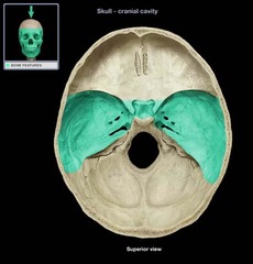
Middle cranial fossa

formed by part of the sphenoid bone and the temporal bone
<p>formed by part of the sphenoid bone and the temporal bone</p>
31
New cards

Orbital surface (sphenoid bone)

posterior surface of the orbit

<p>posterior surface of the orbit</p>
32
New cards

Lateral pterygoid plates

project inferiorly from the greater wings; attachment site for chewing muscles, lateral

<p>project inferiorly from the greater wings; attachment site for chewing muscles, lateral</p>
33
New cards

Medial pterygoid plates

project inferiorly from the greater wings; attachment site for chewing muscles, medial

<p>project inferiorly from the greater wings; attachment site for chewing muscles, medial</p>
34
New cards

Hamulus

a hook-like end that serves as a site for muscle attachment located on medial pterygoid processes

(name is also in the hand but for skull is "Pterygoid hamulus")

<p>a hook-like end that serves as a site for muscle attachment located on medial pterygoid processes</p><p>(name is also in the hand but for skull is "Pterygoid hamulus")</p>
35
New cards

Ethmoid bone

contributes to the anterior cranial fossa, forms part of the nasal septum and the nasal cavity, contributes to the medial wall of the orbit

has...

- Crista gali

- Cribriform plate

- Orbital plate

- Perpendicular plate

<p>contributes to the anterior cranial fossa, forms part of the nasal septum and the nasal cavity, contributes to the medial wall of the orbit</p><p>has...</p><p>- Crista gali </p><p>- Cribriform plate </p><p>- Orbital plate </p><p>- Perpendicular plate</p>
36
New cards

Crista galli

"rooster's comb"; a superior projection that articulates into the frontal bone and attaches to the dura mater, helping to secure the brain within the skull

<p>"rooster's comb"; a superior projection that articulates into the frontal bone and attaches to the dura mater, helping to secure the brain within the skull</p>
37
New cards

Cribriform plates

located lateral to the crista galli; form a portion of the roof of the nasal cavity and the floor of the anterior cranial fossa

<p>located lateral to the crista galli; form a portion of the roof of the nasal cavity and the floor of the anterior cranial fossa</p>
38
New cards

Orbital plates (ethmoid bone)

lateral surfaces of ethmoid bone that contribute to the medial surfaces of the orbits

<p>lateral surfaces of ethmoid bone that contribute to the medial surfaces of the orbits</p>
39
New cards

Perpendicular plate (ethmoid bone)

inferior projection that forms the superior portion of the nasal septum

<p>inferior projection that forms the superior portion of the nasal septum</p>
40
New cards

Nasal bones

small rectangular bones forming the bridge of the nose

<p>small rectangular bones forming the bridge of the nose</p>
41
New cards

Lacrimal bones

each forms part of the medial orbit in between the maxilla and the ethmoid bone

<p>each forms part of the medial orbit in between the maxilla and the ethmoid bone</p>
42
New cards

Maxillae

keystone facial bones because they articulate with all other facial bones

has...

- Frontal process

- Lacrimal notch

- Zygomatic process

- Alveolar process

- Palatine process

- Sinus

<p>keystone facial bones because they articulate with all other facial bones</p><p>has...</p><p>- Frontal process</p><p>- Lacrimal notch</p><p>- Zygomatic process </p><p>- Alveolar process </p><p>- Palatine process </p><p>- Sinus</p>
43
New cards

Frontal process (maxillae)

forms part of the lateral aspects of the bridge of the nose, articulates with frontal bone

<p>forms part of the lateral aspects of the bridge of the nose, articulates with frontal bone</p>
44
New cards

Zygomatic process (maxillae)

articulation process for zygomatic bone

<p>articulation process for zygomatic bone</p>
45
New cards

Alveolar process (maxillae)

inferior margin of maxillae; contains sockets in which the teeth lie

<p>inferior margin of maxillae; contains sockets in which the teeth lie</p>
46
New cards

Palatine process

forms the anterior hard palate; meet anteriorly in the intermaxillary suture

<p>forms the anterior hard palate; meet anteriorly in the intermaxillary suture</p>
47
New cards

Lacrimal notch (maxilla)

articulation with lacrimal bones

<p>articulation with lacrimal bones</p>
48
New cards

Sinus (maxilla)

large opening in medial portion of maxillae, partially covered by palatine bones

<p>large opening in medial portion of maxillae, partially covered by palatine bones</p>
49
New cards

Zygomatic bone

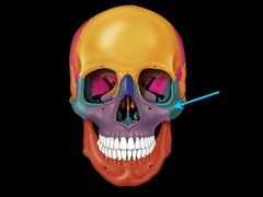
commonly called the cheekbones, each forms part of the lateral orbit

has...

- Frontal process

- Temporal process

- Orbital surface
<p>commonly called the cheekbones, each forms part of the lateral orbit</p><p>has...</p><p>- Frontal process </p><p>- Temporal process </p><p>- Orbital surface</p>
50
New cards

Frontal process (zygomatic bone)

articulates zygomatic bone with frontal bone

<p>articulates zygomatic bone with frontal bone</p>
51
New cards

Temporal process (zygomatic bone)

short extension from the zygomatic bone that forms the anterior portion of the zygomatic arch and articulates with the temporal bone

<p>short extension from the zygomatic bone that forms the anterior portion of the zygomatic arch and articulates with the temporal bone</p>
52
New cards

Orbital surface (zygomatic bone)

lateral surface of eye orbits

<p>lateral surface of eye orbits</p>
53
New cards

Palatine bones

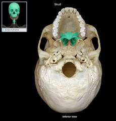
forms the posterior hard palate, a small part of the nasal cavity, and part of the orbit

has...

- Horizontal plate
<p>forms the posterior hard palate, a small part of the nasal cavity, and part of the orbit</p><p>has...</p><p>- Horizontal plate</p>
54
New cards

Horizontal plate (palatine bone)

forms the posterior hard palate

<p>forms the posterior hard palate</p>
55
New cards

Vomer

thin, blade-shaped bone that forms the inferior nasal septum

has...

-Ala

<p>thin, blade-shaped bone that forms the inferior nasal septum</p><p>has...</p><p>-Ala</p>
56
New cards

Ala (vomer)

two wing-like bone extensions on the top border of the vomer bone

<p>two wing-like bone extensions on the top border of the vomer bone</p>
57
New cards

Mandible

the lower jaw bone, which articulates with the temporal bone to form the only freely movable joints in the skull (temporomandibular joints)

has...

- Body

- Angle

- Ramus

- Coronoid process

- Condylar process

- Mandibular condyle

- Alveolar process

- Mental and mandibular foramen

<p>the lower jaw bone, which articulates with the temporal bone to form the only freely movable joints in the skull (temporomandibular joints)</p><p>has...</p><p>- Body </p><p>- Angle </p><p>- Ramus </p><p>- Coronoid process </p><p>- Condylar process </p><p>- Mandibular condyle </p><p>- Alveolar process </p><p>- Mental and mandibular foramen</p>
58
New cards

Mandibular body

horizontal portion that forms the chin

<p>horizontal portion that forms the chin</p>
59
New cards

Mandibular angles

posterior points where the ramus meets the body

<p>posterior points where the ramus meets the body</p>
60
New cards

Mandibular ramus

vertical extensions of the mandibular body

<p>vertical extensions of the mandibular body</p>
61
New cards

Coronoid process (mandible)

"crown-shaped" portion of the ramus for muscle attachment

<p>"crown-shaped" portion of the ramus for muscle attachment</p>
62
New cards

Condylar processes (mandible)

articulate with the mandibular fossa of the temporal bones

<p>articulate with the mandibular fossa of the temporal bones</p>
63
New cards

mandibular condyle

Articulation point of the mandible with the mandibular fossa of the temporal bone

<p>Articulation point of the mandible with the mandibular fossa of the temporal bone</p>
64
New cards

Alveolar process (mandible)

superior margin of the mandible; contains sockets in which the teeth lie

<p>superior margin of the mandible; contains sockets in which the teeth lie</p>
65
New cards

Mental foramina

paired openings on the mandibular body; transmit blood vessels and nerves to the lower lip and skin of the chin

<p>paired openings on the mandibular body; transmit blood vessels and nerves to the lower lip and skin of the chin</p>
66
New cards

Mandibular foramina

located on the medial surface of each ramus; passageway for the nerve involved in tooth sensation

<p>located on the medial surface of each ramus; passageway for the nerve involved in tooth sensation</p>
67
New cards

Hyoid bone

U-shaped bone located in the throat above the larynx; supports the tongue

<p>U-shaped bone located in the throat above the larynx; supports the tongue</p>
68
New cards

Vertebral body

rounded central weight-bearing portion of the vertebra, which faces anteriorly in the human vertebral column

<p>rounded central weight-bearing portion of the vertebra, which faces anteriorly in the human vertebral column</p>
69
New cards

Intervertebral disc

mass of fibrocartilage between adjacent vertebrae

<p>mass of fibrocartilage between adjacent vertebrae</p>
70
New cards

Vertebral arch

composed of two pedicles and two laminae

<p>composed of two pedicles and two laminae</p>
71
New cards

Vertebral pedicle

project posteriorly from the body of the vertebra and provide side protection from the spinal cord and nerves

<p>project posteriorly from the body of the vertebra and provide side protection from the spinal cord and nerves</p>
72
New cards

Vertebral lamina

flattened posterior portion of the vertebral arch

<p>flattened posterior portion of the vertebral arch</p>
73
New cards

Vertebral foramen

opening enclosed by the body and vertebral arch; a passageway for the spinal cord

<p>opening enclosed by the body and vertebral arch; a passageway for the spinal cord</p>
74
New cards

Intervertebral foramen

lateral opening formed from superior and inferior notches on the pedicles that allow for spinal nerves to leave the spinal cord

<p>lateral opening formed from superior and inferior notches on the pedicles that allow for spinal nerves to leave the spinal cord</p>
75
New cards

Transverse processes

two lateral projections from the vertebral arch

<p>two lateral projections from the vertebral arch</p>
76
New cards

Spinous process

single medial and posterior projection formed at the junction of the two laminae

<p>single medial and posterior projection formed at the junction of the two laminae</p>
77
New cards

Superior articular process

knowt flashcard image
78
New cards

Inferior articular process

knowt flashcard image
79
New cards

Superior and inferior articular facets

smooth, cartilage-coated surfaces on the bony projections (processes) of adjacent vertebrae that lock together to form facet joints

Think of your spine like a stack of Lego bricks.

Articular facet just means a smooth, flat "kissing" spot where one bone touches another bone.

Superior means top (facing up).

Inferior means bottom (facing down).

How They Fit Together

The inferior articular facet of a vertebra on top points downward.

The superior articular facet of the vertebra right below it points upward.

They click or stack together like puzzle pieces or small hinges.

<p>smooth, cartilage-coated surfaces on the bony projections (processes) of adjacent vertebrae that lock together to form facet joints</p><p>Think of your spine like a stack of Lego bricks.</p><p>Articular facet just means a smooth, flat "kissing" spot where one bone touches another bone.</p><p>Superior means top (facing up).</p><p>Inferior means bottom (facing down).</p><p>How They Fit Together</p><p>The inferior articular facet of a vertebra on top points downward.</p><p>The superior articular facet of the vertebra right below it points upward.</p><p>They click or stack together like puzzle pieces or small hinges.</p>
80
New cards

Cervical Region has...

Transverse foramen

Dens

Bifed

81
New cards

Transverse foramen

only found in the cervical vertebrae and allow passage of the vertebral artery, vein, and nerve

<p>only found in the cervical vertebrae and allow passage of the vertebral artery, vein, and nerve</p>
82
New cards

Dens

process only in C2 (axis) that allows rotation of C1 (atlas)

cervical region

<p>process only in C2 (axis) that allows rotation of C1 (atlas)</p><p>cervical region</p>
83
New cards

Bifid

forked process found on C5 that locks into spinous process of C6

cervical region

<p>forked process found on C5 that locks into spinous process of C6</p><p>cervical region</p>
84
New cards

Transverse facet

facets found on transverse processes of thoracic vertebrae for articulation with tubercle of ribs

<p>facets found on transverse processes of thoracic vertebrae for articulation with tubercle of ribs</p>
85
New cards

Costal facets

facets lateral and posterior to the body of the vertebrae for articulation with head of ribs

<p>facets lateral and posterior to the body of the vertebrae for articulation with head of ribs</p>
86
New cards

Sacral foramina (superior and inferior)

openings found on either side of the sacral crest that allow for passage of blood vessels and nerves

<p>openings found on either side of the sacral crest that allow for passage of blood vessels and nerves</p>
87
New cards

Sacral crest

formed by fused spinous processes and run down center of posterior sacrum

<p>formed by fused spinous processes and run down center of posterior sacrum</p>
88
New cards

Sacral canal

continuation of the vertebral canal in the sacrum, ending at sacral hiatus

<p>continuation of the vertebral canal in the sacrum, ending at sacral hiatus</p>
89
New cards

Sacral promontory

anterior border of S1 in the sacrum

<p>anterior border of S1 in the sacrum</p>
90
New cards

cervical region

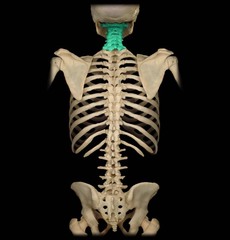
the first 7 vertebrae, comprising the neck

has axis and atlas

c1-c7
<p>the first 7 vertebrae, comprising the neck</p><p>has axis and atlas</p><p>c1-c7</p>
91
New cards

thoracic region

The thoracic region of the human spine contains 12 vertebrae, labeled T1 through T12.

<p>The thoracic region of the human spine contains 12 vertebrae, labeled T1 through T12.</p>
92
New cards

lumbar region

L1-L5

<p>L1-L5</p>
93
New cards

sacral region

The sacral region consists of five individual vertebrae (S1 through S5) that fuse together into a single, wedge-shaped bone called the sacrum during late adolescence and early adulthood (typically by age 30).

Situated at the base of the spine, slotted between the two hip bones (ilia) in the posterior pelvis.

<p>The sacral region consists of five individual vertebrae (S1 through S5) that fuse together into a single, wedge-shaped bone called the sacrum during late adolescence and early adulthood (typically by age 30). </p><p>Situated at the base of the spine, slotted between the two hip bones (ilia) in the posterior pelvis.</p>
94
New cards

Coccyx

four vertebrae fused together to form the tailbone

<p>four vertebrae fused together to form the tailbone</p>
95
New cards

atlas

C1

<p>C1</p>
96
New cards

axis

c2

<p>c2</p>
97
New cards

cervical vertebrae

knowt flashcard image
98
New cards

thoracic vertebrae

Heart-Shaped Body: The main weight-bearing body is roughly heart-shaped, larger than cervical vertebrae but smaller than lumbar vertebrae.

Costal Facets / Demifacets: Smooth articular surfaces on the sides of the vertebral body where the heads of the ribs attach.

Transverse Costal Facets: Found on the transverse processes of vertebrae T1 through T10 to articulate with rib tubercles.

Long Spinous Processes: The spinous processes are long, slender, and point sharply downward/inferiorly.

No Transverse Foramina: Unlike cervical vertebrae, thoracic vertebrae do not have holes in their transverse processes

<p>Heart-Shaped Body: The main weight-bearing body is roughly heart-shaped, larger than cervical vertebrae but smaller than lumbar vertebrae.</p><p>Costal Facets / Demifacets: Smooth articular surfaces on the sides of the vertebral body where the heads of the ribs attach.</p><p>Transverse Costal Facets: Found on the transverse processes of vertebrae T1 through T10 to articulate with rib tubercles.</p><p>Long Spinous Processes: The spinous processes are long, slender, and point sharply downward/inferiorly.</p><p>No Transverse Foramina: Unlike cervical vertebrae, thoracic vertebrae do not have holes in their transverse processes</p>
99
New cards

lumbar vertebrae

Location: Directly below the rib cage (thoracic spine) and above the triangular sacrum at the base of the spine.

Curve: Features a natural inward curvature known as a lordotic curve (shaped like a backward "C").

Structure: Consists of a large, kidney-shaped vertebral body, a protective bony arch (laminae and pedicles), a triangular spinal foramen, and sturdy spinous and transverse processes.

<p>Location: Directly below the rib cage (thoracic spine) and above the triangular sacrum at the base of the spine. </p><p>Curve: Features a natural inward curvature known as a lordotic curve (shaped like a backward "C"). </p><p>Structure: Consists of a large, kidney-shaped vertebral body, a protective bony arch (laminae and pedicles), a triangular spinal foramen, and sturdy spinous and transverse processes.</p>
100
New cards

Vertebrosternal ribs

ribs 1-7, connect directly to sternum by their own costal cartilages

<p>ribs 1-7, connect directly to sternum by their own costal cartilages</p>



