biochem 1 - week 1/2
 Call Kai
 Learn
 Practice Test
 Spaced Repetition
 Match
 Flashcards
 Knowt Play
1/51

Earn XP
Description and Tags
cells and transport
52 Terms
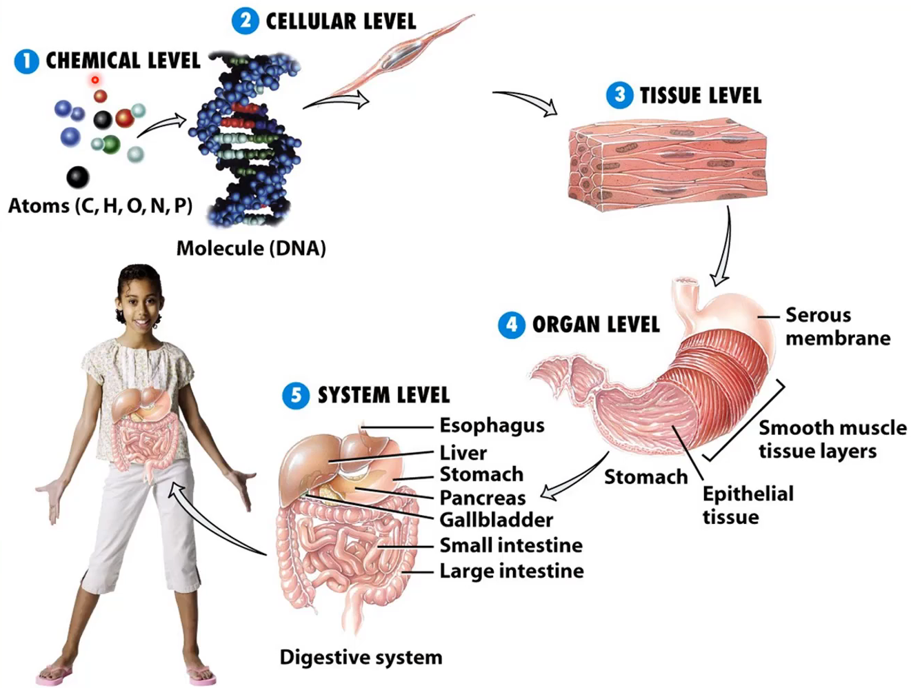
Multicellular organisms and levels
chemical → cellular → tissue → organ → system
Cell theory
All organisms are made up of one or more cells
Cells are the basic units of structure and function in all organisms
All cells come from pre-existing cells
Viruses are not organisms or cells. Why?
Cannot replicate itself
Needs a host cell for making virus particles
Has either DNA or RNA whereas cells have both DNA and RNA
Basic cell functions
Cells separate their internal environment from their external environment to maintain homeostasis (plasma membrane)
Cell must perform the chemical processes of life (cytoplasm)
Cells must store information and pass it to the next generation (DNA)
Cells must be able to build proteins (ribosomes)
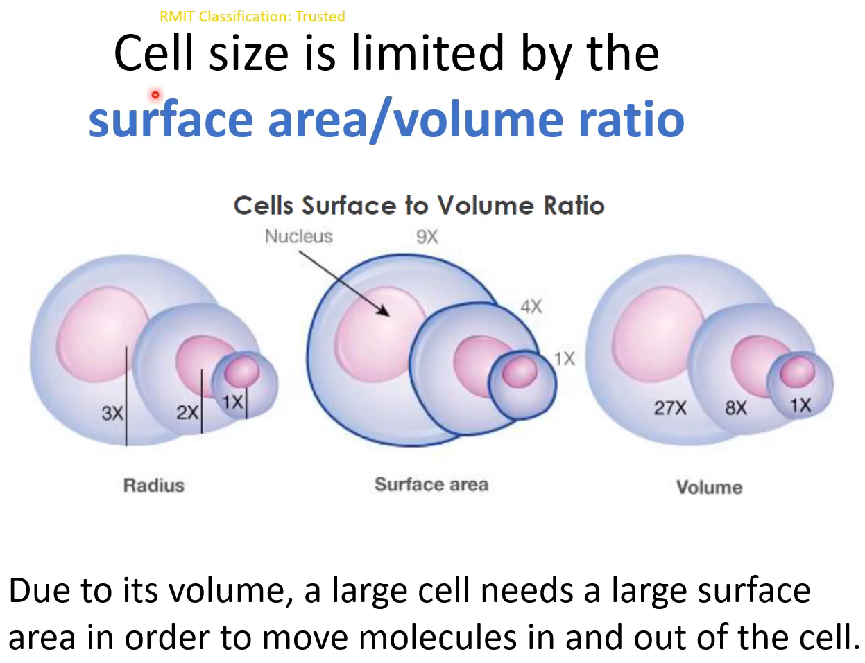
Cells: Surface area to volume ratio
types of cells
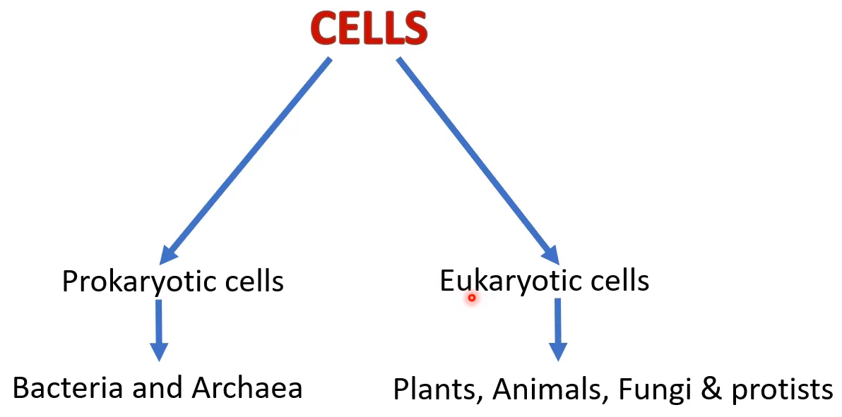
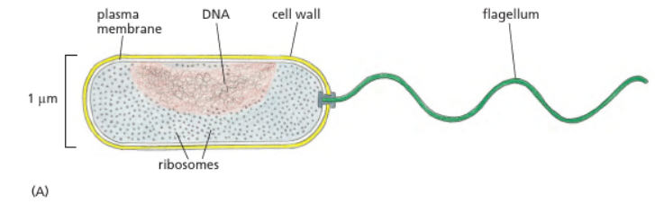
Prokaryotic cells
Unicellular organisms with no true nucleus or membrane-bound organelles
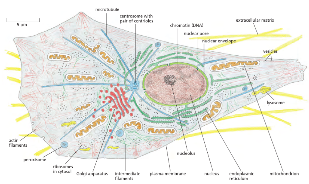
Eukaryotic cells
Have a true nucleus and membrane-bound organelles
Endosymbiosis
Most biologists believe mitochondria and chloroplast developed from prokaryotic cells. This is because:
Both mitochondria and chloroplasts have their own circular DNA and they are similar to the prokaryotic plasmids
They both have ribosomes and the ribosomal structure is more similar to the prokaryotic ribosomes
Their size is similar to prokaryotic cell size
They can reproduce autonomously
nucleus
Information centre of the cell
DNA is stored here
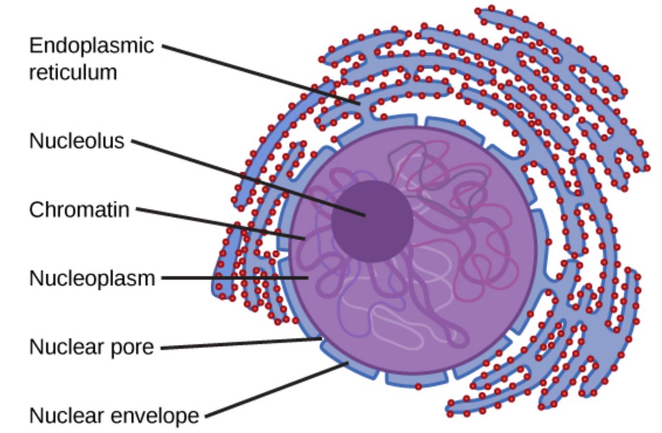
Enclosed by nuclear envelopes formed by 2 membranes called the inner and outer membranes
Nuclear envelopes have pores. Molecules cross between the cytoplasm and the nucleus through the nuclear pores
The inner region of the nuclear envelope is covered by a rope-like structural protein network called nuclear lamina. The nuclear lamina is an intermediate filament (one of the cytoskeleton organelles)
DNA and proteins (histones) are tightly packed to form chromatin fibres. These are tightly packed, coiled, condensed and visible during mitosis. The condensed form being chromosomes
The nucleolus is responsible for synthesising ribosomal RNA and assembling the proteins that are required to form ribosomes
mitochondria
Involved in cellular respiration
Cellular respiration provides chemical energy in the form of ATP
Similar in size to bacteria
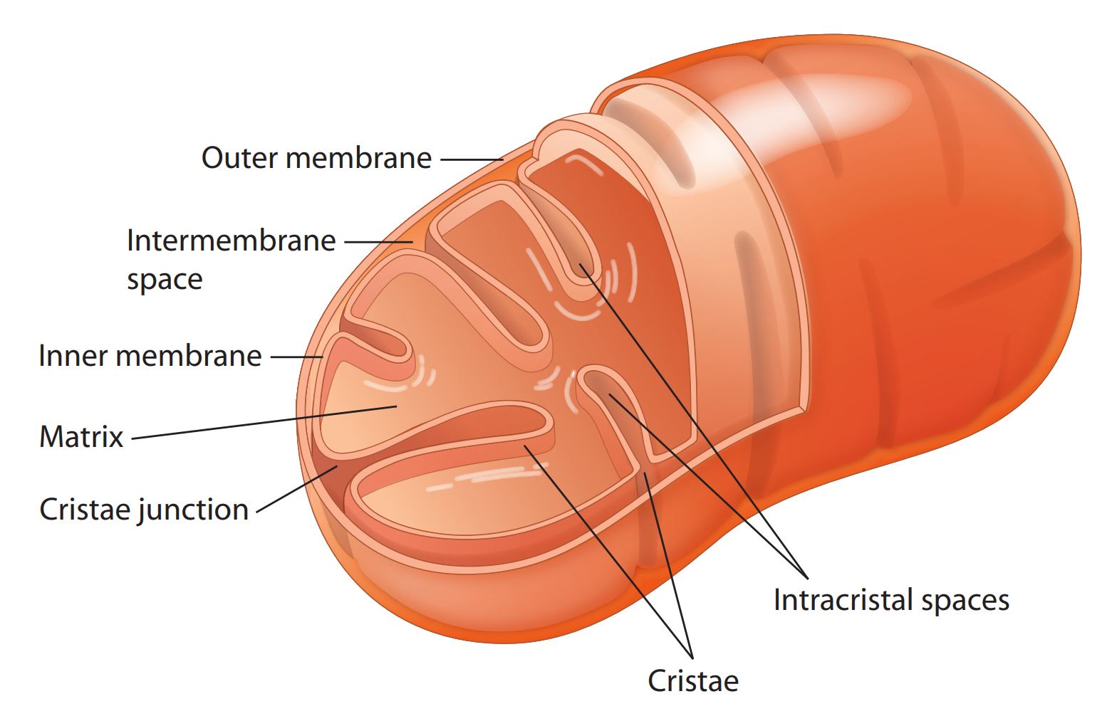
Surrounded by double membranes - the inner membrane folds inwards and forms cristae which provide more space (surface area) for a series of chemical reactions to occur to generate ATP
Mitochondria contain circular DNA, RNA and ribosomes
Mitochondrial DNA is inherited only from the mother to the child
endomembrane system
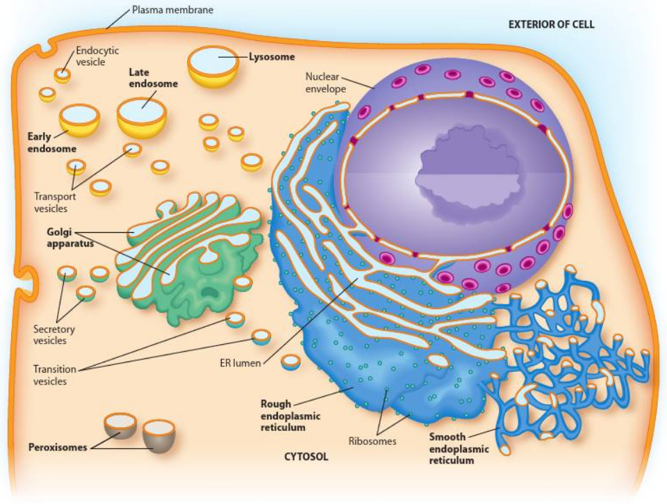
Includes the endoplasmic reticulum (ER), Golgi apparatus and transport vesicles (endosomes and lysosomes)
Associated with the outer nuclear envelope and the plasma membrane. The outer nuclear membrane connects with the lumen of the rough ER
Ribosomes are non-membrane-bound organelles that bind to the outer region of the ER membrane and allow proteins to be synthesised within the lumen of the ER
Protein modification and folding also occur here. Proteins are then packaged into vesicles and moved from the ER to the Golgi apparatus
Smooth ER is important for the synthesis of lipids. Lipids are also packaged into vesicles and moved from the ER to the Golgi apparatus
Inside the Golgi apparatus, proteins are lipids undergo further modification before sorting and packaging for dispatch. Molecules packed within the transport vesicles move to the final destination either inside or outside the cell
peroxisomes
Breaking down fatty acids, alcohols and amino acids (cell metabolism)
protect the cell against hydrogen peroxide and some toxic molecules (detoxification)
synthesis of lipid molecules, including cholesterol and bile acids
lysosomes
contain hydrolytic enzymes and are involved in the breaking down of biomolecules such as carbohydrates, proteins, nucleic acids and lipids
responsible for autophagy, the degradation of the cell’s own components
responsible for cell’s digestion
cytoskeleton
inner framework of cells
provides structure and shape of cells
participates in movement
regulates cell division
microtubules (type of cytoskeletal organelle)
made up of tublin proteins
alpha and beta tublin dimers (subunits) are polymerised and form protofilaments
13 protofilaments form a dynamic tube structure which is a microtuble
originate from centresomes
a pair of centrioles if found within the centrosome and the microtuble structure grows from this region
unbranched structures
essential for vesicular transport and cell division
microfilament (type of cytoskeletal organelle)
thinnest molecules and highly dynamic structures
responsible for shape changes, adhesion and cell migration
made of actin molecules
intermediate filaments
bigger than microfilaments and smaller than microtubles
most stable cytoskeleton molecules and involved in structural or tension-bearing roles in animal cells
Keratin is an example mainly found in epithelial tissues and is the main molecule found in hair
Vimentin, another example, is found in connective tissues and mesenchymal cells (non-adherent)
the fluid mosaic model
‘fluid’ because it is a dynamic and fluid structure
lipids and proteins move within the membrane
‘mosaic’ because it comprises phospholipids, proteins, cholesterol and carbohydrates
membrane provides the boundary for organelles
membrane that surrounds the different types of organelles can have different types and amounts of lipids and proteins
semi-permeable and selects the molecules that can cross the boundary
membrane functions
Covers the cell and provides a boundary to the organelles, allowing them to have separate internal environments
protects cellular contents and controls the content of the cytoplasm by regulating which molecules can enter into and exit from the cytoplasm (transportation)
serves as a site for specific biochemical reactions
contain protein receptors, receive signals, transfer messages to cells and involve cell-to-cell and cell-to-extracellular communication and adhesion
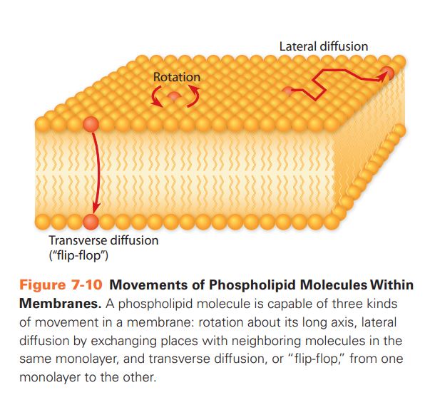
types of lipid movement in plasma membranes
phospholipid (class of membrane lipid)
Most abundant lipids found in the cell membrane
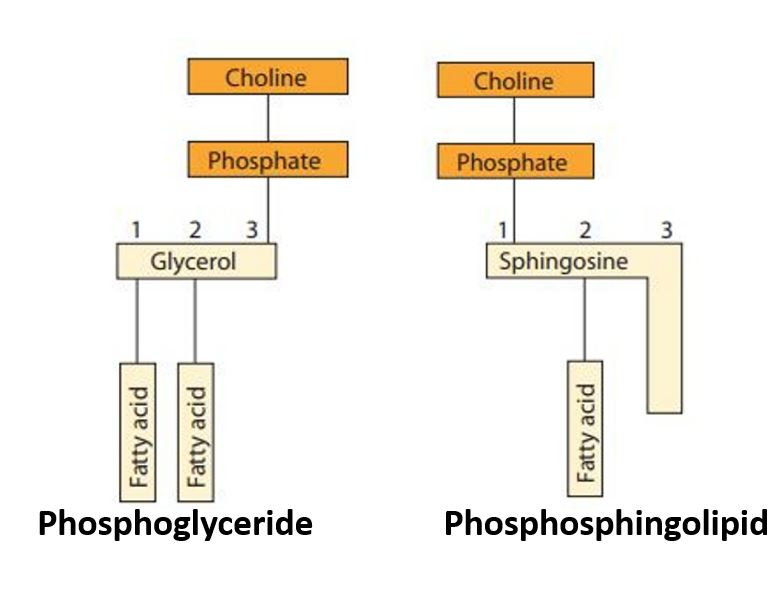
has a glycerol or sphingosine backbone
Phospholipids with glycerol backbones are called phosphoglycerolipids (phosphophingolipids)
Glycerol is a 3-carbon alcohol molecule, and sphingosine is a derivative of the amino acid serine with a fatty acid attached
the backbone is attached to nonpolar fatty acid chains and a negatively charged phosphate molecule
the phosphate group contains a charged or polar head group (e.g. choline)
Amphipathic molecules because they contain a polar head and nonpolar tails (fatty acids)
the nonpolar fatty acid part provides a hydrophobic barrier, and the rest of the molecules which have hydrophilic properties interact with the aqueous environment found both internally and externally
glycolipid (class of membrane lipid)
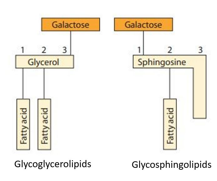
carbohydrates (glyco) are attached to the lipid molecule
have either a glycerol backbone or a spingosine backbone
Depending on the backbone, glycolipids can be further classified as glycoglycerolipids and glycosphingolipids
only found on the outer surface of the plasma membrane, and they are involved in a number of cellular functions, including cell communication, cell recognition, and protection
sterols (class of membrane lipid)
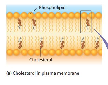
Most eukaryotic cells contain sterols
The main sterol in animal cells is cholesterol, a 4-ring amphipathic molecule
Cholesterol acts as a fluidity buffer, maintains fluidity and stabilises the membrane structure
absent in inner mitochondrial membranes
membrane proteins
plasma membrane contains integral, peripheral and lipid-anchored proteins
play major roles in molecular transport systems, provide structural attachments for the cytoskeleton, serve as enzymes, connect cell junctions and are involved in cell signalling and cell recognition
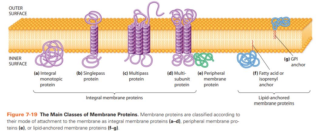
Integral membrane proteins (class of membrane protein)
Amphipathic molecules inserted or embedded in the plasma membrane
Non-polar regions (hydrophobic regions) are embedded within the interior part of the membrane structure
Polar regions (hydrophilic regions) are found outside the lipid bilayer and face the aqueous phases
can be classified as integral monotropic proteins (protein only embedded on one side of the membrane) and transmembrane proteins (protein spanning across the membrane and hydrophilic region protruding from the membrane on both sides)
Some transmembrane proteins cross the membrane once (single-pass), and some several times (multi-pass)
Mutli-pass transmembrane proteins are the gateways to the cell that allow polar molecules, ions, nutrition, waste products, hormones, drugs and other larger molecules to cross the membrane
hard to separate from the membrane due to the strong hydrophobic affinity towards the bilayer; hence, strong detergents are required to solubilise and isolate the integral membrane proteins
peripheral membrane proteins (class of membrane protein)
found on the exterior or interior surfaces of the membrane and are loosely attached to either integral membrane proteins or phospholipid molecules
does not penetrate the lipid bilayer
Provide structural support to the cell by connecting the cytoskeleton and extracellular matrix
Carry out enzyme activities that are involved in cell communication
interact via weak electrostatic forces and hydrogen bonds, thus can be easily separated from the membrane by altering the pH or ionic strength
lipid-anchored membrane proteins (class of membrane protein)
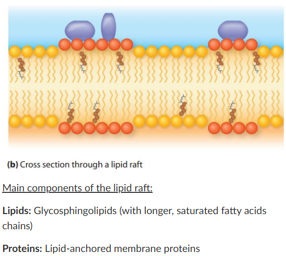
located on one of the surfaces of the lipid bilayer and are covalently attached to the lipid molecules, such as glycosphingolipids
Some regions of the plasma membrane contain a higher number of glycospingolipids and cholesterol; these regions are called lipid rafts and are found outside
Because the fatty acid chains found in sphingolipids are longer, the membrane region is raised out
Lipid rafts contain lipid-anchored membrane proteins that are involved in cell signalling
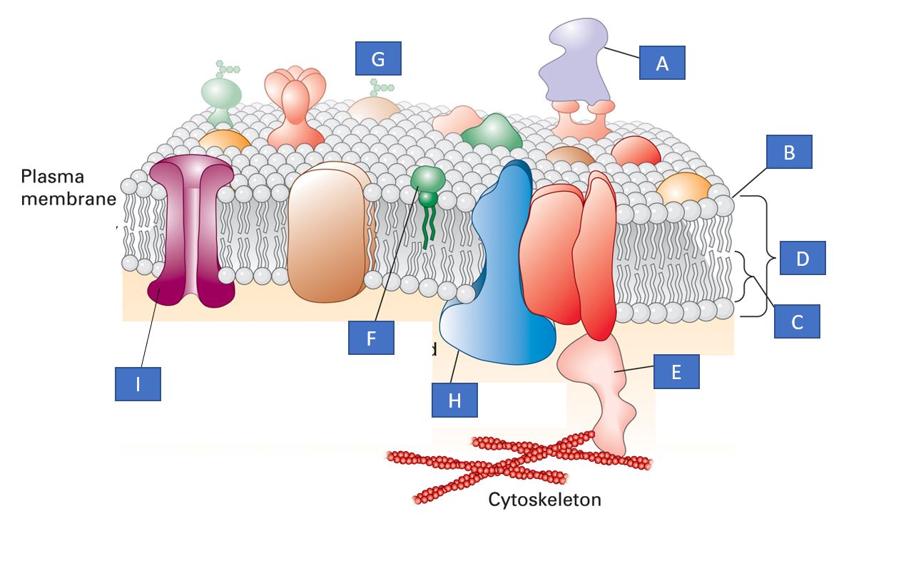
Label the diagram
A and E are peripheral membrane proteins
B is the hydrophilic phospholipid head
C is the hydrophobic fatty acid chain of the phospholipid molecule
D is the phospholipid bilayer
F is a lipid-anchored membrane protein
G is a glycoprotein (found outside the cell and involved in cellular communication)
H and I are transmembrane proteins
passive transport
When molecules cross the cell membrane from a region of higher concentration to one of lower concentration, energy is not required
Diffusion is a passive transport mechanism that will occur until the concentration on both sides of the semipermeable membrane reaches equilibrium
Sometimes, water crosses the membrane from a lower concentration to a higher concentration to reach equilibrium, this is because charged molecules do not normally cross the cell membrane, which is called osmosis
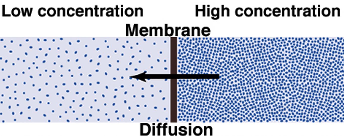
simple diffusion (passive transport)
the unassisted net movement of a solute from the higher concentration region to the lower concentration region
does not require energy and occurs until the concentration on both sides becomes equal (until it reaches equilibrium)
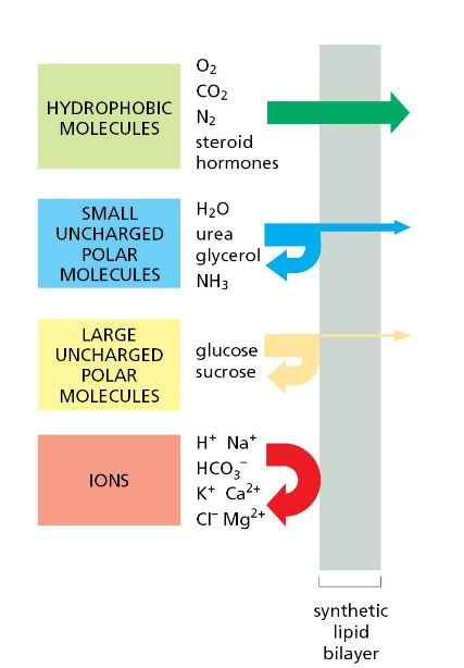
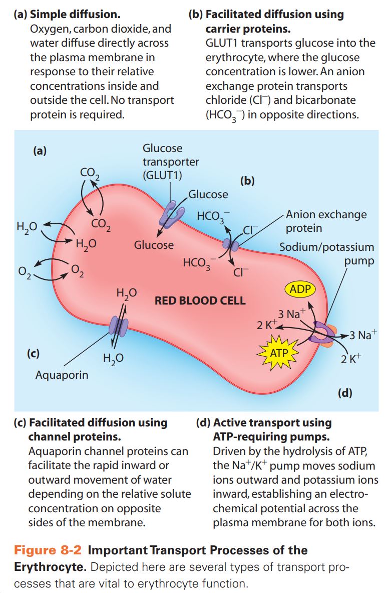
small hydrophobic (non-polar) molecules such as oxygen and carbon dioxide can cross the membrane very quickly (green arrow in the figure)
Some molecules, such as charged molecules (e.g. sodium ions, potassium ions and chloride ions), are impermeable to the lipid membrane (red arrow in the figure)
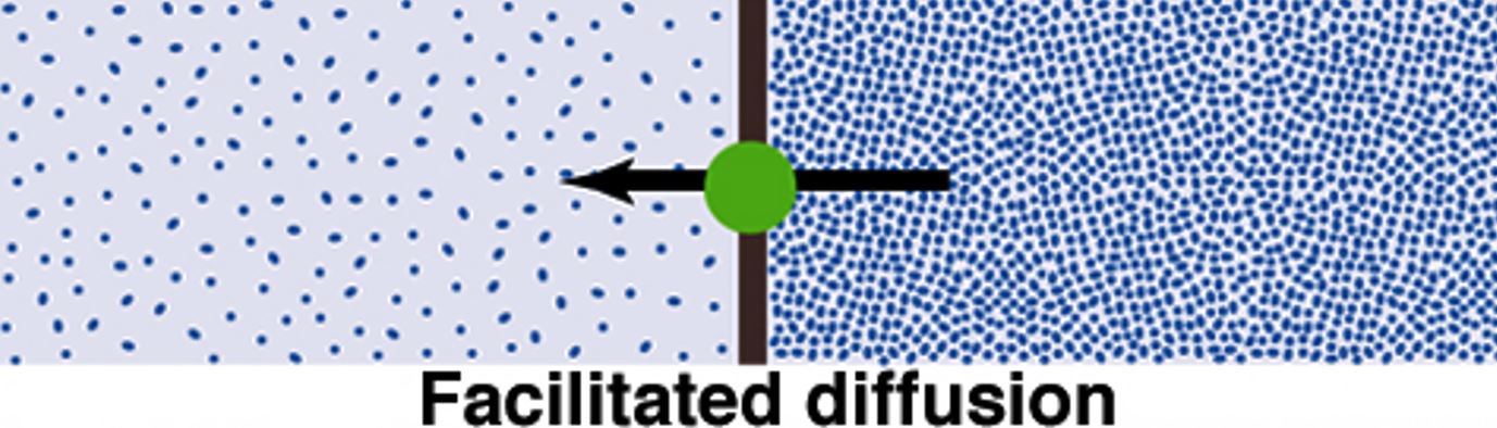
facilitated diffusion
Some molecules and ions (e.g. sodium ions, potassium ions and chloride ions) are impermeable to the lipid membrane
These molecules and ions can cross the biological membrane via transmembrane proteins
If these molecules and ions move from a higher concentration to a lower concentration, they do not need energy
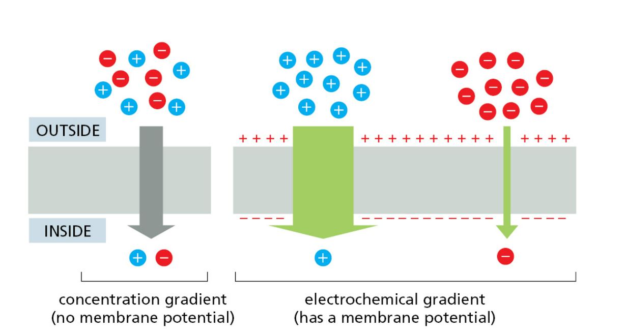
The rate depends on the molecules’ concentration and electrochemical gradient as polar uncharged molecules move entirely on the concentration gradient, but for charged molecules, the concentration gradient and electrochemical gradient drive movement (see figure)
transporter-mediated and channel-mediated
These are two types of transmembrane proteins that regulate the facilitated diffusion process
Transporter-mediated diffusion usually transports uncharged polar molecules, whereas channel-mediated mainly transports charged molecules
Water movement is facilitated by a special type of channel protein called aquaporin
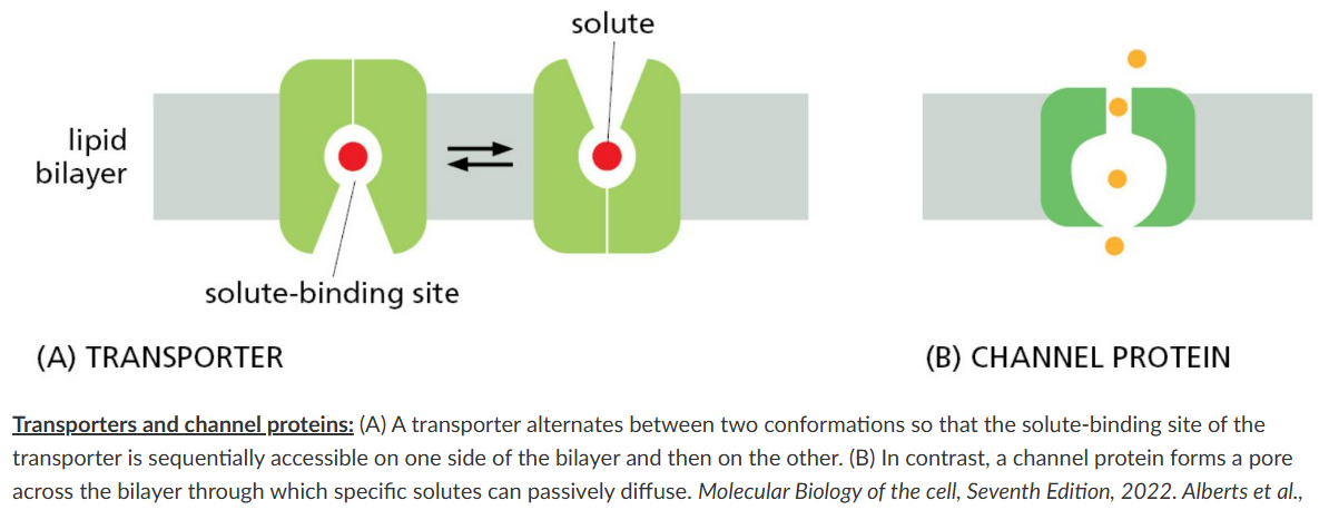
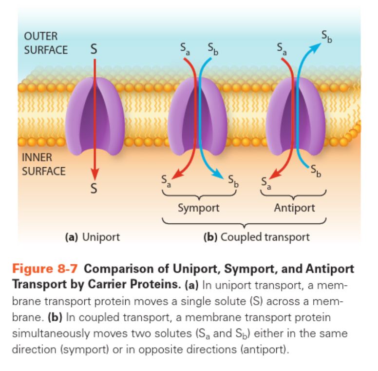
uniport (type of transporter)
a single substance is moved in one direction (e.g. the glucose transporter (GLUT) transports glucose into the cell via facilitated diffusion)
co-transport/coupled transport (type of transporter)
two types of substances are moved. This can be symport (two different substances move in the same direction) or antiport (two different substances move in the opposite direction)
An anion exchanger (Cl-/HCO3- exchanger) is an example of a coupled transporter (passive and facilitate diffusion)
Coupled transporters are also involved in the active transport process
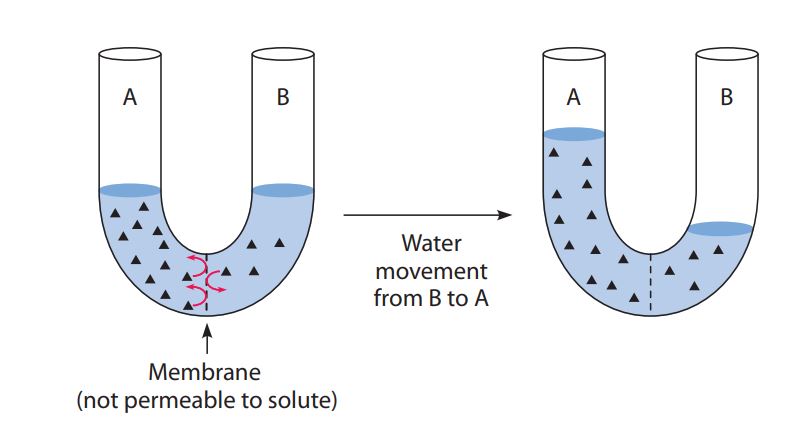
osmosis
Water moves across the membrane in response to the differences in solute concentration on both sides, as seen in the figure
Water movement occurs from the lower concentrated solution to the higher concentration to equalise the solute concentration
In the cell, the osmotic movement of water is related to the relative osmolarity (total solute concentration) of the cytoplasm vs the extracellular solution
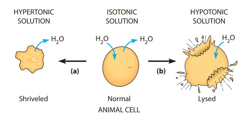
Relative osmolarity: tonicity
Hypertonicity: Higher concentration of a solution with non-permeable solutes outside the cell. Water leaves the cell, and the cell shrinks
Hypotonicity: Lower concentration of a solution with non-permeable outside the cell. Water enters the cell, and the cell swells and may burst (if animal cell)
Isotonicity: Same concentration of a solution. No net movement of water
active transport
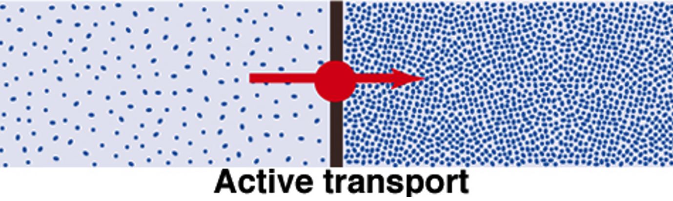
When molecules cross the membrane against the concentration gradient, the cell uses energy
moves the solute away from the equilibrium (opposite of diffusion)
an uphill transport system that transports large, polar molecules or ions, which requires a transporter and energy
active transport functions
Allows the uptake of essential nutrients from the extracellular fluid even though their concentration inside the cell is already high
Allows waste materials to be removed from the cell even when the concentration outside the cell is higher than inside
It helps the cell to maintain constant nonequilibrium intracellular concentrations of specific inorganic ions such as Na+, K+ and Ca2+
direct (primary) active transport (type of active transport)
occurs against the electrochemical gradient and is mainly regulated by ATP hydrolysis (ATP hydrolysis transporters include P-type, V-type, F-type and ABC-type transporters)
ion pumps are the most important primary active ATP hydrolysis transporters. They contain ATPase, which hydrolyses the ATP and provides the energy to the pump to transport the specific ions.
Sodium, potassium, calcium and magnesium ions are transported through the membrane against the concentration gradient using the primary active transport mechanism
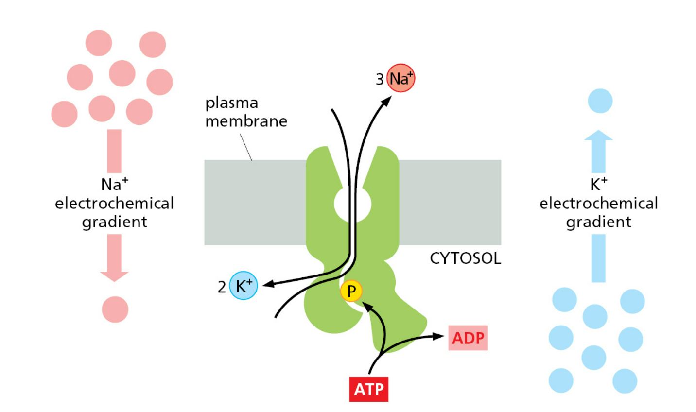
a sodium/potassium (Na+/K+) pump of a P-type pump (a P-type ATPase). This maintains the electrochemical potential of sodium and potassium ions across the plasma membrane in animal cells.
The diagram illustrates how the Na+/K+ pump, a P-type ATPase (transmembrane proteins drawn in green), transports Na+ outside of the cell and K+ inside the cell using the energy generated by ATP hydrolysis
Indirect (Secondary) Active Transport (type of active transport)
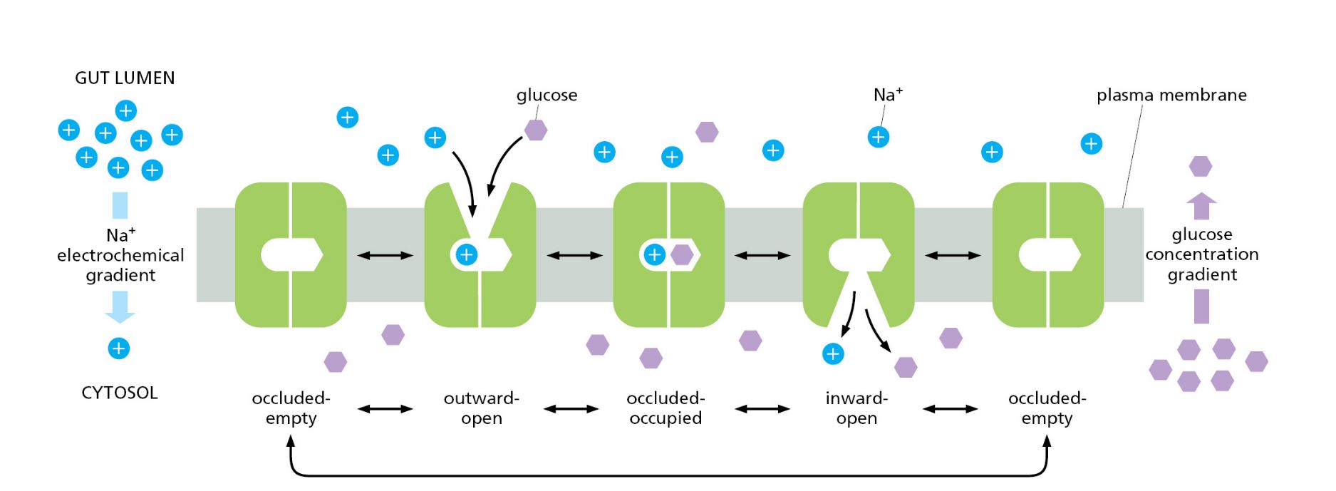
The transporter proteins couple the movement of ions down the concentration gradient and the movement of another molecule against the concentration gradient
The energy stored in the electrochemical gradient of an ion when it moves toward the concentration gradient (downhill) is used to transport another molecule against the concentration gradient (uphill)
In animal cells' plasma membrane, Na+ is the usual ion that moves downhill (outside to inside) towards the electrochemical gradient. It provides energy to drive the second molecule against the concentration gradient (uphill). This means the electrochemical gradient offers a large driving force for the active transport of a second molecule
The figure illustrates Na+/Glucose couple transport (symport). Na+ moves towards the electrochemical gradient (downhill) and provides energy to drive glucose together (uphill or against the concentration gradient)
bulk transport
Large particles or quantities of smaller particles use this mechanism to cross the membrane using energy (active transport)
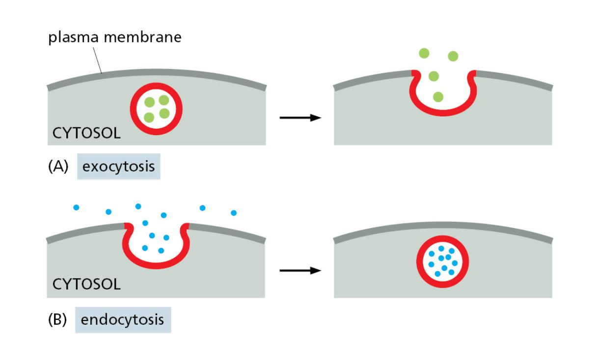
uses the membrane to enclose the molecules, form vesicles, and cross the boundary by fusing with the membrane (exocytosis) or pinching off from the membrane (endocytosis)
endocytosis (type of bulk transport)
transports larger molecules or large quantities of smaller molecules inward (inside the cell) by enclosing them
when the particles reach the surface of the plasma membrane, the membrane folds inwards and forms a bud (pocket) around the particles. The bud then pinches off and lets the particles trapped within the vesicles
phagocytosis (type of endocytosis)
Large particules such as cells, cellular debris, or some pathogens can be engulfed by the cell
Once the cell engulfs the target particle, the pocket pinches off from the membrane and forms a membrane-bound compartment called the vesicle or vacuole
Later, this compartment fuses with a lysosome as they possess enzymes that help break down the particle into simple molecules such as amino acids and sugars
pinocytosis (type of endocytosis)
extracellular fluid and solutes dissolved in that fluid enter the cell by endocytosis
occurs continuously
Some nutrients in the surroundings may be taken up into the cell
receptor-mediated endocytosis (type of endocytosis)
Receptor proteins (transmembrane proteins) on the cell membrane interact with the specific target molecules, and then the membrane folds inwards, forms a bud and pinches off from the membrane
Takes large amounts of specific molecules from the surroundings that are relatively low concentrations in the extracellular
Cells take cholesterol (which is carried by LDL) by this process. Some unwanted particles, such as viruses, also use this mechanism to enter the cell
exocytosis (type of bulk transport)
Cells release molecules such as extracellular particles, proteins, mucus and waste products. These molecules are packed in vesicles, and the vesicle fuses with the plasma membrane, and the particle is released into the external environment
constitutive exocytosis (type of exocytosis)
supplies the plasma membrane with newly made lipids and proteins. It enables the cell to expand before cell division and replace old proteins and lipids
It also carries soluble proteins for secretion
Entry into the constitutive pathway does not require a signal and operates continually
regulated exocytosis (type of exocytosis)
operates in cells that are specialised for secretion (e.g. hormones and mucus)
a signal stimulates the vesicles to fuse with the plasma membrane and release their contents
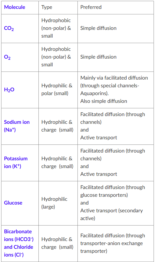
molecules and preferred transport system
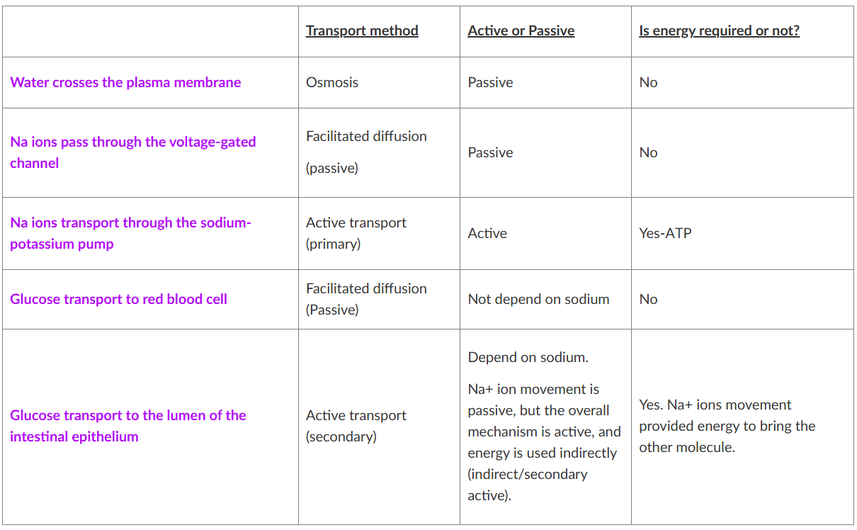
transport mechanisms
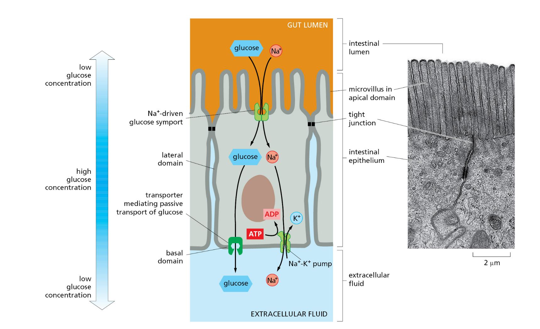
transport mechanisms: glucose transport from the lumen of the intestine to the blood
illustrates the cellular transport of glucose across an intestinal epithelial cell
Explain glucose transport from the intestinal lumen to extracellular fluid (glucose moves to the blood)
Glucose is pumped into the cell through the apical domain of the membrane by a Na+ -powered glucose symporter (couple transporter and secondary active)
glucose passes out of the cell (down its concentration gradient) by passive movement through a glucose uniporter in the basal and lateral plasma membrane domains
The Na+ gradient driving the glucose symport is maintained by the Na+/K+ pump in the basal and lateral plasma membrane domains, which keeps the internal concentration of Na+ low