Biology A-Level: Cell Structure
 Call Kai
 Learn
Learn Practice Test
 Spaced Repetition
 Match
Match Flashcards
 Knowt Play
1/46

There's no tags or description
Looks like no tags are added yet.
Name | Mastery | Learn | Test | Matching | Spaced | Call with Kai | Chat |
|---|
No analytics yet
Send a link to your students to track their progress
47 Terms
What is the general method when making temporary preparations of cellular material suitable for viewing with a light microscope?
Obtain a thin sample of the tissue or cells.(Thin samples allow light to pass through the specimen.)
Place the specimen on a clean glass slide.
Add a drop of water or an appropriate solution.
Add a stain if necessary. (Stains increase contrast and make structures easier to see.)
Carefully lower a coverslip over the specimen at an angle.
This helps prevent air bubbles.
Remove excess liquid with filter paper.
Place the slide on the microscope stage.
Begin with the lowest-power objective lens.
Focus using the coarse adjustment, then use the fine adjustment.
Increase magnification if necessary and refocus.
How do you prepare a slide using a liquid specimen?
Add a few drops containing the liquid sample to a clean slide using a pipette.
Lower a coverslip over the specimen and gently press down to remove air bubbles.
Coverslips protect the microscope lens from liquids and help to prevent drying out.
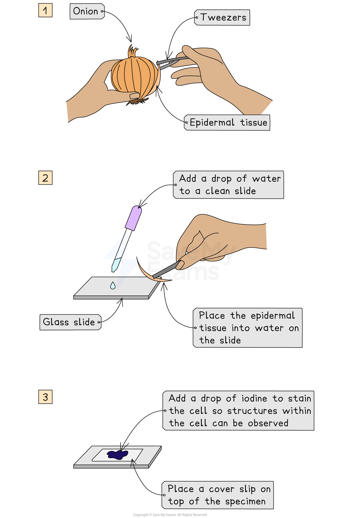
How do you prepare a microscope slide using a solid specimen?
Use scissors or a scalpel to cut a small sample of tissue, and peel away or cut a very thin layer of cells from the tissue sample.
The preparation method always needs to ensure that samples are thin enough to allow light to pass through.
Place the sample onto a slide.
A drop of water may be added at this point
Apply iodine stain.
Gently lower a coverslip over the specimen and press down to remove any air bubbles.

How do you prepare a slide using human cells?
Brush teeth thoroughly with normal toothbrush and toothpaste.
This removes bacteria from teeth so they don't obscure the view of the cheek cells
Take a sterile cotton swab and gently scrape the inside cheek surface of the mouth for 5-10 seconds.
Smear the cotton swab on the centre of the microscope slide for 2-3 seconds.
Add a drop of methylene blue solution.
Methylene blue stains negatively charged molecules in the cell, including DNA and RNA
This causes the nucleus and mitochondria to appear darker than their surroundings
Place a coverslip on top.
Lay the coverslip down at one edge and then gently lower the other edge until it is flat
This reduces bubble formation under the coverslip
Absorb any excess solution by allowing a paper towel to touch one side of the coverslip.
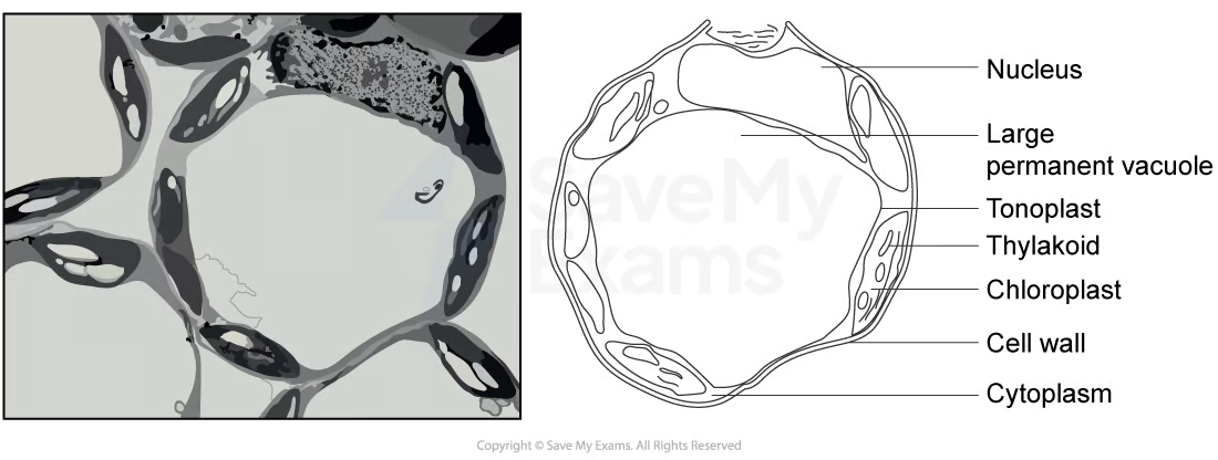
How do we draw draw cells from microscope slides and photomicrographs?
It should be made with a sharp pencil, without shading, with a title, magnification/scale, correct proportions, and neat labels with straight, non-crossing lines, and connect directly to the part of the drawing being labelled.
What is magnification?
The number of times larger an image of an object is than the real size of the object.
What is resolution?
The ability to distinguish between two objects very close together.
What is the difference between resolution and magnification, with reference to light microscopy and electron microscopy?
Magnification is the number of times larger the image is compared with the actual specimen, whereas resolution is the ability to distinguish two points that are close together as separate. Light microscopes have lower magnification and resolution because they use visible light, while electron microscopes have much higher magnification and resolution because they use electrons with a shorter wavelength, allowing smaller cell structures to be distinguished.
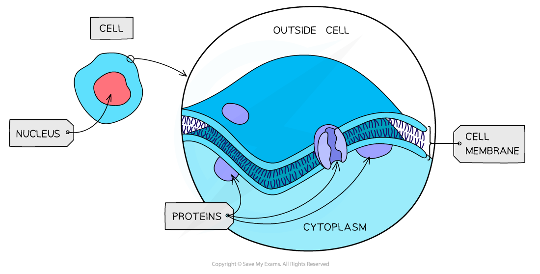
What is the structure of a cell surface membrane?
It has a very thin phospholipid bilayer containing proteins. It forms the outer boundary of the cytoplasm.
What is the function of a cell surface membrane?
Its partially permeable boundary controls the exchange of materials between the internal cell environment and the external environment. It signals cells through receptor proteins, cell recognition, and cell adhesion.
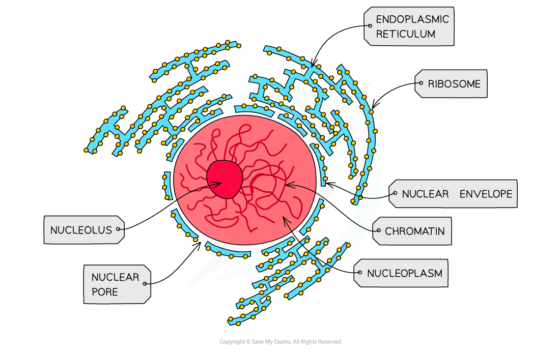
What is the structure of the nucleus, nuclear envelope, and nucleous?
Its usually the largest organelle. It contains chromatin and is surrounded by a double nuclear envelope containing nuclear pores. The outer membrane may be continous with rough ER.
What is the function of the nucleus, nuclear envelope, and nucleous?
It stores DNA, controls cell activities through gene expression, and is the site of DNA replication and transcription.
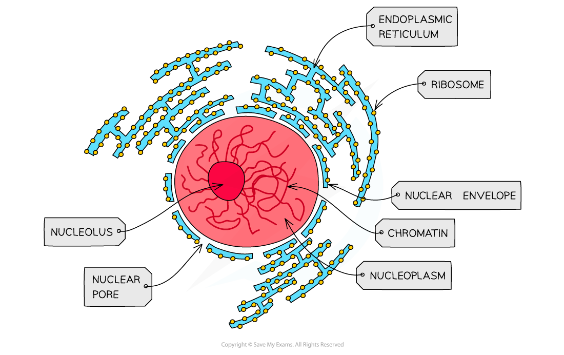
What is the structure of the nucleous?
It is a dense, non-membrane bound region inside the nucleus containing genes for rRNA and associated proteins.
What is the function of the nucleous?
It synthesises rRNA and assembles ribosomal subunits.
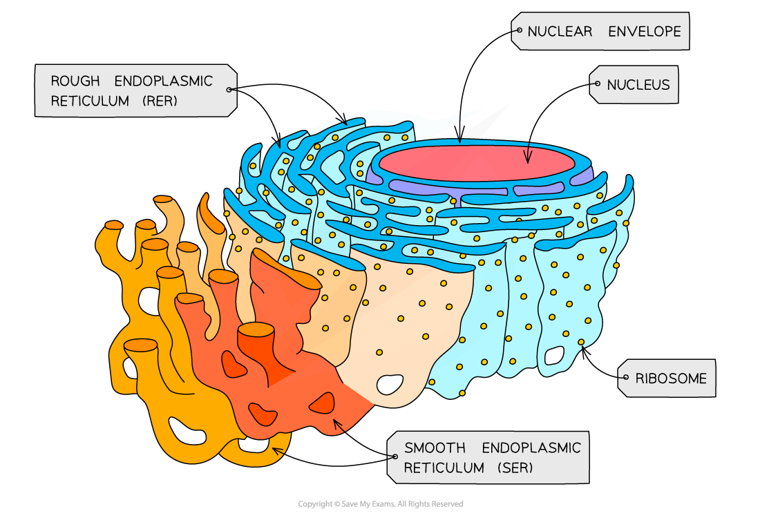
What is the structure of the rough endoplasmic reticulum (RER)?
It is an interconnected system of flattened membrane sacs called cisternae. Ribosomes are attached to the cytosolic surface; its lumen is continuous with the nuclear envelope.
What is the function of the rough endoplasmic reticulum (RER)?
Its role is to process proteins that are produced on the ribosomes. It synthesizes them for secretion, membranes and lysosomes; transports proteins in vesicles to the Golgi Body.
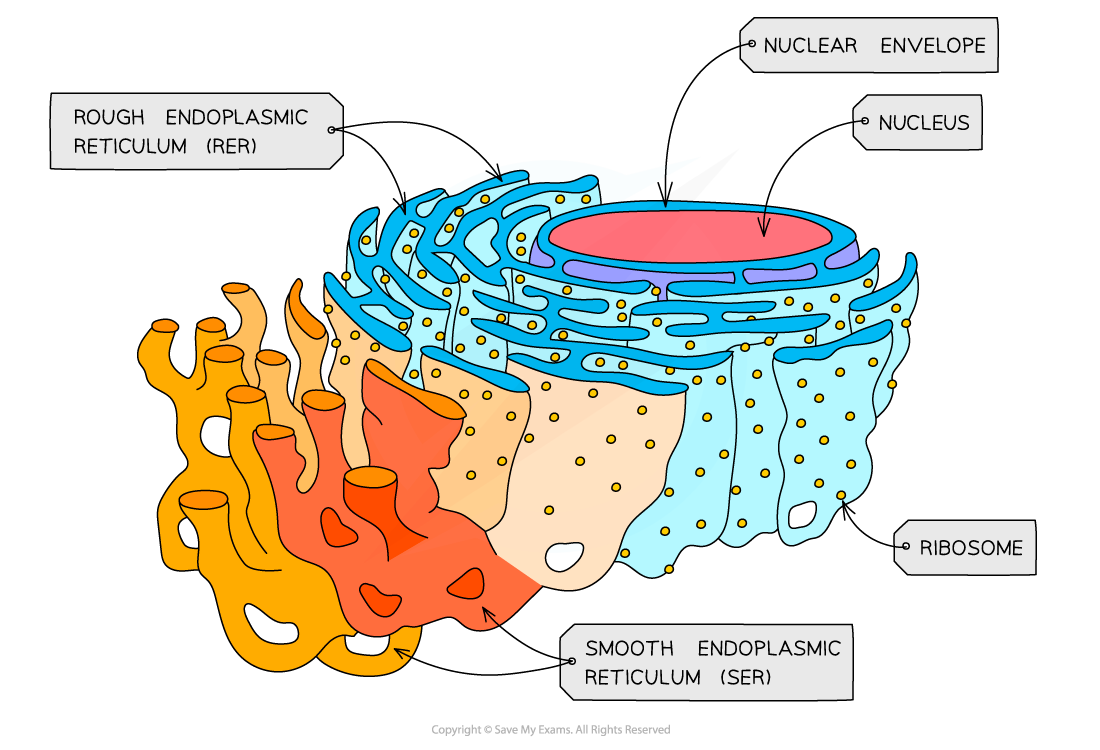
What is the structure of Smooth endoplasmic reticulum (SER)?
It is a branching network of membrane tubules with NO attached ribosomes.
What is the function of the Smooth endoplasmic reticulum (SER)?
It produces lipids, and steroid hormones such as oestrogen and testosterone. It contributes to membrane production, detoxifies substances, and stores and releases calcium ions in specialized cells.
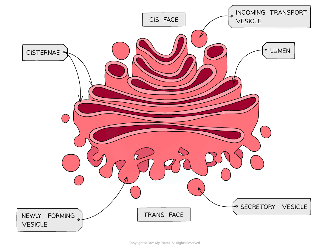
What is the structure of the Golgi Body (the Golgi Apparatus/Golgi Complex)?
A series of flattened, curved membrane bound cisternae with a forming face, recieving vesicles.
What is the function of the Golgi Body?
It modifies proteins and lipids and packages them into vesicles. It produces secretory vesicles & lysosomes and processes polysaccharides in plant cells.
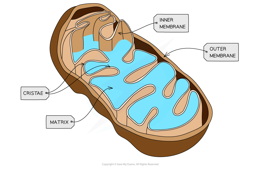
What is the structure of the mitochondria?
It is a double membrane organelle. The inner membrane is folded into cristae and encloses the matrix. The matrix contains enzymes, 70S ribosomes, and small circular DNA.
What is the function of the mitochondria?
Aerobic respiration. Krebs cycle reactions occur in the matrix; oxidative phosphorlyation and ATP synthesis occur on the inner membrane.
Circular DNA and 70S ribosomes allow synthesis of some mitochondria proteins.
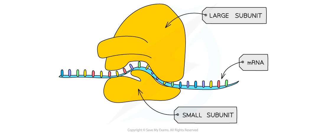
What is the structure of ribosomes?
They are non-membrane-bound particles made of rRNA and protein, with a large and small subunits.
What is the function of the ribosomes?
It is the site for polypeptide or protein synthesis. Free ribosomes mainly produce cytosolic proteins. RER-bound ribosomes produce proteins for secretion, membranes, or lysosomes.
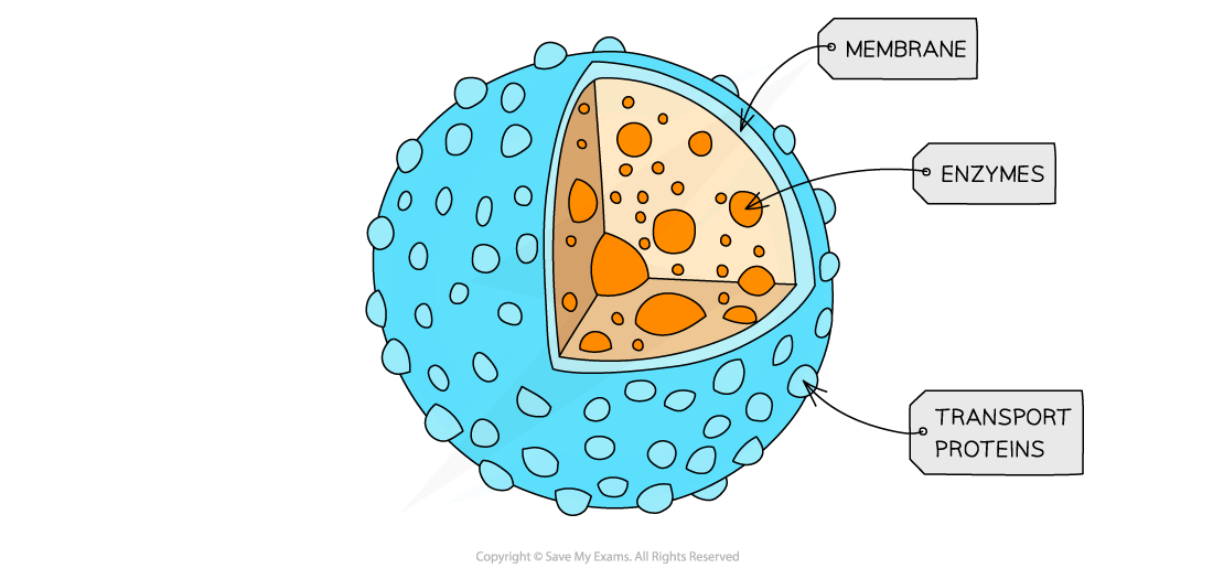
What is the structure of lysosomes?
A small, single-membrane vesicle containing hydrolytic enzymes and maintained at an acidic internal pH.
What are the functions of lysosomes?
It digests material taken in by endocytosis, destroys pathogens in phagocytcic cells, breaks down worn-down organelles by autophagy, and recycles useful monomers.
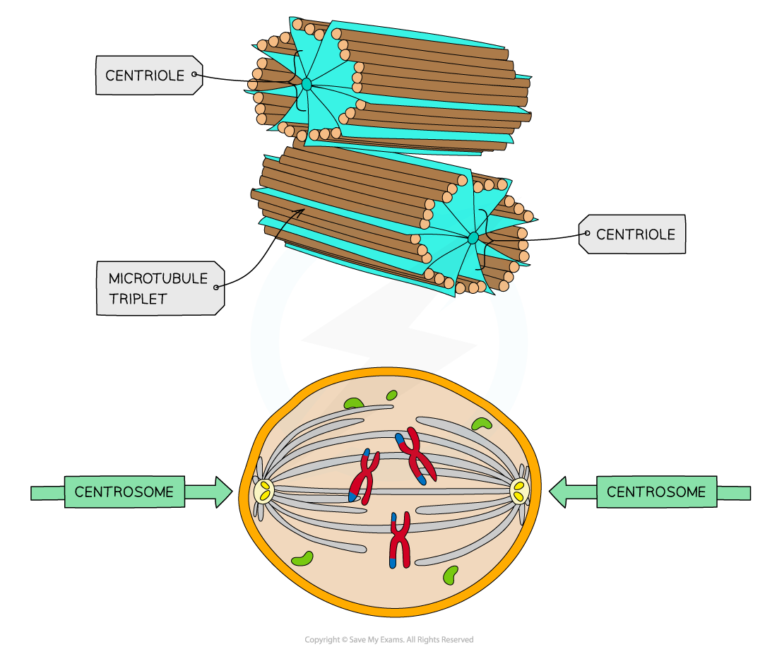
What are the structure of centrioles?
Short cylinders usually present as a perpendicular pair in the centrosome. Each centriole contains nine triplets of microtubules arranged in a ring.
What is the function of centrioles?
They organize microtubules, helping form the spindle during nuclear division and act as basal bodies from which cillia develop.
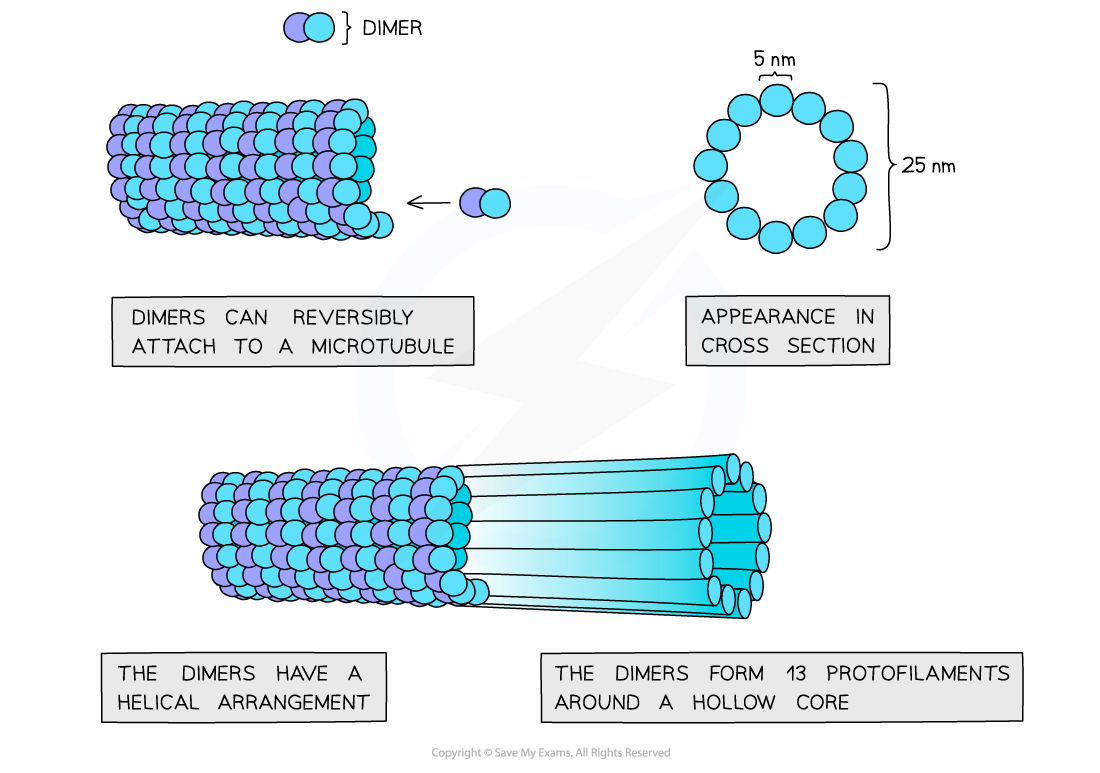
What is the structure of the microtubules?
It is a hollow tube made of tubulin and part of the cytoskeleton. The cytoskeleton is used to provide support and movement of the cell.
What is the function of microtubules?
They maintain cell shape, move vesicles and organelles using motor proteins, seperate chromosomes in the spindle, and form the structural core of cillia.
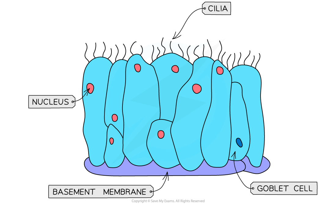
What is the structure of cillia?
They are hair-like projections made from microtubules and covered by the cell membrane.
What is the function of cillia?
They beat rhythmically to allow the movement of substances across the cell surface.
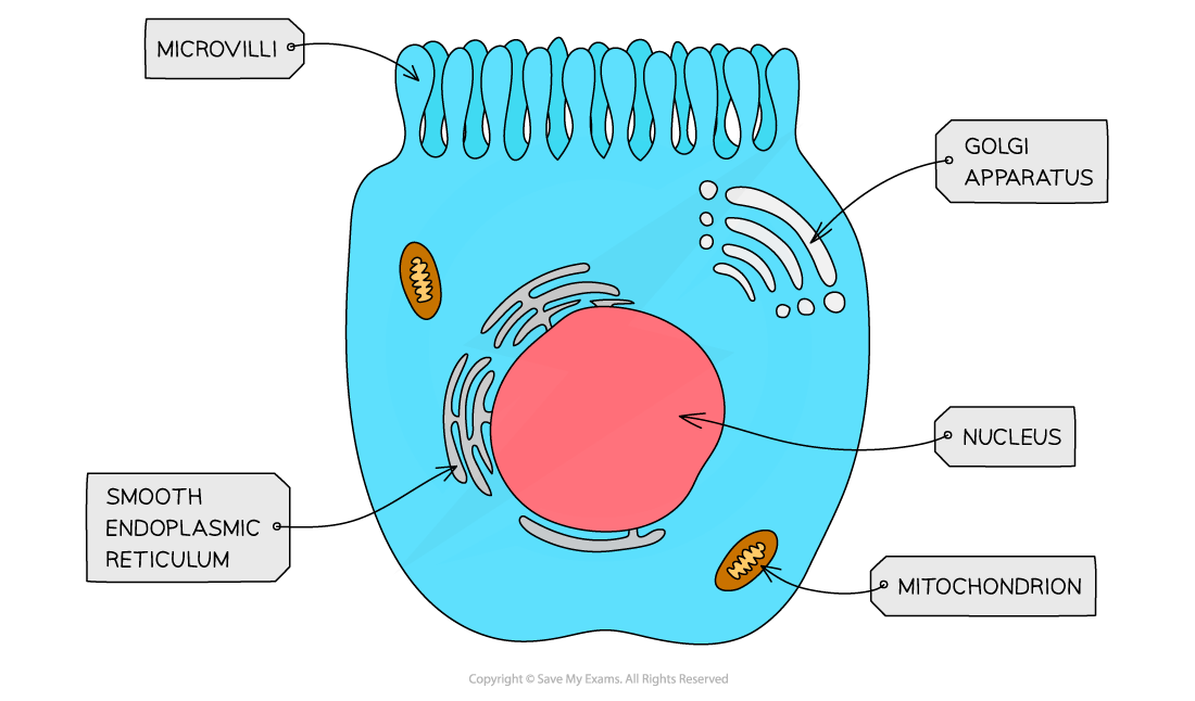
What is the structure of microvilli?
It has numerous narrow folds of the cell surface membrane supported by bundles of actin microfilaments.
What is the function of microvilli?
They increase the surface area for absorption or secretion.
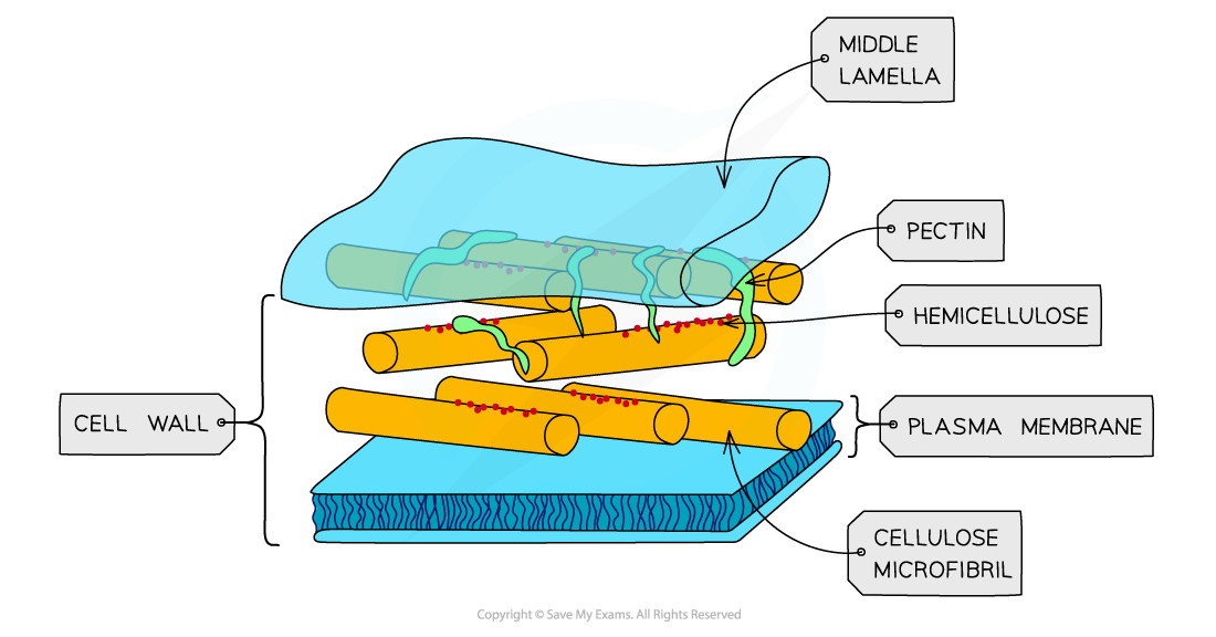
What is the structure of a cell wall?
A freely permeable layer outside the cell surface membrane, composed mainly of cellulose microfibrils embedded in a matrix of other polysaccharides.
What is the function of the cell wall?
It provides mechanical strength, maintains cell shape, prevents bursting when water enters, supports the plant allowing turgor pressure, and permits movement of water through the apoplast.

What is the structure of a chloroplast?
It is a double-membrane organelle containing thylakoid membranes stacked into grana and connected by lamellae, where chlorophyll absorbs light energy for photosynthesis. It also contains circular DNA and ribosomes, which are used to make proteins needed for chloroplast replication and photosynthesis.
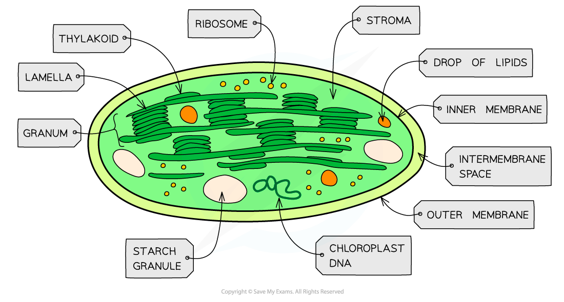
What is the function of chloroplast?
Site of photosynthesis. Light dependent reactions occur on thylakoid membranes, carbon fixation occurs in the stroma. Circular DNA and 70S ribosomes allows synthesis of some chloroplast proteins.

What is the structure of plasmodesmata?
It’s narrow cytoplasmic channels through plant cell walls. Each is lined by cell surface membrane.
What is the function of a plasmodesmata?
It connects the cytoplasm of neighbouring cells to form the symplast; permit cell to cell movement of water, ions, sugars, and signalling molecules.
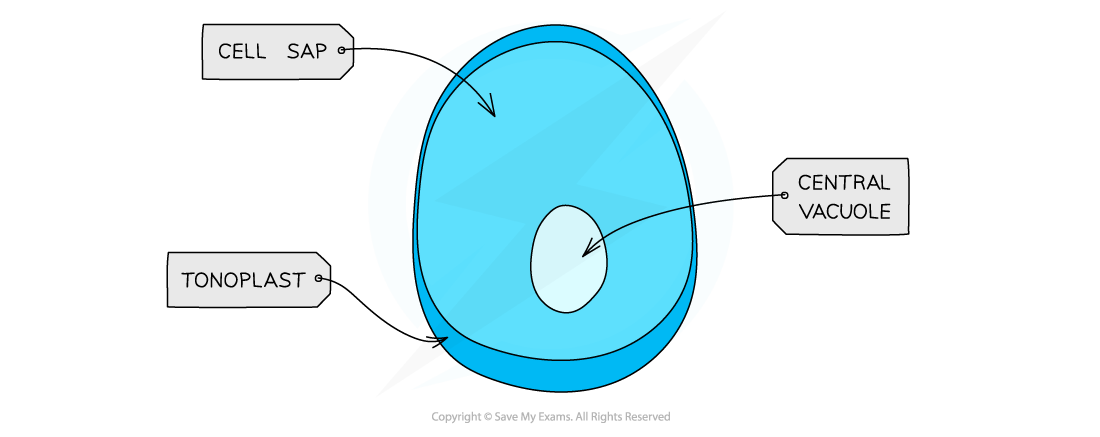
What is the structure of a large permanent vacuole?
It is a large fluid-filled compartment containing cell sap. It is bounded by a partially permeable membrane called the tonoplast.
What is the function of a large permanent vacuole?
They store cell sap, provide additional structural support to cells, contributes to water potential control, and isolates potentially harmful substances.
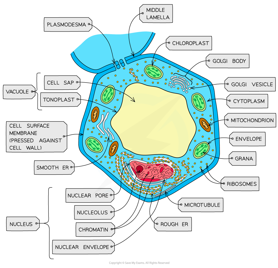
How are the structure of typical plant and animal cells different?
Plant cells are generally larger and more regular and additionally have a cellulose cell wall, large permanent vacuole, chloroplasts and plasmodesmata, while animal cells uniquely have centrioles and microvilli.
But they both share the cell membrane, cytoplasm, nucleus, mitochondria, ER, Golgi bodies, vesicles, lysosomes, ribosomes and microtubules.
What do cells use ATP from respiration for?
Energy-requiring processes, such as in anabolic reactions to build larger molecules from smaller molecules, to move substances across the cell membrane in active transport, or to move substances within the cell, and in animals, energy is required for muscle contraction and in the conduction of nerve impulses.
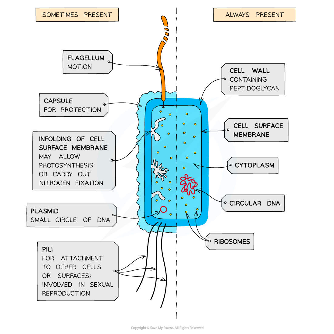
What are the key structural features of a prokaryotic cell found in a typical bacterium?
They are always unicellular.
Their cells are very small, usually 1-5 micrometers in diameter.
Their cell walls are made of peptidoglycan.
Their genetic material or DNA is free in the cytoplasm and is circular.
Their ribosomes are smaller at (70 S).
They lack membrane-bound organelles.
What is the difference of the structure of a prokaryotic cell found in a typical bacterium to the structures of a typical eukaryotic cells found in plants and animals?
A prokaryote has a size of 0.5-5 micrometers while the size of an eukaryote is up to 100 micrometers.
A prokaryote has a circular chromosome in the cytoplasm and is not associated with proteins while an eukaryote has linear chromosomes in the nucleus and are associated with histone proteins.
In prokaryotes, their cells divide through binary fission and no spindle fibres are involved while in eukaryotes, cells are divided through mitosis or meiosis and chromosomes are separated by spindle fibres.
Prokaryotes ribosomes are structurally smaller (70 S) in comparison to those found in eukaryotic cells (80 S).
A prokaryotes organelles are not bound by a membrane while eukaryotes have multiple membrane-bound organelle such as in the a nucleus, mitochondria, and chloroplasts.
In a prokaryote, the cell wall is made up of peptidoglycan while an eukaryote their cell wall is made of cellulose in plants, or chitin in fungi.
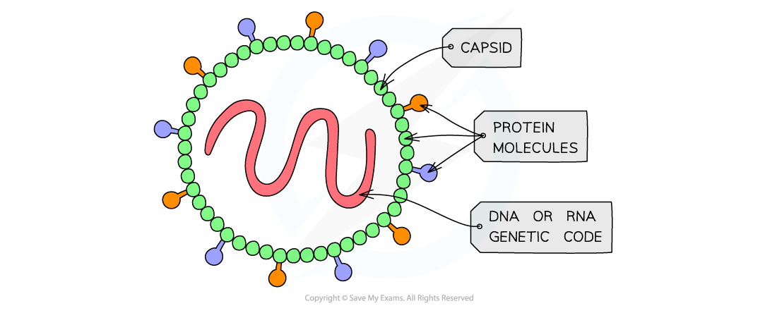
What are the key features of viruses?
They are non-cellular structures, consisting of a DNA or RNA nucleic acid core surrounded by a protein capsid, with some having a phospholipid envelope, and they can only reproduce by infecting living cells.