dev bio module 8: ectoderm
 Call Kai
 Learn
 Practice Test
 Spaced Repetition
 Match
 Flashcards
 Knowt Play
1/51

Earn XP
Description and Tags
**watch and add last 20 min of 4/7 lecture
52 Terms
neurulation
the formation of the neural tube, can overlap with when gastrulation starts
ectoderm includes:
surface ectoderm, neural crest, neural plate/neural tube
DPP
homolog to BMP
is inhibited by Dorsal on the ventral side in drosophila
specifies surface ectoderm in drosophila
surface ectoderm derivatives
mouth epithelium (tooth enamel), lens, cornea, epidermis, hair, nails, sebaceous glands, olfactory epithelium
high BMP (=DPP homolog)
neural crest derivatives
facial cartilage, dentin, pigment cells, adrenal medulla, peripheral NS (nerves, neurons outside of CNS)
neural plate derivatives
brain, spinal cord, motor neurons, retina
no BMP

stage 1: Early neurula
neural folds = border between surface ectoderm and neural plate

stage 2 neurula
neuropores
neural groove: canal folding into itself

BMP
Bone Morphogenic Signaling
paracrine signal: signals to other cells (not self)
cell proliferation
cell migration
induces surface ectoderm formation in vertebrates
BMP signaling
part of TGFb family of receptors (transforming growth factor beta)
1. BMP ligan binds to TGFb receptors
2. receptors dimerize
3. causes SMAD to become phosphorylated —> turns ON
4. ON SMAD enters nucleus and starts transcription (TF)

how to inhibit BMP signaling?
inhibit SMAD phosphorylation
receptor mutation
overexpress TGFb ligand
mutate binding seq of SMAD
Noggin
inhibitor of TGFb/sequesters BMP —> inhibits BMP signaling pathway
Chordin
inhibitor of TGFb/sequesters BMP —> inhibits BMP signaling pathway
SOX TFs
they can…
activate neural plate gene programs
or inhibit BMP signaling
SOX represses the expression of ligands or receptors - does not interact directly with BMP
external neural plate —> neurulation —→ __
internal neural tube
Primary neurulation
neural plate invaginates and forms neural tube
formation of the anterior of the tube = rostral side
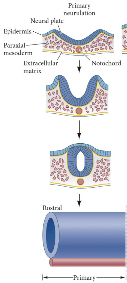
secondary neurulation
ingression:
cells in ectoderm ingress
they detach and undergo E→M transition
they condense into a tube with no gap
cavitation
posterior of the tube formation = caudal side

junctional neurulation
formation of the middle of the neural tube

Neurulation 1: ELONGATION
growth in the anterior-posterior axis directions
3 ectoderms:
surface ectoderm
progenitors of the neural crest
neural plate
Axial elongation: cells in the neural plate are dividing in A-P direction

Neurulation 2: FOLDING
medial hinges (on top of notochord): stiff rod - axis of folding
embryo grows towards midline
notochord (mesodermal derivative) induces invagination:
notochord signals neural plate —> cell differentiation in neural plate—> formation of medial hinge

Neurulation 3: ELEVATION
elevation of either sides of the neural plate,
neural crest progenitors + surface ectoderms move towards each other
notochord dragged towards ventral side
formation of the neural groove
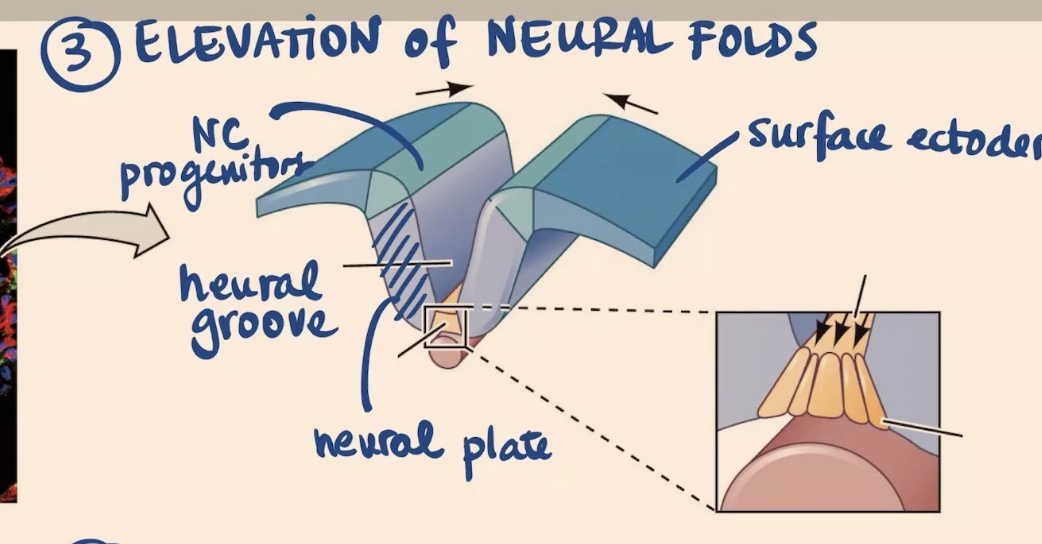
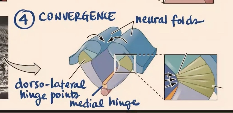
Neurulation 4: CONVERGENCE
formation of 2 new hinges:
1. medial hinge
2. dorso-lateral hinge points
Neurulation 5: CLOSURE/FUSION
surface ectoderm is connected
neural crest cells are located most dorsally in the tube —> will migrate
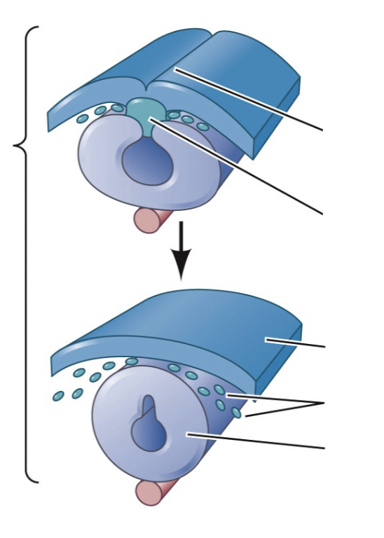
neural crest cells vs neural crest
neural crest = transient structure
neural crest cells = permanent structure
regulation of hinge point formation
pre-neurulation
precursors of notochord activate noggin and chordin
chordin/noggin inhibit BMP in neural plate area (BMP is expressed in surface ectoderm
neurulation
notochord expresses shh —> shh inhibits noggin
noggin presence = bend (expressed in dorso-lateral hinge points and in neural plate (pre-neurulation)
high BMP presence = prevents bending (noggin needed to inhibit BMP in hinge areas)
for hinges to form…
apical side of cells shrink smaller than basal side of cells
defects in neural tube closure
exencephaly: brain expands outside of skull cavity
anencephaly: most brain does not develop
spina bifidia: gap in spine vertebrae
paralysis
Hedgehog signals
dessert Hh —> spermatogenesis
Indian Hh —> gut, cartilage, bone
Sonic Hh —> morphogen
sonic Hh
specification of motor neurons NT
vertebrae formation
feather formation
hair
patterning of the hand
shh mutation
cyclops: born with one eye
corn lily: alkaloid
SHh secretion
nucleus (mRNA) —> rough ER (translation) —> membrane (secretion)
translation
autoprotylitic cleavage (auto terminal exposed)
modify: adds cholesterol and palminic acid
transported to membrane vesicles
secretion methods (5)
Shh secretion methods
multimers formation
complex with lipoproteins
exovesicle formation
detaining from membrnae as monomers —→ needs protein channel
cytonemes (actin)

Shh secretion signaling
Shh OFF
“patched” receptor inhibits “smoothened” receptor which internalizes
Gli connected to microtubule
PKA ON cleaves and phosphorylated Gli
half Gli enters nucleus and inhibits transcription
Shh ON
Shh binds patched receptor and deactivates it
smoothened receptor can now travel up to top of cilium
smoothened activates Gli and inhibits PKA
whole Gli enters nucleus and activates transcription

Cadherins
Ca2+ dependent addition molecules, transmembrane proteins that anchor to the cytoskeleton
E cadherins = early embryo + epithelium
N cadherins = neural tissue
E cadherins
attraction towards the same type —> closure
fusion happens at different points (mouse @ points 1,2,3
Zipper closure
neuropores: normal gaps where neural tube is not yet closed
Dorso-ventral regionalization of neural tube 1
newly formed Neural Tube
2 main signaling centers: epidermis (BMP4,7), notochord (shh)
shh from floor plate in gradient manner vs BMP from roof plate in gradient manner
DV regionalization of NT 2
formation of secondary signalling centers
BMP and shh = paracrine , gradient
BMP: feedback loop (autocrine)
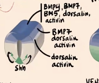
DV regionalization of NT 3
dorsal interneurons: processors and integrators of info from sensory neurons
ventral motor neurons: sends axons from spinal cord to muscles, controls voluntary/reflex movements
AP regionalization of NT
very dependent on Hox genes
3 primary brain vesicles
prosencephalon
mesencephalon (midbrain)
rhombencephalon (hindbrain)

5 secondary brain vesicles
telencephalon: cerebral hemispheres
diencephalon: eye cap, retina
mesencephalon: midbrain
metencephalon: pons, cerebellum
myencephelon: medulla oblongata
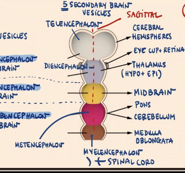
4 flexures
flexures: curvatures points in the anterior NT
sagittal flexure: between the cerebral hemispheres from dorsal view
cephalic flexure
pontine flexure
cervical flexure

Neural crest
comes from neural crest progenitor cells next to surface ectoderm that ingresses and delaminates
ectodermal derivative
transient
EMT epithelial to mesenchymal transition + extensive migration
A→P
D→V
neural crest as 4th germ layer?
gives rise to
peripheral nervous system PNS,
endocrine and paraendocrine derivatives,
pigment cells,
facial cartilage and bones,
connective tissue
specification of neural crest cells
depends on the expression of the same morphogens (BMP, Wnt) at different concentrations and for diff periods of time
not variations of BMP or wnt - its the same morphogen

cranial placodes
they are thickenings in the surface ectoderm/border of surface ectoderms
sensory organs
1. olfactory: olfactory epithelium
2. lens: eye lens
3. trigeminal: sensory neurons of trigeminal nerve
4. otic: inner ear (cochlea, vestibular apparatus)
5. epibranchial: nerves (vagus, facial, etc)
Cranial NCC
craniofacial mesenchyme → bone, cartilage, pigment cells, glia, connective tissue
pharyngeal arches and pouches → thymus, odontoblasts, middle ear and jaw bones
cardiac NCC
muscular and connective wall of large arteries of heart
septum that separates pulmonary circulation for aorta
melanocytes, neurons, cartilage, connective tissue
trunk NCC
ventrolateral migration: sensory neurons of dorsal root ganglia + sympathetic ganglia, adrenal medulla, nerve clusters around the aorta
dorsolateral migration → pigment cells
vagal & sacral NCC
parasympathetic ganglia of the gut
early inter-species NCC transplant expts
created hybrid Quck or Duail skull structures
showed NCC autonomous properties - not environment dependent - the NCC have some cell pre-determined cell fate
how the test if NCC can be multipotent?
fluorescing every step of NCC differentiation (single cell resolution)
reporter line: multiple cassettes with diff color reporters
can perform tissue specific deletions with flox + cre driver lines (LoF)
1. lineage tracing
2. conditional gene deletion: creating mutation in the cells that express cre → cut at lox sites → stop codons → color expressed