Exam Two - Molecular Biology of Neuron
 Call Kai
 Learn
 Practice Test
 Spaced Repetition
 Match
 Flashcards
 Knowt Play
1/123

There's no tags or description
Looks like no tags are added yet.
124 Terms
Gap junction hemi-channels
M1-4 transmembrane domains → form connexin
6 connexins need to come together to form a hemi-channel
Hemichannels at pre- and post-synaptic membrane = align adjacent to each other. ⇒ form gap junction
Gap junction characteristics
Have large diameter = allow non-selective ion passage.
Symmetrical synapse → cannot define pre- and post-synaptic membrane (no PSD)
Comparisons between channels & ion selectivity
Voltage-gated cation channels: tetramers (4 subunits) = small, selective.
Transmitter-gated ion channels (ligand-binding): 5 protein subunits forming central pore.
Gap junctions: 6 connexins → greater diameter = even less selectivity
Gap-junction Transmission: Electrical synapse
Transmission can be bi-directional; permit direct passive flow.
Transmission is fast ⇒ 0.2 ms in electrical vs. 2 ms in chemical
Electrical activity is synchronized
Electrical synapses do not have gain: induced response is always smaller than original signal in the source neuron.
Response always the same sign as source (excitation cannot produce inhibiting response)
Evidence for electrical synapse
Action potential in crayfish: first discovered in crayfish nervous system.
Found in neural systems that require fastest possible response (e.g. defensive reflexes)
Synchronization of electrical activity among population of neurons like brainstem neurons (e.g. generating rhythmic electrical activity for breathing)
Measured membrane potential in pre- and post-synaptic neuron:
Action potential & EPSP response in post-synaptic neuron shows minimal delay (<0.1 ms)
Action potential in hippocampal interneurons = immediate post-synaptic response
Chemical synaptic transmission: cycle
Vesicle docks:
Cytoskeleton guides vesicle to pre-synaptic membrane (directly adjacent to PSD & receptors).
SNARE complexes form to pull membranes together
SNARE proteins: SNARE complex proteins all contain coil domains called “SNARE motif” → Tight binding
Synaptobrevin (on synaptic vesicle membrane)
On pre-synaptic membrane: Syntaxin & SNAP-25
Entering Ca2+ binds synaptotagmin
Ca2+-bound synaptotagmin catalyzes membrane fusion
With calcium binding: Synaptotagmin inserts into phospholipid = interact with membrane = release NTs as vesicle fuses
Removal of Ca2+ from the cleft by astrocytes; recycling at level of pre-synaptic button.
EXPERIMENT: Otto Loewi’s evidence for chemical neurotransmission
Hypothesized that chemical NT must be released for communication of nerve impulses.
Isolated frog hearts: 1 incubated in beaker w/ fluid (continue to beat)
Stimulate the vagus nerve = heartbeat of heart 1 slows down.
If NT is released =
Attachment of heart 1 to heart 2 → chemical NT released into fluid of beaker 1 that is connected to beaker 2 by a tube = affects heart 2 by slowing down its heart-beat.
Evidence of a secretory ligand that then inhibits heart 2.
Neurotransmitter: Acetylcholine (parasympathetic innervation)
Small vs. large-molecular transmitters
Small-molecule neurotransmitters: contained in small clear-cored vesicles (40-60 nm in diameter)
Acetylcholine
Purines (ATP, GTP, adenosine)
Amino acids (excitatory: glutamate, aspartate; inhibitory: GABA, and glycine)
Biogenic amines (catecholamines [dopamine], indoleamine [serotonin], imidazoleamine)
Neuropeptides: more than 100 peptides, usually 3-30 amino acids long.
Contained in large dense-cored vesicles (90-250 nm in diameter)
Vesicle synthesis: small-molecule transmitters/vesicles
Synthesis of enzymes occurs in the cell body
Slow axonal transport of enzymes into terminal
Precursors (broken down after NTs have been released) are transported back into terminal from cleft.
Synthesis & packaging of NT at pre-synaptic button (axon terminal)
Vesicle synthesis: large dense-core vesicles
Large dense-core vesicles budded off from Golgi & synthesis of NT precursors and enzymes at the cell body.
Transport down microtubule tracks (fast) ⇒ 400 mm/day
Enzymes modify precursors to produce peptide NTs
EXPERIMENT: Difference in rate of release = differences in spatial arrangement of vesicles
Low-frequency stimulation:
Localized increase in Ca2+ ⇒ causes preferential release of small clear-core vesicles to be released (they are closer to the membrane)
Therefore, small-molecule NTs “act” faster.
High-frequency stimulation = more diffuse increase in Ca2+ concentration
Both types of transmitters (clear-core & dense-core) are released
Therefore, release of vesicles depends on concentration of Ca2+
EXPERIMENT: Testing calcium dependence of vesicle release
Voltage-clamp & set presynaptic membrane potential to -25 mV
Observed inward presynaptic calcium current
Post-synaptic membrane response observed.
Block Ca2+ channel ⇒ reduce presynaptic calcium current → no postsynaptic membrane response.
Synaptic vesicle cycle
Recycling: Vesicles formed through clathrin-dependent endocytosis.
Empty vesicles merge into early endosome
Undergo NT uptake
Trafficked onto the active zone (zone of pre-synaptic membrane w/ vesicles aligned/docked & primed by ATP)
Primed vesicles will be the first to fuse with the membrane.
Cav is located adjacent to primed vesicles.
Synaptic vesicle cycle: Protein-protein interactions
Protein-protein interactions are essential for synaptic vesicle cycle to occur.
Can make antibodies to label these proteins.
SNARE Protein: syntaxin, snap-25, synaptobrevin ⇒ synaptic vesicle proteins
Synaptotagmins on the membrane
Piccolo & Bassoon: active zone proteins
MINT proteins: bind to synaptic vesicle proteins (neurotransmission) & interact w/ Alzheimer’s disease proteins.
Synaptic vesicle pools
Readily-releasable pool
1-2% docked at active zone → released <1 second with fast recycling
Recycling pool
10-20% scattered
Reserve pool
80-90% scattered → 10s of seconds-minutes; slow recycling (minutes)
Bulk of vesicle cluster.
EXPERIMENT: Order of synaptic vesicle release
Can tag synaptic vesicles w/ fluorescent tag ⇒ detect fluorescence changes in extracellular media.
Over time, if you continuously excite pre-synaptic neuron ⇒ observe sequence: RRP released first, then followed by the others. ⇒ will eventually depleting the pool
Katz’s model system: Neuromuscular Junction
Model system: frog’s neuromuscular junction model: neuron’s synaptic button ⇒ release ACh ⇒ bind to ACh receptors on post-synaptic muscle membrane = contraction.
Large junction = easy to visualize
Stimulate the axon w/ current & record post-synaptic membrane potential of muscle cell.
End-plate potential (EPP) ⇒ transient depolarization of post-synaptic membrane potential in response to action potential
If large enough → can trigger action potential.
EXPERIMENT: Katz’s seminal study - spontaneous membrane potential changes
Even without stimulation, there are spontaneous changes in post-synaptic membrane potential ⇒ Miniature end plate potentials (MEPPS)
Experiment 1: Pre-synaptic modification ⇒ Removed calcium (responsible for NT release) ⇒ no more MEPPs in muscle cell
Experiment 2: Post-synaptic modification ⇒ Curare (ACh receptor inhibitory) ⇒ no more MEPPs in muscle cell
EXPERIMENT: Katz’s quantal mechanism of NT release
Hypothesis: MEPPs are spontaneous events that respond to fusion of synaptic vesicles (release of NTs) at rest (w/o action potential) ⇒ Is it caused by one molecule of ACh or by many molecules?
Mathematician: Proposed that 10,000 ACh/vesicle = quanta
Experiment 3: Vary the concentration of acetylcholine (1 vs. 10000 molecules)
Finding:
Each vesicle = one quantum of transmitter amounting to several thousand molecules.
Each quantum produces post-synaptic potential of fixed size (quantal amplitude)
Neuron’s response is the sum of individual quanta
Action potential accelerates and synchronizes release of quanta.
EXPERIMENT: Quanta and synaptic vesicle fusion
Quanta result from fusion of individual synaptic vesicles with the plasma membrane.
Stimulate the pre-synaptic membrane⇒ can see vesicles fusing with plasma membrane.
Positive linear correlation between number of quanta released into synapse & number of vesicles fusing.
Modes of NT release: Evoked
Evoked synchronous release: initiated within milliseconds after action potential
Evoked asynchronous release: sets in w/ delay after action potential.
Normally negligible: hidden by synchronous release or synaptotagmin suppresses async release.
Experiment: Knockout of synaptotagmin-1 (essential for evoked synchronous release) ⇒ can still visualize asynchronous release; therefore, synaptotagmin-1 is not essential for asynchronous release.
Purpose: May shape neural function at circuit level; may maintain synaptic vesicles.
Modes of NT release: Spontaneous
Spontaneous “mini” release: exocytosis of a single vesicle that is independent of action potentials but dependent on Ca2+
Minis (mEPSC) are also observed in WT neurons
Fusion machinery (proteins) are also unique to evoked synchronous release.
Synapsins
Maintaining reserve pool
Mobilization of synaptic vesicles:
Synapsin is the “highest” protein in the brain that can be phosphorylated → regulated by this post-translational modification.
Synapsin phosphorylation
Dephosphorylated synapsin I (inactive): bound to synaptic vesicles & binds them to the nerve terminal cytoskeleton (actin) ⇒ holds at reserve pool
Phosphorylated synapsin I ⇒ release from vesicles = free vesicles from cytoskeleton & make them available for exocytosis.
Phosphorylation can occur through secondary cascades
G-protein activated in response to NT binding → activates adenylyl-cyclase (AC = secondary messenger) → cAMP kinase → phosphorylation of synapsin I
Experiment: Synapsin triple knockout mice
Must knockout all three synapsins (6 alleles) to observe clear phenotype, as they compensate for each other.
Finding:
Viable and fertile
However: seizures and behavioral deficits.
Experiment: Electron micrograph of synaptic vesicles in synapsin KO
Electron micrograph of pre-synaptic excitatory synapse (asymmetric: have pre-synaptic vesicles & thick PSD; inhibitory synapse has a finer PSD)
Observed mean number of synaptic vesicles at increasing distance from active zone: 100, 200, 300, 400, 500 nm
KO: Further away = reduction in # of SV ⇒ likely affecting the reserve pool.
Not much changes in docked vesicles compared to WT.
Experiment: Reserve pool depletion in synapsin KO
Experimenter can keep stimulating the neuron ⇒ triple KO will deplete in reserve faster
Plotted # of stimuli & normalized EPSC current to see where depletion occurs. ⇒ TKO curve drops faster at around 50 stimuli.
Depression time constant ⇒ faster depression in triple KO.
RIM Proteins
Docking of synaptic vesicles at pre-synaptic membrane
Are adapter protein that links synaptic vesicles to Cav.
Rab3: connection point between RIM and synaptic vesicle.
RIM-BP and Munc13 are also involved in complex.
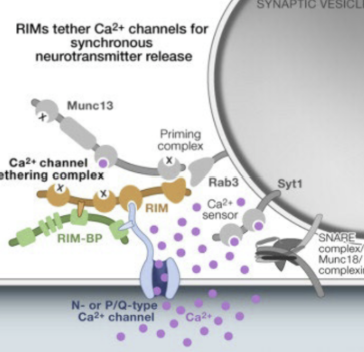
EXPERIMENT: Conditional double-knockout RIM
Conditional double-knockout (RIM1&2): number of vesicles docked at active zone is significantly reduced.
However, vesicles per button have not changed.
Experiment:
Small EPSC & IPSC from cDKO
Also examines mEPSC: frequency is reduced.
Syntaxin
Part of SNARE complex
N-terminus: HA,B,C domains → SNARE motif → trans-membrane region (TMR)
Has open and closed conformation:
Auto-inhibited: folds upon itself (HA-C folds on SNARE motif).
Open conformation: can bind to SNAP-25 & Synaptobrevin
Responsible protein: Munc18 ⇒ binds well to closed Syntaxin & must dissociate for syntaxin to open.
EXPERIMENT: Munc-18 binding
Method: Western blot
Syntaxin is tagged with GST (bait) that sticks to beads
Make mutated versions of various amino acids of syntaxin (e.g. Leucine165 & E166).
Homogenized rat brain tissue: membrane-bound proteins (Munc18, SNAP-25, synaptobrevin) dissolved in mild detergent and can bind the bait.
Beads incubated w/ lysate & washed = release bait & binding partners that can be separated by size on gel
Western blot: Identifies the protein pulled-down.
Findings:
Munc-18 binding is lost when we mutated at 165 & 166
Where Munc-18 could not bind, Snap-25 & synaptobrevin could
Negative controls: Synatogyrin & Rab3A shows no band = no interaction w/ Munc-18
EXPERIMENT: Knock-in mutation of Leucine165 & E166
Knock-in mutation in vivo ⇒ mutated Leucine165 & E166 into Alanine ⇒ mice demonstrates behavioral deficits.
Mechanisms of Endocytosis
Clathrin-dependent endocytosis:
Clathrin bind to one another⇒ triskelion structure ⇒ involved in invagination of the membrane.
Dynamin ring pinches off the membrane
Vesicle (coated by clathrin) are translocated by actin filaments.
Clathrin is removed by Hsc-70/auxilin
Experiment: local vesicle recycling in presynaptic terminals
Treat neurons w/ horseradish peroxidase in extracellular medium.
HRP can be taken up by synaptic vesicles → wash away extracellular HRP → wait 5 minutes → follow where they go.
Process: total 1 minute
Fusion (1 ms) → endocytosis & budding (10-20 seconds) → endosome → budding from endosome → docking → priming → exocytosis.
Alternative method: FM1-43
Voltage-sensitive dyes
Has a lipophilic tail & carries a fluorophore and positive charge.
Treated with HRP in extracellular medium = follow fluorescent protein ⇒ visualize both endocytosis & fusion events.
Neuropathologies: Synaptic transmission
Familial infantile myasthenia: reduces vesicle size
Congenital myasthenic syndromes: impaired vesicle recycling.
LEMS (antibody) can cross BBB & attack presynaptic Ca2+ channels in autoimmune disorders ⇒ Synaptic transmission won’t occur
Experiment Method to Study Synaptic Transmission: Toxins
Toxins that trigger vesicle fusion in pre-synaptic membranes:
Neurexins: initially detected as receptors for alpha-latrotoxin (component of black widow spider venom)
Causes MASSIVE NT release.
Possible hypothesis: Cell-adhesion molecules ⇒ perhaps interfere w/ pre-synaptic membrane = Ca2+ influx independent of Cav.
Toxins that affect SNARE proteins:
Botulinum (BTX ⇒ e.g. botox): inhibits neurotransmitter release
Tetanus toxin: inhibits release of inhibitory transmitters ⇒ hyper-excitability
Synaptic plasticity
Short-term: Few minutes or less => Largely based on pre-synaptic event: changes in amount of NT released & synaptic vesicle pools.
Alterations in Ca2+ signaling = important
Long-term (>30 mins): Largely based on Ca2+ and other intracellular secondary messenger pathways. → takes place at the postsynaptic level.
Short-term Facilitation
Facilitation: enhances NT release by persistent action of Ca2+ ions
When 2(+) APs invade presynaptic terminal within 10 ms of each other → greater amplitude of post-synaptic membrane potential.
Due to prolonged elevation of pre-synaptic Ca2+ levels = more release of vesicles.
Facilitation decays w/ longer the interval between stimuli ⇒ therefore, the pairs must arrive CLOSE together.
Molecular mechanisms of Short-term facilitation
Serotonin released from facilitator interneuron (modulatory) → bind to G-coupled receptor that activates G-alpha-s (Gαs)
Gαs → activates adenylyl cyclase → converts ATP into cAMP
cAMP binds regulatory subunit of PKA → release catalytic subunit that phosphorylates K+ channels = close
Less K+ = AP repolarizes more slowly = Ca2+ channels open longer (increase Ca2+)
More Ca2+ = more Glutamate vesicles released = larger EPSP
Short-term Depression & Experiment
Depression: decreases amount of NT due to depletion of RR-vesicles.
Experiment: consistent stimulation over 5s ⇒ big drop in post-synaptic response.
Dependent on Ca2+:
Lower concentration of Ca2+ {intermediate, 0.1 normal) during stimulation: does not lead to depression → leads to augmentation.
Lowering Ca2+ availability = decreased efficiency of fusion.
More relative amount of NT release = more relative amount of depression.
LTP Mechanisms
CAMKII (involves calmodulin) & insertion of AMPA receptors to modify synapse excitability.
AMPA/NMDA: AMPA responds first to GLU ⇒ leads to Na+ influx → removes Mg2+ plug of NMDA, so GLU binding can allow Ca2+ to come in → Ca2+ binds calmodulin & activates CAMKII → phosphorylates downstream substrates (effector proteins) ⇒ inserts additional AMPA receptors.
Morphology of dendritic spines (mushrooms)
Long-term plasticity model: Hippocampus
Unidirectional information movement
Entorrhinal cortex (EC) sents axons along perforant-path → synapse on dentate gyrus → sends axons to synapse w/ Ca3 region (pyramidal neuron)
Ca3 synapse on Ca1 neuron → back into EC
Slice physiology matters in study of LTP
Experiment: Hippocampus induction of LTP
Stimulate Ca3 and record EPSP in Ca1 neuron
High-frequency stimulation (100 Hz) for long period of time: long-term potentiation ⇒ Ca1 recording enhanced for long period of time (hours)
Low-frequency stimulation (1 Hz): lead to long-term depression ⇒ EPSP decreased over time
LTP characteristics
Input specific: to a specific synapse
Pathway 1: active ⇒ synapse strengthened
Pathway 2: inactive ⇒ synapse not strengthened
Associative:
Pathway 1 has strong stimulation & Pathway 2 has weak stimulation ⇒ however, if they fire at the same time = can strengthen both synapses.
Molecular Mechanisms of Long-term Facilitation
Repeated or strong serotonin exposure
cAMP stays high so catalytic PKA move into nucleus → phosphorylates CREB
CREB (transcription factor) turns on gene expression
Output: New gene expression and structural change
C/EBP transcription factors continues to “switch on” genes.
Ubiquitin hydrolase degrades PKA regulatory subunit → PKA stays active even w/o cAMP
Proteins for synaptic growth build new synaptic connections between sensory and motor neurons.
Long-term depression
Reduction of receptors through endocytosis → decrease postsynaptic response.
LTD: Hippocampus
1 Hz stimulation
Internalization of AMPA receptors ⇒ Ca2+-dependent.
Glutamate activates AMPA = Na+ influx & NMDA = Calcium influx ⇒ Calcium activates protein phosphatases ⇒ dephosphorylate substrates ⇒ internalization of AMPA receptors.
LTD: Cerebellum
AMPA receptors endocytosed ⇒ BUT DIFFERENT PROCESS
Glutamate activates metabotropic glutamate receptors (GPCR) → activates G-alpha-q → activates phospholipase C & DAG ⇒ increase Ca2+
Ca2+ → activates PKC → phosphorylates substrate proteins → long-term depression through internalization of AMPA receptors
Associative activation: Presynaptic terminal of parallel fiber & climbing fiber = synapses must be activated at the same time for LTD to occur.
Parallel fiber = DAG from mGluR; Climbing fiber = large Ca2+ influx ⇒ strong activation of PKC.
Kandel’s Model System: Siphon (Aplysia) - Behavioral
Non-noxious model system: Gill contracts every time the siphon is “tickled”
Over time, gill response decreases significantly (habituation)
Noxious stimuli (inflict pain): shock the tail = muscle contracts.
Touch siphon + shock = siphon becomes more sensitive to pain (sensitization)
Experiment:
With single tail shock = response spikes compared to no shock but eventually goes away → short-term sensitization
4 trains of tail shocks or 4 trains/day for 4 days ⇒ amplified reflex lasts longer
Stimulus pattern determines whether effect is long/short-term.

Kandel’s Model System: Siphon (Aplysia) - Molecular system
Siphon skin’s sensory neuron → synapses to motor neuron → causes gill contraction when it fires (excitation at neuromuscular junction)
Interneuron connects to motor neuron ⇒ over time, inhibits motor neuron response to non-noxious stimulus.
EPSP decreases over time.
However, stimulate the tail → activate modulatory interneuron (serotonin) → activates synapse between motor neuron & gill = causes firing.
Restores EPSP in motor neuron (4th panel)

Endocannabinoids
Plant-derived: THC
Endogeneous:
Anandamide (AEA)
2-AG
Affect neural functions: euphoria, appetite stimulation, sedation, etc.
G-protein coupled receptors: CB1 (pre-synaptic terminals or axons), CB2
CB1: Post-synaptic (making of messengers)
Glutamate binds to mGluRs & ionotropic receptors post-synaptically → activates G-alpha-q (Gaq)
Gaq activated = PLC-beta is activated = cuts PIP2 = DAG and IP3 → increase in Ca2+
Ca2+ & DAG stimulate enzymes converting membrane precursor into AEA or 2-AG = diffuse into cleft.
Recycling: Transporter takes endocannabinoids back into post-synaptic cell & enzyme breakdown into precursors
CB1: Pre-synaptic (localization & effect)
AEA or 2AG binds to CB1 pre-synaptically → activates the G-alpha-i pathway → inhibits adenylyl cyclase (AC) to decrease ATP, cAMP, PKA
Additionally, negative feedback regulation to NT release: Beta-gamma subunits can activate Kv and inhibit Cav ⇒ inhibits NT release
Experiment: Depolarization-induced (glutamate) suppression of inhibition (inhibit GABA-ergic neurons)
Requires increase in postsynaptic calcium ⇒ associated w/ decrease in IPSC frequency and not amplitude; GABA has no effects.
Increase in Ca2+ after innervation by glutaminergic afferent ⇒ production of 2AG ⇒ binds to CB1 in GABA-ergic afferent ⇒ inhibition of NT release.
Experiment:
Depolarizing pulse to post-synaptic neuron → transient decrease in IPSC response
Treat with AM281 (antagonist of CB1) → maintained IPSC
Experiment: LTP & Cannabinoids
LTP inhibited by CB1 receptor blockade
100 Hz stimulation (Theta-burst stimulation: TBS) = LTP observed
Can also apply reagents that enhance LTP (chem-LTP): adenylyl cyclase activator (e.g. forskolin) and phosphodiesterase inhibitor in presence of low Mg.
Both LTPs can be inhibited by CB1 competitive receptor NESS-0327 OR CB1 receptor inverse agonist
Homeostatic Plasticity & Experiment w/ Toxins
Negative feedback mechanisms that neurons use to offset excessive excitation (excitotoxicity due to excess Ca2+) or inhibition by adjusting synaptic strength.
Experiment: culture neurons
Treat with TTX for long period of time (hours): Blocks voltage-gated Na+ channels = reduces network activity.
Compensatory mechanism: insert more receptors at the postsynaptic site.
Treat with Bicuculline: Blocks ionotropic GABA-A receptors (inhibitory)
Compensatory mechanism: endocytosis of receptors.
Arc protein
Mediates homeostatic synaptic scaling of AMPA receptors
Regulated by neuronal activity in primary culture
Bicuculline = increased activation of neurons = increase Arc = decrease in surface AMPA receptors.
TTX = decreased network activity = decreased Arc expression.
Highly expressed post-synaptically.
Homeostatic Plasticity: Glial cells
Blocking network activity w/ TTX → decrease Glutamate from neurons → glia sense activity loss and release TNF-alpha (soluble factor) = scaling up excitatory synapses.
Experiment: TNF-a
TNF-a mediates synaptic scaling during activity blockade.
Treat with TNF-alpha & chronic treatment w/ TTX = increase in AMPA-receptors surface expression.
mEPSCs:
Chronic activity blockade = increase in mEPSC amplitude due to AMPA-r increase .
Conditioned medium from TTX-treated cultures applied to untreated neurons = causes increase in mEPSC amplitude.
To confirm that TNF-alpha is contained in the conditioned media (CM) ⇒ treat CM w/ sTNFR.
sTNFR bind TNF-alpha ⇒ no TNF-receptor activation ⇒ no increase in mEPSCs amplitude.
Case study: H.M.
Seizures from bicycle accident in youth ⇒ bilateral lesion of medial temporal lobe, MTL (contains the hippocampus = important for learning & memory).
Severe anterograde amnesia: unable to establish new memories (consolidation of “recent memory”)
Temporally graded retrograde amnesia: memories created prior to the event are lost.
In-tact:
Working memory: temporary storage & manipulation of information, operates over a few seconds.
Procedural memory.
Preserved older memories (remote memory ⇒ stored at level of cerebral cortex)
Therefore, hippocampus functions as temporary storage for new information, but LTM/permanent storage (consolidation) depends on distributed cortical networks.
Spatial Learning & Hippocampus
Paradigm - Morris Water Maze: mouse placed in opaque pool at random start point → locate platform. ⇒ latency SHOULD decrease over time as the animal learns spatial cues.
Hippocampal lesion: mouse can no longer build/use a spatial map of the maze & surrounding environments. → path not direct to platform like controls.
Synthesis of place & grid cells in rodent brain
Grid cells in EC recognize head direction of animal → GPS system w/ place cells in hippocampus (form synapses) → causes firing of place cells.
Grid cells ⇒ how far & what direction it has moved.
Place cells ⇒ where it is.
Experiment: O-Keefe’s Place Cells
O’Keefe’s Experiment: Hippocampus as spatial reference map
Electrodes placed on rat’s hippocampus to record single neuron firing (A, B, C, etc.) as rat moves around rectangular box. Curtain hides external cues.
Finding:
Neuron fires strongly at A, and less or not at all at other positions.
Therefore, subpopulation of neurons sensitive to spatial position ⇒ activated upon specific directionality.
O’Keefe discovered “place cells” located in hippocampus
Experiment: Moser & Moser “Grid Cells”
Identified nerve cells in EC that were activated & form hexagonal grids that react to unique
Form coordinate system = allows for spatial navigation
Learning & consolidation: synaptic level
Very fast; morphological changes = necessary for initial stabilization of memories in hippocampal circuits (e.g. growth of new synaptic connections, restructuring of existing synaptic connections = e.g. spine morphology)
Synaptic activation
Activation second messenger systems: e.g. PKA, CAMKII
Transcription factors & protein synthesis ⇒ e.g. CREB
Learning & consolidation: system level
Slow: Gradual process of reorganization of brain regions that support memory.
⇒ Over time, connections between hippocampus & cortical modules weaken as objects are stored. Circuit between cortical modules become strengthened to store remote memory (refer to figure.)
Sleep is important for memory consolidation over time ⇒ strengthens cortical consolidation.
PFC and Remote Memory
Recent memory: HPC strongly linked to cortical modules (hub); PFC weakly engaged ⇒ recall is hippocampus-dependent
Remote memory: cortical modules + PFC strongly interconnected; hippocampus connections weak ⇒ recall is hippocampus-independent
PFC inhibits the hippocampus as the memory matures ⇒ memory maintained by the cortical network (systems consolidation)
Behavioral paradigm: contextual fear conditioning
Pavlovian condition where animal learns association between distinctive place [context] and aversive event [shock].
Single training experience = freezing behavior even w/o electrical shock response in the future.
Experiment: Contextual fear memories & hippocampus
Lesions to hippocampus either 1, 7, 14 or 28 days after training.
1 day: no longer freezes = has not “learned” behavior
28 days: same freezing profile as control
Long-term memory located in the cortex
Finding: HPC lesions produce temporally-graded retrograde amnesia for contextual fear memories in rats.
Behavioral paradigm: 8-arm radial maze
W/ novel objects on some arms.
Animal will explore the arms & usually gravitate towards the novel object.
Test recent memory (5 days): animal can go back to the arm w/ the object.
Test for remote memory after long retention interval:
Same context, same baited arm: animal can re-learn route.
Different context, different baited arm = back to recent memory —> benefit of switching back & forth
Tracing activity in 8-arm maze paradigm
Can inject radioactive 2-DG intravenously:
Retention testing → brain removal & sectioning → autoradiographs.
Finding:
Hippocampal level: lighting up for recent memory
Remote: anterior cingulate cortex (ACC) lights up; HPC no longer lights up.
When switching back to recent memory w/ the same paradigm: HPC lights up again.
NOW: Can do C-Fos staining & immunostain for activity.
Opsins
Light-gated ion channels
Sensory photoreceptors in green algae controlling photoaxis (movement towards light)
Can be expressed onto neurons
Functions:
Channelrhodopsin: blue light & allows Na+ ions to enter ⇒ spiking activity in neuron.
Halorhodopsin: responds to yellow light & allows Cl- ions to enter. ⇒ inhibition
Channelrhodopsin specificity: Plasmid design
Contains ChR & fluorophore (see which cells are expressing ChR)
Also has promoter: can be specifically selected
Example: CAMKII promoter ⇒ only expressed in CAMKII+ excitatory neurons
Example: GAD65 promoter: only in inhibitory GABA-ergic neurons.
WPRE, RRE ⇒ viral components
Neurons are post-mitotic = difficult to express DNA readily. HOWEVER, viruses can infect neurons well.
Culture = lenti-virus
Rat: adenovirus as packaging systems.
Experiment: Characterization of ChR
Hippocampal neurons labeled w/ GFP & co-expressed CAMKII.
Shining blue light = inward current response throughout the whole duration of the light being on.
Increase in Hz of light ⇒ increased in frequency of AP responses.
Rhodopsin: Halorhodopsin in hippocampal CA
Inject viruses (eNpHR) into hippocampus at different Bregma sites (intersection between frontal & parietal bones of skull)
Halorhodopsin expressed in CA region of hippocampus.
Finding:
Turning yellow light on through double cannula guide = no firing (decreased frequency) ⇒ creates temporary lesion
Experiment: CA1 inhibition & contextual fear acquisition/retrieval
CA1 optogenetic inhibition blocks contextual fear acquisition & retrieval
Mice placed in cage & pure tone is paired with foot shock = induce fear conditioning in controls.
In mice w/ halorhodopsin:
Light on during testing (in same chamber) = no longer freezing
Light off = mice w/ halorhodopsin can learn fear conditioning well.
Experiment: Auditory-cue fear memory
Design:
Fear-conditioned mice placed in pyramid-shaped cage w. smooth floor (different set-up).
2.9 kHz tone was sounded for 2.5 minutes.
Freezing behavior was measured over 2.5 minutes
No difference between control & eNpHR mice w/ light on or off
Experiment: eNpHR & amygdala
Paradigm: Mice in open field
Presence of virus w/ halorhodopsin = not much change compared to control ⇒ no motor deficits to hinder movement.
Finding: eNpHR expression in basal lateral amygdala = inhibit amygdala neurons = loss of contextual & auditory-cue freezing response (memory acquisition)
Anterior cingulate cortex & remote memory
Anterior cingulate cortex optogenetic inhibition disrupts remote (1 month) but not recent (1 day) fear memory recall.
Expression eNpHR in ACC in one day = freezing response normal
Expression of eNpHR in one month = loss of learning response.
Decreased # of c-FOS-expressing cells.
Acetylcholine cycle
Glucose -> pyruvate -> Acetyl CoA
Acetyl CoA -> acetylcholine by choline acetyltransferase.
Vesicular ACh transporter = packages ACh into NTs.
Once released into the cleft, binds to acetylcholine receptors in postsynaptic cells.
Can be broken down in the synaptic cleft -> acetylcholinesterase breaks acetylcholine into acetate & choline
Choline can be recycled by Na+/choline transporter to pre-synaptic cleft.
UNIQUE PROPERTY COMPARED TO GLU/GABA: bcz astrocytes are not primary function to break down ACh.
Some early drugs for Parkinson’s inhibit acetylcholinesterase = increase availability in synapse.
Receptors
Ligand-gated ion channels: fast, use concentration gradient.
Pentameric: ion pore = larger ⇒ ion selectivity decreases.
G-protein-coupled receptors: Rely on G-protein subunits that activate downstream messengers (G-alpha-i/s/q) = modulate ion channels.
Lefkowitz & Kobilka (2012 Nobel Prize):
~200 functionally known receptors, ~600 functionally unassigned receptors (orphan receptors)
Sensory (taste, smell) & hormone receptors
G-protein receptors have unique pockets ⇒ can bind drugs (Account for 60% of all prescription drugs.)
Structure of G-coupled proteins
7-transmembrane segments w/ C & N terminal
Heterotrimeric: 3 distinct proteins
Alpha: binds GTP (active) and GDP (inactive) → Ga is an GTPase.
Beta-gamma: functional unit = governs particular channels.
E.g. activation of Kv channels and inactivation Cav.
Binding of GTP to Ga causes Ga-GTP to dissocitate from Beta-Gamma
Hydrolysis of GTP → GDP = “turns off” Ga (allows By to rejoin and resets the cycle.
Toxins to G-protein
Cholera toxin: activates G-proteins
Pertussis toxin: inhibits G-protein
Summary of G-alphas: Ga (toxin sensitive)
Gαs: CTX sensitive (activated by Cholera toxin)
Adenylyl cyclase activation → increased PKA ⇒ implicated in facilitation
Read out: increased cAMP
Gαi: Sensitive to PTX (inactivated by pertussis toxin)
Inhibits adenylyl cyclase → decrease in PKA activation → decrease cAMP
Seen in: presynaptic CB1-coupled G-protein.
Activates K+ (efflux), inhibits Ca2+ ⇒ overall hyperpolarizing effect.
Examples: GABAb, opioid, mGluR2-4.
Summary of G-alphas: Gaq
Gαq:
Activates phospholipase C (PLC) ⇒ Breaks down PIP2 into DAG
DAG (diacylglycerol) increases IP3 → increase Ca2+
Implicated in: LTD in cerebellum; post-synaptic mGluRs’ G-protein (endocannabinoids synthesis pathway)
Increased Ca2+ = read-out
Examples: mGluR1,5; muscarinic M1 & 3, etc.
Nicotinic acetylcholine receptors
Ligand-gated
5-subunits (pentameric): can be homomeric or heteromeric
Only alpha subunits bind to ligand
Delta & gamma subunits highly expressed at neuromuscular junctions.
Structure: Ligand binding pocket, Gate, Central pore
Assembly of nicotinic acetylcholine receptor
Each subunit: Large N-terminal + 4 transmembrane segments + C terminal. ⇒ must come together to form functional channel.
Heteromeric: increases diversity of nicotinic-ACh channel.
Alpha-4-Beta-2 (a42b23) ⇒ 2 alpha-4, 3 beta-2
Subunit composition and stoichiometry can influence:
Receptor desensitization: e.g. a7 = lower ACh affinity, rapid desensitization (closes fast)
Ligand affinity: e.g. a42b23 has higher affinity to ACh & nicotine
Conductance
Conformational states of nicotinic acetylcholine receptors
Fast activation: high-affinity binding of ligand = opens quickly
Closed: at rest or low-affinity for agonist
Desensitized state: channel closed & unresponsive to ligand
nAChR Toxin: Alpha-bungarotoxin
Structure: alpha-sheets
Experiment: a-Bungarotoxin inhibits muscle nAChR expressed in Xenopus oocytes
10 nM - 50 nM of a-Bungarotoxin ⇒ significant depression of inward current (Na+ or Ca2+)
Higher concentration (50 nM) = complete inhibition.
Lynx-1 Protein
Endogenous; morphologically similar to alpha-bungarotoxin. → similar inhibitory effects.
Lynx genes belong to ly-6/PLAUR superfamily ⇒ prototoxins: ancestors to non-venomous alpha-neurotoxins.
Modulators in governing control points in nicotinic receptor signaling pathways.
In-vivo:
Highly expressed in brain; In-situ hybridization: MC of olfactory bulb, CA3, layers 5&6 of cerebral cortex
Experiment: Lynx1 coexpression w/ nAChR
Lynx1 enhances desensitization when co-expressed w/ nicotinic receptors
Apply ACh to a4b2 receptor → inward current & prolonged current
Presence of ACh + lynx 1
Accelerated onset of desensitization
Slows recovery of desensitization = molecular brake.
Reduced agonist sensitivity
Experiment: Lynx1 knockout
Paradigm: monocular deprivation (close one eye for several days) = high plasticity in young mammals, but restricted in adults.
In younger mice: the closed eye sends little activity so visual cortex neurons shift their responses toward the open eye (take over closed eye’s cortical territory).
Knock-out Lynx1 in 60-day-old adult = exhibited plasticity that matches 30-day-old juvenile WT mice (after 4 days of MD).
WT adult mice do not show this plasticity.
Usally, Lynx1 inhibits nAChRs, so less cholinergic signaling, so less plasticity
Homomeric a7 nACHRs & Schizophrenia
Cholinergic neurons release ACh → binds to A7 in GABA-ergic interneurons = excite interneuron.
Interneuron synapses to pyramidal cell = inhibits the cell = diminishes Glu
Schizophrenia patients have abnormalities in filtering sensory stimuli that underlie hallucinations/delusions.
Some studies have shown that schizophrenia patients have the greatest rate & intensity of cigarette smoking.
Nicotine (in cigs) = transiently improves their sensory inhibition.
Schizophrenia patients & sensory stimuli filtering
Experiment: Cerebral evoked P50 potentials to repeated sounds (e.g. background sounds) over time are inhibited in controls but not in schizophrenia patients.
Normal: decrease in response because the pyramidal cell is normally inhibited by the excited interneuron.
Mutated A7: pyramidal cell activated
Myasthenia Gravis
Autoimmune disease ⇒ produces antibodies that bind to nAChR ⇒ unable to bind ligands
Results: profound weakness in muscles, diplopia, drooping of corners of mouth/eye (sorrowful appearance)
Plasmapheresis can help alleviate symptoms.
Muscarinic ACh Receptors
GPCRs:
Types: M1-5
M1, M3, M5: G-alpha-q ⇒ stimulatory = increases in Ca2+
Predominantly post-synaptic ⇒ always excitatory
Increase PLC → increases Ca2+ levels
Can co-localize with glutamate AMPAR & NMDAR
Can enhance NMDA receptor activity: cross-signaling between GPCR & other ion channels
M2, M4: G-alpha-i ⇒ inhibitory = decreases in Ca2+
EXPERIMENT: M1,3,5 & NMDA Receptor activity
Can enhance NMDA receptor activity: cross-signaling between GPCR & other ion channels
Experiment: Activation of M1 muscarinic receptor potentiates NMDA receptor currents in hippocampal cells.
Drug: carbachol (CCH) = cholinergic agonist that activates muscarinic acetylcholine receptor.
Stimulation of M1 receptor = potentiation effect in NMDA
Treatment with atropine: nonspecific mAChR inhibitor to M1,3,5 ⇒ inhibits potentiated response.
Treatment with M1-selective antagonist: decrease in % potentiation ⇒ not seen for M4-selective antagonist.
M2, M4: Localization
Presynaptic: negative feedback ⇒ “stop” releasing NTs.
At cholinergic terminals:
M2 mAChRs = autoreceptors that suppress ACh release & inhibit cholinergic NT at select CNS synapses.
At non-cholinergic neurons:
M2 mAChRs are heteroreceptors: control release of other NTs (e.g. dopamine)
Experiment:
Field EPSP & LTP effects (over 60 minutes)
Presence of CCH
WT: good potentiation response
M2 KO: deficits in LTP response.
Ionotropic glutamate receptors
Tetrameric (4 subunits), non-selective cation channels.
NMDA receptors: Heteromers → at least 5 different forms (NR1 + NR2A-D come together)
Non NMDA
AMPA: GluR1-4
Kainate: low affinity or high affinity
Delta: orphan receptors
Do not exhibit agonist-induced ion channel currents; unresponsive to glutamate
May play a role in synaptogenesis