Radiology Ch. 19, 6, 22
 Call Kai
 Learn
Learn Practice Test
 Spaced Repetition
 Match
Match Flashcards
 Knowt Play
1/58

There's no tags or description
Looks like no tags are added yet.
Name | Mastery | Learn | Test | Matching | Spaced | Call with Kai | Chat |
|---|
No analytics yet
Send a link to your students to track their progress
59 Terms
paralleling
Also know as extension cone paralleling technique, right-angle technique, and long-cone technique
perpendicular
The central ray of the x-ray beam is directed _____ to the film and the long axis of the tooth
object-receptor distance
Must be increased to keep the receptor parallel with the long axis of the tooth
target-receptor distance
Must be increased to ensure that only the most parallel rays will be directed at the tooth
beam alignment device
A device used to position the receptor in the mouth and retain the receptor in position during exposure
size 1 receptor
Used in the anterior region
Long portion in a vertical direction
size 2 receptor
Used in posterior region
Long portion in the horizontal direction
maxillary right to left and mandibular left to right
Exposure sequence for paralleling anterior teeth
shallow palate, bony growths, and mandibular premolar region
3 modifications in paralleling technique
shallow palate
Tilting of the bite-block results in a lack of parallelism between the receptor and the long axis of the tooth
Two cotton rolls can be used, one placed on each side of the bite-block
maxillary tori
The receptor must be placed on the far side of the torus and then exposed
mandibular tori
The receptor must be placed between the tori and the tongue and then exposed
5-15
In order to modify a radiograph for a shallow palate, the vertical angulation can be increased by how much degrees?
accuracy, simplicity, and duplication
What are the 3 advantages of paralleling
receptor placement and discomfort
2 disadvantages of paralleling
sharpness
the capability of the x-ray receptor to reproduce the distinct outlines of an object
penumbra
lack of image sharpness is present in a radiograph
focal spot, film composition, and movement
What are the 3 factors influencing sharpness
sharper image
Smaller focal spot=
magnification
Results from the divergent paths of the x-ray beam as they radiate from the focal spot
less
A longer PID and target-receptor distance results in ____ image magnification
increase
An increase in object-receptor distance results in an _____ in image magnification
distortion
A variation in the true size and shape of object being radiographed
distortion
Results from unequal magnification of different parts of the same object
object-film alignment
The object and film must be parallel to each other or there will be distortion
x-ray beam angulation
The x-ray beam must be directed perpendicular to the tooth and film or there will be distortion
unexposed receptor
The image is completely clear
Failure to turn on the x-ray machine or malfunction of x-ray machine
film exposed to light
The image is completely black
Turn off all lights in the darkroom except the safelight
overexposed film
Image appears dark
Excessive exposure time, kilovoltage, or milliamperage
overexposed film
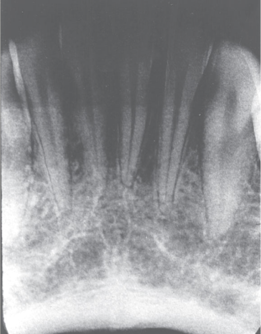
underexposed receptor
The image appears light
Check the exposure time, kilovoltage, and milliamperage
absence of apical structures
The receptor was not positioned in the patient’s mouth to cover the apical regions of the teeth
absence of apices

underexposed receptor

dropped receptor corner
The occlusal plane appears tipped or tilted
The edge of the receptor was not placed parallel to the incisal- occlusal surfaces
dropped corner
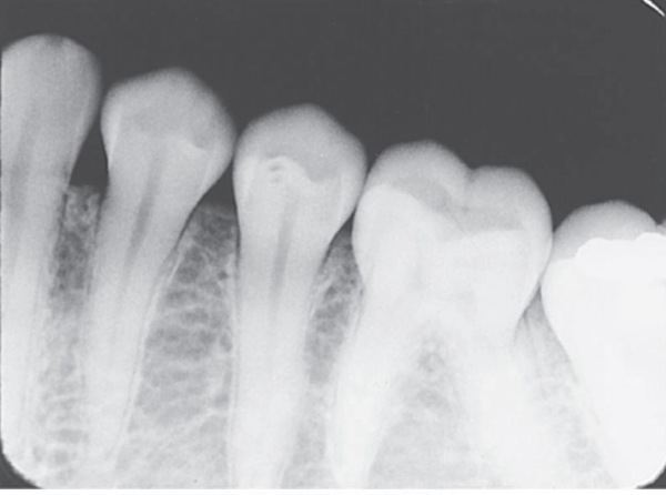
angulation
Overlapped contacts
The central ray was not directed through interproximal spaces
overlapped contacts

foreshortening
Short teeth with blunted roots
Excessive vertical angulation
foreshortening

elongation
Long, distorted teeth
The vertical angulation was insufficient.
elongation

cone cut
A clear/white area appears on the image
The PID was not directed to the center of the receptor
cone cut

receptor too posterior
Distal surfaces of the canines are not visible on the pre-molar bitewing image
receptor too anterior
Third molar regions are not visible on molar bitweing image
film bending
Images appear stretched and distorted
Check film placement before exposure
film creasing
A thin radiolucent line appears on the image
film bending

film creasing

phalangioma
The patient’s finger was positioned in front of the receptor
phalangioma

failure to remove partial
Any removable appliance must be removed from the patient’s mouth prior to exposure of radiographs
failure to remove partial

thyroid collar

double exposure
The receptor was exposed twice in the patient’s mouth
double exposure

reversed film
Light images with a herringbone pattern appear on the radiograph
reversed film
