Anatomy 2300 Unit 4: The Special Sense Organs Pt1
 Call Kai
 Learn
 Practice Test
 Spaced Repetition
 Match
 Flashcards
 Knowt Play
1/30

There's no tags or description
Looks like no tags are added yet.
31 Terms
Eye layers (superficial to deep)
The eyeball has three layers (tunics).
From superficial to deep they are the fibrous tunic, the vascular tunic (uvea), and the sensory tunic (retina).
The lens and the internal cavity sit inside these layers. The special sense of sight uses photoreceptors (rods and cones) as its receptor type.
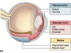
Fibrous Tunic (made up of?)
The most superficial, protective layer of the eyeball. It is made of the sclera and the cornea.

Sclera (What attaches to it?)
The white of the eye, forming the tough outer covering over most of the eyeball. The extrinsic eye muscles attach to it.

Cornea (Function?)
The clear, anterior part of the fibrous tunic. Light passes through it, and it refracts (bends) the light entering the eye.

Vascular Tunic (Other name? Made up of?)
(uvea): The middle layer, made of the choroid, ciliary body, iris, pupil, and lens.

Iris (Function? Para. & Sym.?)
The colored part of the eye, in the anterior vascular tunic in front of the lens. It changes the pupil's diameter.
Parasympathetic stimulation (CN III) contracts its circular muscles and constricts the pupil.
Sympathetic ("fight or flight") stimulation contracts its radial muscles and dilates the pupil.

Pupil (Function?)
The central opening of the iris, where light enters the eye.

Lens (Function?)
A structure behind the iris and pupil, held in place by the suspensory ligament. It focuses light onto the retina by changing shape, which the ciliary body controls.

Ciliary Body (Function? Far vs Near vision?)
A muscular part of the vascular tunic around the lens.
It changes the shape of the lens. When the ciliary muscles are relaxed, the lens is set for far vision (far accommodation). When they contract, it is set for near vision (near accommodation).

Choroid (Function? Highly what?)
The posterior part of the vascular tunic, lying deep to the sclera.
It is highly pigmented, so it absorbs light, and it is highly vascularized.

Sensory Tunic (Other name? Contains?)
The deepest layer of the eyeball, also called the retina. It contains the photoreceptors that detect light, and the optic nerve (CN II) leaves from it.

Retina (Made up of?)
The sensory tunic lining the back (deep side) of the eyeball.
It is made of photoreceptors, and the axons of its ganglion cells form the optic nerve (CN II).

Internal Cavity (Two segments?)
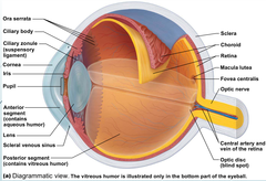
The space inside the eyeball, divided by the lens into two segments.
The anterior segment is in front of the lens and is filled with aqueous humor. (Replaced often)
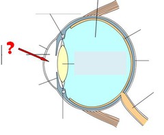
The posterior segment is behind the lens and is filled with vitreous humor. (Not replaced)
Aqueous humor (replaced? viscous?)
The liquid filling the anterior segment, in front of the lens. It is replaced often.
Vitreous humor (replaced? viscous?)
The gelatinous material filling the posterior segment, behind the lens. It is not replaced.

Lacrimal gland (Tear flow? Supplied by?)
The tear-producing gland, located at the upper lateral side of the eye.
It is supplied by parasympathetic fibers of CN VII. Tears flow across the eye toward the medial corner.

Nasolacrimal duct (Function?)
The duct that drains tears from the eye into the nose.
Tears enter through the lacrimal punctum and canaliculus, then pass into the lacrimal sac. From there the nasolacrimal duct carries them down to the inferior meatus of the nasal cavity.

Photoreceptors (Made up of? Function?)
(rods and cones): The sensory receptors for sight, located at the back of the retina. They convert light into an electrical signal, which is the first step of the visual pathway.

Ganglion cells (Function? Leave eye as?)
The retinal neurons on the inner side of the retina that receive the signal from the photoreceptors.
Their axons leave the eye together as the optic nerve (CN II).
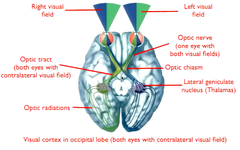
optic nerve (How does it travel?)
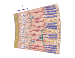
The optic nerve (CN II) carries visual information from the retinal ganglion cells in the retina to the brain.
It travels from the back of the eye to the optic chiasm, where some fibers cross, and then continues toward the thalamus and visual cortex for processing.
Optic chiasm (nasal and peripheral?)
The point where the two optic nerves meet, before the optic tracts.
It is a partial crossover: fibers from the nasal side of each retina cross to the opposite side, and these carry peripheral vision.
Optic tract (Carries information to? Which side?)
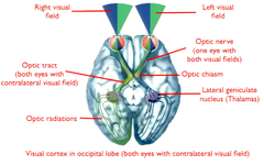
The continuation of the fibers after the optic chiasm, carrying them toward the thalamus.
Each tract carries information from the contralateral (opposite) visual field. The right tract carries the left visual field, and the left tract carries the right visual field.
Detail the visual pathway in order
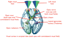
Light path through the eye: Light passes through the cornea, aqueous humor, pupil, lens, and vitreous humor to reach the retina.
Photoreceptors: They convert the light into an electrical signal.
Ganglion cells: Their axons leave the eye as the optic nerve (CN II). Each optic nerve carries information from both the right and left visual fields.
Optic chiasm: A partial crossover, where nasal retinal fibers carrying peripheral vision cross to the opposite side.
Optic tract: Each tract carries the opposite (contralateral) visual field.
Thalamus: Most optic tract fibers synapse here.
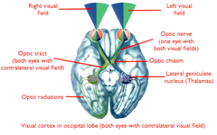
Primary visual cortex: Fibers from the thalamus (the optic radiations) project to the primary visual cortex in the occipital lobe. The right cortex integrates the left visual field, and the left cortex integrates the right visual field.
Extrinsic Eye Muscles
Six muscles that move the eyeball. They originate on the surface of the orbit (bone) and insert on the sclera.
A way to remember the nerves is LR6 SO4 AO3: the lateral rectus is CN VI, the superior oblique is CN IV, and all others are CN III.

Superior rectus muscle
On the top of the eyeball. It elevates the eye ("look up").
Action: Elevation of eyeball
Innervation: Oculomotor nerve (CN III)

Inferior rectus muscle
On the bottom of the eyeball.
Action: Depression of eyeball
Innervation: Oculomotor nerve (CN III)

Medial rectus muscle
On the nose side of the eyeball.
Action: Adduction of eyeball
Innervation: Oculomotor nerve (CN III)
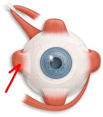
Lateral rectus muscle
On the outer side of the eyeball.
Action: Abduction of eyeball
Innervation: Abducens nerve (CN VI)

Superior oblique muscle
Passes through the trochlea on the medial side of the orbit.
Action: Depression & abduction of eyeball
Innervation: Trochlear nerve (CN IV)

Inferior oblique muscle
Runs along the bottom of the eyeball.
Action: Elevation & abduction of eyeball
Innervation: Oculomotor nerve (CN III)

Trochlear
A pulley-shaped structure in the orbit of the eye that guides the tendon of the superior oblique muscle, helping the eye move downward and outward.