Exam 2 Content Flashcards - BIOC 384
 Call Kai
 Learn
Learn Practice Test
 Spaced Repetition
 Match
Match Flashcards
 Knowt Play
1/46

There's no tags or description
Looks like no tags are added yet.
Name | Mastery | Learn | Test | Matching | Spaced | Call with Kai | Chat |
|---|
No analytics yet
Send a link to your students to track their progress
47 Terms
What is the function of gel electrophoresis?
tool used to separate, identify, and compare proteins based on charge, size, and abundance
SDS-PAGE and IEF are foundational techniques and can be combined with 2D PAGE to enable deep proteomic analysis of complex samples
M4T1
What is polyacrylamide gel electrophoresis (PAGE)?
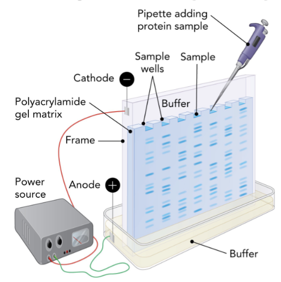
PAGE uses a polyacrylamide gel matrix & electric field to separate proteins by charge & size
gel is positioned between 2 buffer chambers connected to an external power supply with a cathode (- charge) side and anode (+ charge) side
negatively charged cathode that attracts positively charged proteins (cations)
positively charged anode that attracts negatively charged proteins (anions)
M4T1
What is SDS-PAGE?
uniform negative charge
uses sodium dodecyl sulfate (SDS) to denature proteins and give negative charges
proteins then migrate toward the anode at rates inversely proportional to their molecular mass
separation based on pore size
in vertical SDS-PAGE systems, small proteins migrate faster than large proteins through polyacrylamide matrix
pore size of gel is adjusted by changing acrylamide concentration
low % gel resolves large proteins
high % gel resolves small proteins
M4T1

How can SDS-PAGE be used to estimate molecule mass?
by comparing the migration distances of unknown proteins to molecular mass markers, you can estimate apparent molecular mass
log/mass vs migration distance plot provides a standard curve to interpret results
M4T1

How can SDS-PAGE be used to visualize proteins?
by staining
blue dye binds to basic amino acids and stain proteins in gels
M4T1

What is IEF and its function?
IEF separates proteins by their isoelectric point (pl), the pH at which they have no net charge
pH = pl
gel slices can be cut out and proteins extracted for used in biochemical assays
M4T1
What is 2D PAGE and its function?
2 dimensional gel electrophoresis
2D PAGE combined IEF (1st dimension) and SDS-PAGE (2nd dimension) to separate proteins by pl and molecular mass
M4T1

What is Edman degradation?
allows residue-by-residue identification for short peptides
phenylisothiocyanate (PITC) is used to label the N-terminus amino acid
after cleavage with trifluoroacetic acid, the labeled residue is removed as a thiazolinone derivative and converted to a PTH-amino acid for identification via chromatography
proteins are digested into smaller, overlapping fragments using proteases like trypsin (cleaves after R and K) and chymotrypsin (cleaves after Y, W, F, L, M)
these overlapping peptides are sequenced individually and their overlap is used to reconstruct the full protein sequence
key concept: protein sequencing method that uses chemical labeling and cleavage, in combination with amino acid standards, to identify N-terminal amino acids sequentially
M4T2
What is mass spectrometry?
determines peptide mass to charge (m/z) ratios from which molecular masses are calculated
data is matched to predicted peptide masses from genomic databases
enables accurate protein identification
key concept: measures the mass-to-charge ratio (m/z) of molecules, which is used to deduce the molecular mass of peptide fragments through computational analyses of the spectral data to predict the identity of proteins
M4T2

What is Tandem mass spectrometry (MS/MS)?
uses two spectrometers
first selects specific tryptic fragments
second analyzes sub-fragments after collision-induced dissociation
compares experimental spectra to in silico spectra from genome databases to get precise peptide identifications
M4T2

What is electrospray ionization spectrometry (ESI)?
ESI emits peptide-laden droplets from high-voltage capillary resulting in solvent evaporation that creates highly charged gas phase
ESI is less harsh that MS/MS which allows analysis of intact proteins & peptides from complex biological samples
M4T2

What is MALDI-TOF mass spectrometry?
matrix-assisted laser desorption/ionization
paired with time of flight molecular mass analysis (MALDI-TOF) to analysis biological samples embedded in a light-absorbing matrix
used to design treatment strategies for patients with life threatening bacterial infections
M4T2
What is X-ray crystallography?
uses a focused x-ray bean directed at a protein crystal which is diffracted by electron-dense atoms
creates patterns captures on detectors
computational analysis transforms diffraction data into 3D electron density maps used to model protein structure
M4T3

What are ribbon representations?
pioneered by Jane Richardson to illustrate protein secondary structures (alpha helices, beta strands)
based on atomic coordinates, they highlight structural motifs and domain organization
M4T3
What is NMR spectroscopy?
NMR exploits nuclear magnetic properties of isotopes like 1H, 15N, and 13C
protein samples in solution are subjected to radiofrequency pulses in a strong magnetic field, which reveals interatomic distances and bonding patterns
M4T3
What is cryo-electron microscopy (cryo-EM)?
provides 3D structures from electron beam images of flash frozen proteins
recent advances enable atomic resolution reconstructions of large complexes without crystallization
requires 5 steps
M4T3
What are the major classes of functional proteins?
metabolic enzymes
structural proteins
transport proteins
cell signaling proteins
genomic caretaker proteins
M4T4
What is the function of metabolic enzymes?
catalysts for life
catalyze biochemical reactions essential for energy conversion and macromolecule synthesis/degradation without altering delta G or Keq
specificity of enzymes come from unique active site encironments shaped by amino acid side chains
M4T4
What is the function of structural proteins?
cellular architecture
form cytoskeletal structures involved in cell migration, chromosomal segregation, and muscle cell contraction
maintain cell shape and form networks of scaffolding in cytoplasm & extracellular matrix
ex. cytoskeletal proteins, including actin, tubulin, and intermediate filaments support cell structure, motility, and intracellular transport
M4T4
What is the function of transport proteins?
membrane gatekeepers
span membranes & function as selective pores
transport proteins are embedded in membranes & facilitate movement of molecules across lipid bilayers
two main types: passive transporters and active transporters
M4T4
What is the function of cell signaling proteins?
information flow
respond to changes in extracellular environment by undergoing conformational changes
membrane receptors like G protein-coupled receptors & receptor tyrosine kinase initiate intracellular signals in response to extracellular ligand binding
growth hormone receptors, like erythropoietin receptor, dimerize & activate intracellular kinases that mediate a phosphorylation cascade
M4T4
What is the function of genomic takecare proteins?
DNA protection
ensure genomic stability by facilitating DNA replication, repair, recombination, and gene transcription
DNA polymerases, ligase, topoisomerase, RecA, and ReBCD complex protect DNA from environmental damage & from DNA replication errors
M4T4
Why is the globin heme group important?
O2 binding to heme triggers structural transitions that regulate affinity & enable efficient O2 storage (myoglobin) or delivery (hemoglobin)
O2 reversibly binds to an iron atom contained in porphyrin ring of the heme but only when Fe is in the +2 oxidation state
heme is needed because none of the AA side chains in globin is ideally suited for reversible binding of oxygen to protein
M4T5
Describe the structure of myoglobin and hemoglobin?
myoglobin is a monomer with 1 heme
hemoglobin is a tetramer with 4 heme
all globin subunits share the globin fold which is made up of eight alpha helices
tetrameric hemoglobin is a dimer of a heterodimer enabling cooperative binding
M4T5

Describe the heme coordinate of the histidine residues.
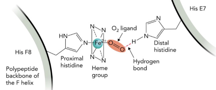
Fe2+ in the heme forms 6 coordination bonds
4 in plane with porphyrin
2 perpendicular
one axial bond connects to His F8 (proximal histidine)
other binds O2, stabilized by His E7 (distal histidine)
M4T5
Describe the conformational changes on O2 binding in hemoglobin.
O2 binding reduces the Fe2+ radius, allowing it to move into the heme plane
this shifts His F8 and tilts the F helix causing conformational changes
in hemoglobin, the movement in iron leads to large quaternary shifts
M4T5

Describe ligand binding equilibria and Kd.
binding of O2 to the heme Fe2+ of the hemoglobin and myoglobin is reversible & structure of protein changes under different conditions and results in altered affinities for oxygen ligand
reversible binding of ligands is a common feature of many biomolecules
P + L ←→ PL
P: concentration of protein
L: concentration of ligand
PL: concentration of protein-ligand complex
low Kd = high affinity for L
high Kd = low affinity for L
M4T5

Describe the structural changes in Hb upon O2 binding.
conformational changes within one Hb subunit is response to O2 binding affects the quaternary structure of the entire hemoglobin protein
the O2 bound form of hemoglobin is oxyhemoglobin and is R state (relaxed) conformation, whereas the form of hemoglobin without bound O2 is deoxyhemoglobin and in in T state (tense) conformation
helices reposition in the alpha & beta subunits when hemoglobin goes from the T state (deoxy) to R state (oxy)
this movement involves the breaking and reforming of many noncovalent interactions between adjacent alpha and beta subunits including hydrogen bonds
M4T5

What is the Bohr effect?
describes how addition to a drop of O2 concentration in tissues compared to lungs, there is also a decrease in Ph and in the tissue (7.2) compared to the lungs (7.6)
lower pH leads to protonation of His146 in the B1 subunit, which forms an ionic bond with Asp84, stabilizing the T state
second ionic bond with Lys40 in the a2 subunit positions His146 properly, which contributes significantly to the pH sensitive conformational change
M4T6

Describe how CO2 transport affects tissue pH.
primary by product of aerobic respiration is CO2, which is hydrated by carbonic anhydrase to form highly soluble bicarbonate (HCO3-)
in addition to forming bicarbonate from CO2 + H2O, carbonic anhydrase produces H+, which helps reduce the pH in tissues
M4T6

Describe carbamate formation on N-term valine.
bicarbonate generates a carbamate group on the N-terminal residue (Val) of all 4 hemoglobin subunits
this reversible rxn releases H+ that contributes to the Bohr effect & stabilizes the T state in tissues
M4T6

How does 2,3-BPG stabilize the T state conformation?
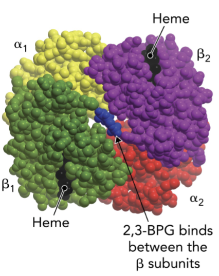
2,3-BPG is a negative heterotropic regulator that binds the central cavity of deoxyglobin, stabilizing the T state
interacts with His2, Lys82, and His143 on both beta subunits
these ionic interactions stabilize the T state, lower O2 affinity, and promote O2 unloading in tissues, especially under low O2 conditions
only 1 2,3-BPG molecule binds per tetrameter, but its effect is sufficient to reduce O2 binding to all subunits
M4T6
How do changes in O2 affinity mediate O2 delivery?
2,3-BPG levels remain constant in tissues & lungs
O2 affinity is lower in tissues due to low O2 concentration and the pH dependence of Bohr effect
function 2,3-BPG is to maintain T state stability, maximizing O2 release when hemoglobin encounters low pO2 and high CO2/H+ tissues
fetal hemoglobin has high O2 affinity bc His143 → Ser143 in y subunits eliminates two positive charges
this promotes more R state hemoglobin in the fetus, enhancing O2 transfer from mother to fetus across the placenta
M4T6

M4T6
M4T6
M4T6