[4] Evaluating the Deviation
 Call Kai
 Learn
 Practice Test
 Spaced Repetition
 Match
 Flashcards
 Knowt Play
1/119

There's no tags or description
Looks like no tags are added yet.
120 Terms
What is strabismus, and what components of binocular vision can it affect?
Strabismus is a misalignment of the eyes that represents a breakdown in one or more components of normal binocular vision. Associated abnormalities may be:
Sensory
Motor
Both sensory and motor
What are the three major types of ocular deviation (ocular misalignment)?
Heterophoria: Latent deviation controlled by fusion and revealed when fusion is disrupted.
Heterotropia: Manifest deviation present despite binocular viewing (may be constant or intermittent).
Fixation disparity: Small residual misalignment despite maintained fusion.
What are the key requirements for normal binocular vision?
Normal binocular vision requires:
Healthy eyes and visual pathways
Accurate monocular fixation by each eye
Proper neuromuscular coordination of intra- and extraocular systems
Accurate bifoveal fixation
Normal retinal correspondence (foveae serving as corresponding points)
Similarity of the two retinal images
Sensory fusion (unification of the two ocular images)
Which sensory and motor factors are essential for maintaining binocular single vision?
Motor factors:
Accurate monocular fixation
Proper neuromuscular coordination
Bifoveal alignment/fixation
Sensory factors:
Normal retinal correspondence
Similar retinal image size/clarity
Sensory fusion of the two images
What is fixation disparity, and how does it differ from a tropia or phoria?
Fixation disparity is a small ocular misalignment present under fused (associated) viewing conditions where the retinal images still fall within Panum's fusional area, so diplopia does not occur.
Fusion is maintained
Usually < 30 minutes of arc
Smaller than a phoria or tropia
Considered a residual alignment error during binocular viewing
Under what viewing conditions is fixation disparity measured, and what tests are used?
Fixation disparity is measured under associated (fused) viewing conditions, meaning fusion is maintained during testing.
Common tests:
Borish Card
Saladin Card
Wesson Card
Disparometer
Bernell Box
What is a heterophoria (phoria), and when is it measured?
A heterophoria is a latent ocular deviation that is normally controlled by fusion.
It is measured only when fusion is disrupted (dissociated viewing conditions).
Key distinction:
Phoria = latent deviation + fusion broken
Fixation disparity = tiny residual deviation + fusion maintained
How is a heterophoria quantified, and what are the common testing methods?
Heterophorias are measured in prism diopters (Δ).
Common tests:
Alternating Cover Test
Von Graefe
Maddox Rod
Modified Thorington
Compare fixation disparity and heterophoria.
Feature | Fixation Disparity | Heterophoria |
|---|---|---|
Fusion present? | Yes | No (disrupted) |
Viewing condition | Associated | Dissociated |
Size | Very small (<30 min arc) | Larger latent deviation |
Units | Minutes of arc | Prism diopters (Δ) |
Symptoms | Can contribute to asthenopia | May decompensate and cause symptoms |
Example tests | Borish, Saladin, Wesson | Cover test, Von Graefe, Maddox Rod |
What is strabismus, and what are its common synonyms?
Strabismus is a manifest ocular deviation in which binocular fixation is not present under normal viewing conditions. One eye's visual axis fails to intersect the object of fixation.
AKA:
Heterotropia
Tropia
Squint
Manifest deviation
Strab
How is a tropia different from a phoria?
Tropia (heterotropia):
Manifest deviation
Present during normal binocular viewing (associated conditions)
Fusion cannot fully maintain alignment
Detected with unilateral cover test
Phoria (heterophoria):
Latent deviation
Revealed only when fusion is broken (dissociated conditions)
Detected with alternating cover test and other dissociating techniques
High-yield:
Tropia = manifest
Phoria = latent
How is strabismus measured and what tests are commonly used?
Strabismus is measured in prism diopters (Δ).
Common tests:
Unilateral (cover-uncover) Cover Test
Hirschberg Test (corneal light reflex assessment)
Exam pearl: A unilateral cover test detects a manifest deviation (tropia), whereas an alternating cover test reveals a latent deviation (phoria).
What are the major risk factors for developing strabismus?
Major risk factors include:
Prematurity / low birth weight
Maternal smoking during pregnancy
Family history
Neurologic or CNS disorders
Examples of neurologic associations:
Cerebral palsy
Down syndrome
Which refractive errors are commonly associated with esotropia and exotropia?
Hyperopia → Esotropia
Excessive accommodative effort can drive accommodative convergence.
Anisometropia and astigmatism → Exotropia
Reduced binocular sensory input can predispose to exodeviation.
High-yield association:
Hyperopia = ET, whereas anisometropia/astigmatism = XT.
What are the major etiologic categories of strabismus?
Strabismus can result from four broad categories:
Optical abnormalities
Sensory (ocular disease) abnormalities
Innervational abnormalities
Anatomical abnormalities
How can optical abnormalities lead to strabismus? Give examples.
Optical abnormalities can disrupt binocular vision and sensory fusion, leading to strabismus.
Examples:
Anisometropia
High refractive error
Mechanism: Unequal or blurred retinal images weaken fusion and may result in ocular misalignment.
How does sensory deprivation from ocular disease influence the type of strabismus that develops?
Sensory ocular disease can cause loss of fusion and subsequent strabismus.
Age-dependent pattern:
Younger children → Esotropia (ET) more likely
Older children and adults → Exotropia (XT) more likely
Exam pearl: Sensory deprivation tends to produce ET in young children but XT in older patients.
What are innervational causes of strabismus?
Innervational strabismus occurs from abnormalities in muscle tone or neural control of eye movements.
Causes include:
Intoxication
Medications
Trauma
Neurologic disease
Congenital innervational abnormalities
Abnormal AC/A ratios (high or low)
High-yield: Think of a defect in the neural command to the extraocular muscles, rather than the muscles themselves.
What anatomical abnormalities can cause strabismus?
Anatomical causes involve structural abnormalities affecting ocular alignment.
Examples:
Extraocular muscle (EOM) abnormalities
Abnormal orbital contents
Abnormal ligaments or tendons
Mechanism: Physical restriction or altered muscle anatomy prevents normal ocular alignment.
What are the key characteristics used to describe a strabismic deviation?
A deviation is characterized by:
Direction (eso, exo, hyper, hypo)
Frequency (constant or intermittent)
Magnitude (size of deviation)
Laterality (unilateral vs alternating)
AC/A ratio
Cosmesis (appearance/significance of the deviation)
Comitancy (whether deviation remains the same in all gazes)
What does "comitancy" mean when evaluating a strabismus?
Comitancy refers to whether the angle of deviation remains constant in all directions of gaze.
Comitant deviation: Same magnitude in all gaze positions.
Incomitant deviation: Magnitude varies with gaze direction, often suggesting neurologic, muscular, or mechanical pathology.
Why are laterality, frequency, and magnitude important when describing a strabismus?
Laterality: Determines whether one eye consistently deviates (unilateral) or fixation alternates between eyes.
Frequency: Indicates whether the deviation is constant or intermittent.
Magnitude: Quantifies the size of the deviation, usually in prism diopters (Δ).
These characteristics help guide diagnosis, prognosis, and treatment planning.
How is strabismus classified by frequency?
Strabismus can be classified as:
Heterophoric (latent deviation, controlled by fusion)
Intermittent strabismus (manifest only part of the time)
Constant strabismus (manifest all of the time)
How is control of intermittent strabismus quantified?
Common methods include:
POTS = Percentage of Time Strabismic
POTT = Percentage of Time Troped
Office Control Scale (commonly used for exotropia)
High-yield: The more frequently the eye manifests a tropia, the poorer the patient's binocular control.
How is the magnitude of a strabismic deviation measured and classified?
Magnitude is measured in prism diopters (Δ) at both distance and near.
Classification:
Micro: 0-5Δ
Small: 6-20Δ
Moderate: 21-40Δ
Large: >40Δ
What are the major directional classifications of strabismus?
Strabismus can be classified by direction as:
Eso-: Eye turns inward
Exo-: Eye turns outward
Vertical: One eye higher or lower than the other
Hypertropia
Hypotropia
Cyclotorsional: Rotational misalignment
Combination: Horizontal + vertical and/or torsional components
What do the terms esotropia, exotropia, and hypertropia mean?
Esotropia (ET): Deviating eye turns toward the nose.
Exotropia (XT): Deviating eye turns away from the nose.
Hypertropia: Deviating eye is higher than the fellow eye.
Naming rule: The deviation is named by the position of the deviating eye.
Left eye inward → Left ET
Left eye outward → Left XT
Left eye higher → Left HT
How is the cosmetic significance (cosmesis) of strabismus graded?
Cosmesis describes how noticeable the deviation appears to an observer.
Common grading:
Excellent
Good
Fair
Poor
At what magnitude does strabismus become cosmetically noticeable?
General guidelines:
< 10Δ → Usually not cosmetically noticeable
10-20Δ → May be cosmetically noticeable
> 20Δ → Usually cosmetically noticeable
Exam pearl: Cosmetic concerns become increasingly likely once the deviation exceeds 20Δ.
What facial and ocular features can make an esotropia appear more cosmetically noticeable?
Factors associated with esotropia appearance:
Negative angle kappa
Wide nasal bridge
Prominent epicanthal folds
Small interpupillary distance (IPD)
What facial and ocular features can make an exotropia appear more cosmetically noticeable?
Factors associated with exotropia appearance:
Positive angle kappa
Narrow nasal bridge
Minimal/absent epicanthal folds
Large interpupillary distance (IPD)
These features can accentuate the appearance of an outward deviation.
What is the AC/A ratio, and what is considered normal?
The AC/A ratio (Accommodative Convergence/Accommodation ratio) describes how much convergence occurs for a given amount of accommodation.
Normal AC/A: 4:1 ± 2
Approximate normal range:
2:1 to 6:1
How are AC/A ratios classified?
Low AC/A: Less convergence than expected for a given accommodative response
Normal AC/A: Approximately 4:1 ± 2
High AC/A: Greater convergence than expected for a given accommodative response
How do cosmesis and AC/A ratio contribute to a strabismus evaluation?
Cosmesis
Assesses how noticeable the deviation is
Influenced by deviation size and facial anatomy
Guides treatment decisions and patient concerns
AC/A Ratio
Assesses the relationship between accommodation and convergence
Helps explain differences between near and distance deviations
Can identify abnormal accommodative-convergence relationships
What should be evaluated during direct observation of a patient with suspected strabismus?
Key observations include:
Eyelid position (ptosis vs pseudoptosis)
Palpebral fissure width and whether it changes
Presence of epicanthal folds
Abnormal head posture
Head turn
Head tilt
Head tip/chin position
Why are eyelids and epicanthal folds important when evaluating strabismus?
They can create the appearance of strabismus or mask a true deviation.
Look for:
Ptosis (true drooping eyelid)
Pseudoptosis (appears droopy but not true ptosis)
Changes in palpebral fissure width
Prominent epicanthal folds
What abnormal head postures should be assessed in strabismus, and why do they occur?
Evaluate for:
Head turn
Head tilt
Head tip (chin up/down posture)
These are often compensatory positions adopted to:
Maintain binocular single vision
Reduce diplopia
Place the eyes in a field of gaze with less deviation
How can an abnormal head posture help localize an extraocular muscle palsy?
Patients frequently adopt a posture that minimizes the deviation and diplopia.
Rule:
The head is often turned toward the field of action of the paretic muscle.
Example:
A patient with a left lateral rectus palsy may turn the face to the left to reduce the need for left gaze and improve binocular alignment.
What information can the Hirschberg test provide about a strabismus?
The Hirschberg test can:
Determine whether strabismus is present
Identify the direction of deviation
Identify laterality (which eye deviates)
Estimate frequency
Estimate magnitude
How does the Hirschberg test work?
The Hirschberg test is an objective assessment of ocular alignment that evaluates the position of the corneal light reflex relative to the center of the pupil.
Symmetric reflexes → aligned eyes
Asymmetric reflexes → ocular deviation present
Why is the Hirschberg test especially useful in young children?
The Hirschberg test:
Requires minimal patient cooperation
Is objective
Does not require verbal responses
Preserves normal binocular viewing conditions
Why is direct observation important before formal strabismus testing begins?
Direct observation helps determine:
Direction of deviation
Laterality
Frequency (constant vs intermittent)
before any formal testing is performed.
Why is cosmesis an important part of strabismus evaluation?
Cosmesis is important because:
It may influence treatment decisions
It is often considered before surgical referral
It commonly represents the patient's or parent's primary concern
The perceived deviation may matter as much as the measured deviation
What is angle kappa (lambda), and under what conditions is it measured?
Angle kappa (lambda) is the relationship between the visual axis and pupillary axis, assessed by observing the position of the corneal light reflex.
Measured under monocular conditions
Usually symmetric between eyes
Most commonly, the reflex is displaced ~0.5 mm nasally
Occurs because the fovea lies temporal to the pupillary axis
Why is angle kappa important when evaluating strabismus?
Although angle kappa is not highly sensitive for measuring eye turns, it can:
Simulate strabismus (pseudostrabismus)
Conceal true strabismus
Exaggerate the appearance of strabismus
High-yield: Always compare binocular Hirschberg findings with each eye's monocular angle kappa before diagnosing a deviation.
What is the normal relationship between angle kappa and the corneal light reflex?
In the typical (positive) angle kappa:
The fovea lies temporal to the pupillary axis.
The corneal light reflex is therefore seen slightly nasal to the pupillary center.
Normal displacement ≈ 0.5 mm nasal.
How can corneal light-reflex displacement be converted to prism diopters?
Approximate Hirschberg conversion:
1 mm displacement ≈ 22Δ
0.5 mm displacement ≈ 11Δ
What is the Hirschberg test procedure?
Patient fixates a penlight/transilluminator binocularly (typically ~50 cm away).
Observe the corneal light reflex in each eye.
Compare the reflex position relative to the center of each pupil.
Determine whether the reflexes are:
Symmetric → alignment likely normal
Asymmetric → possible strabismus
Compare findings with each eye's monocular angle kappa.
How are angle kappa and Hirschberg testing related?
Angle Kappa | Hirschberg Test |
|---|---|
Measured monocularly | Measured binocularly |
Normal physiologic reflex position | Screens for strabismus |
Usually symmetric | Compares reflexes between eyes |
Can mimic or mask strabismus | Used to detect direction and estimate magnitude |
How do you interpret horizontal corneal light reflex displacement during the Hirschberg test?
Compare the displaced reflex to the normal position.
Equal reflexes in both eyes → No strabismus
Temporal displacement of the reflex → Esotropia (ET)
Nasal displacement of the reflex → Exotropia (XT)
How do you interpret vertical corneal light reflex displacement during the Hirschberg test?
Inferior displacement of the corneal reflex → Hypertropia
Superior displacement of the corneal reflex → Hypotropia
What Hirschberg findings correspond to eso-, exo-, hyper-, and hypotropia?
Reflex Displacement | Deviation |
|---|---|
Temporal | Esotropia (ET) |
Nasal | Exotropia (XT) |
Inferior | Hypertropia (HT) |
Superior | Hypotropia |
What is the Krimsky test, and when is it useful?
The Krimsky test is a modification of the Hirschberg test that uses prisms to quantify the magnitude of a strabismus.
Especially useful for:
Young children
Poorly cooperative patients
Patients with severely reduced vision in one eye
Patients in whom cover testing is difficult

Why is the Krimsky test often preferred over Hirschberg alone for measuring deviation size?
The Hirschberg test requires estimating corneal reflex displacement in millimeters, which can be difficult and imprecise.
The Krimsky test improves accuracy by:
Using prisms
Neutralizing the deviation
Determining the prism power needed to align the corneal reflexes

What are the steps of the Krimsky test?
Observe the corneal reflexes (as in Hirschberg).
Place a prism over the fixating eye.
Increase or decrease prism power until the corneal reflexes become symmetric.
The prism power that equalizes the reflexes equals the approximate deviation magnitude.

Where is the prism placed during a Krimsky test?
The prism is placed over the fixating eye and adjusted until the corneal light reflexes are symmetrical.
What endpoint indicates a successful Krimsky neutralization?
The endpoint is reached when the corneal light reflexes are symmetric between the two eyes.
Interpretation:
The prism power required to achieve symmetry is the estimated magnitude of the strabismus in prism diopters (Δ).
What is the Bruckner test and how is it performed?
The Bruckner test is a screening test for ocular misalignment using the red reflex.
Procedure:
Patient fixates an ophthalmoscope light from approximately 1 meter away.
Light illuminates both eyes simultaneously.
Compare the brightness of the two red reflexes.
How are Bruckner test results interpreted?
Compare the red reflexes from the two eyes.
Brighter reflex = deviating eye
Symmetric reflexes suggest normal alignment
High-yield: A brighter red reflex generally comes from the non-fixating/deviating eye because of the altered optical alignment.
In which patients is the Bruckner test most useful?
The Bruckner test is especially helpful in:
Infants
Young children
Patients with developmental or special needs
Patients unable to cooperate with cover testing
What factors improve visualization during the Bruckner test?
Use an ophthalmoscope from about 1 meter
Evaluate both eyes simultaneously
Pupil dilation may improve results
Observe relative brightness of the red reflexes
What are the three major cover tests used in strabismus evaluation, and what does each measure?
Unilateral Cover Test (UCT) → Detects a manifest deviation (tropia).
Simultaneous Prism Cover Test (SPCT) → Measures the manifest component of a deviation.
Alternating Cover Test (ACT) / Prism Alternate Cover Test (PACT) → Measures the total deviation (phoria + tropia).
Why is target selection important during cover testing?
Accurate cover testing requires an accommodative target that keeps the patient actively fixating and accommodating.
Key principle: Ensure the target remains clear throughout testing.
How can you verify that a patient is truly fixating an accommodative target during cover testing?
Ask the patient to:
Read letters on the target
Describe details in a picture
Identify specific features of the fixation object
How should fixation targets be modified for patients with amblyopia?
The target should be adjusted so it falls within the patient's visual threshold.
Use larger optotypes if needed.
Ensure the amblyopic eye can actually see the target.
What fixation targets can be used with digital acuity systems during cover testing?
Digital systems may use:
Cycling picture targets
Animated fixation targets
Movies (especially for young children)
Age-appropriate accommodative letters
What is the purpose of the Unilateral Cover Test (UCT)?
The Unilateral Cover Test (Cover-Uncover Test) is used to detect and characterize a manifest deviation (tropia).
It helps determine:
Direction of deviation
Laterality (unilateral vs alternating)
Frequency (constant vs intermittent)
Approximate magnitude (based on amount of movement)
How is the Unilateral Cover Test performed?
Have the patient fixate binocularly on an accommodative target.
Start in primary gaze (midline).
Cover one eye.
Observe the uncovered eye.
Look for any movement as it takes up fixation.
Then "chase" the deviation by covering the fixating eye and observing the other eye.
How can the Unilateral Cover Test help determine frequency and laterality of strabismus?
Repeat the test multiple times (5-10 repetitions).
This helps determine:
Constant vs intermittent deviation
Unilateral vs alternating fixation
Findings:
Same eye always deviates → unilateral tropia
Either eye can fixate → alternating tropia
Eye turn seen only some of the time → intermittent tropia
Why should the examiner allow time for refixation during a UCT?
The patient must be given enough time for the uncovered eye to:
Detect the target
Acquire fixation
Complete any refixation movement
Failing to wait may cause small or slow tropias to be missed.
What clinical information can be obtained from the Unilateral Cover Test?
Information | Can UCT Determine? |
|---|---|
Tropia present? | ✅ Yes |
Direction? | ✅ Yes |
Laterality? | ✅ Yes |
Constant vs intermittent? | ✅ Yes |
Estimate magnitude? | ✅ Rough estimate |
Phoria? | ❌ No |
What is the Simultaneous Prism Cover Test (SPCT), and what does it measure?
The SPCT neutralizes the movement seen during the Unilateral Cover Test (UCT) and measures the size of the manifest deviation (tropia) only.
How is the Simultaneous Prism Cover Test performed?
Identify the deviating eye during UCT.
Place a prism over the deviating eye.
Simultaneously cover the fixating eye.
Observe the deviating eye's movement.
Increase prism power until there is no movement.
Confirm with reversal (bracketing).
What is the endpoint of the Simultaneous Prism Cover Test?
The endpoint is reached when the deviating eye shows no movement when the fixating eye is covered.
Bracketing (reversal):
Too little prism → eye still moves in original direction.
Neutralizing prism → no movement.
Too much prism → eye moves in the opposite direction.
Example:
5Δ → eye moves out
8Δ → no movement (neutralized)
10Δ → eye moves in (reversal)
Thus the deviation is approximately 8Δ.
How does SPCT differ from UCT and PACT?
Test | Measures |
|---|---|
UCT | Detects presence of a tropia |
SPCT | Measures the manifest deviation (tropia) |
PACT/ACT | Measures the total deviation (tropia + phoria) |
In which patients should SPCT be performed?
SPCT should be performed only when a manifest strabismus (tropia) has already been identified.
Why?
The test is designed to neutralize a visible deviation.
No manifest deviation = nothing to neutralize.
What is the major advantage of the Simultaneous Prism Cover Test?
Because fusion is disrupted only minimally, SPCT measures the size of the manifest deviation alone, without allowing the latent component (phoria) to appear.
Clinical significance: It isolates the true tropic component of the deviation.
What is the primary objective of the Alternate Cover Test (ACT/PACT)?
The Alternate Cover Test measures the total angle of deviation () under fully dissociated (fusion-free) conditions.
This represents:
Tropia + Phoria
The patient's full deviation
How is the Alternate Cover Test performed?
Patient fixates an accommodative target with best correction.
Place prism over the deviating eye.
Alternate the cover rapidly between eyes, ensuring full dissociation.
Observe movement of the eye behind the prism.
Increase prism power until neutralization.
Continue until the first reversal is observed.
What is meant by “full dissociation” during the Alternate Cover Test?
Full dissociation means fusion is completely disrupted by alternately covering the eyes before binocular fixation can be re-established.
Why?
Reveals the full latent deviation.
Prevents fusional vergence from masking the true angle.
How is prism neutralization determined during ACT/PACT?
Use bracketing (reversal):
Increase prism until no movement is seen.
Continue increasing until the first reversal occurs.
Record the highest neutral prism value.
Why should larger deviations be given additional time to refixate during ACT?
Larger angles require larger eye movements to regain fixation.
Allowing sufficient time:
Improves measurement accuracy
Prevents underestimation of deviation
Makes reversal easier to identify
Clinical pearl: The bigger the deviation, the longer you should wait for complete refixation.
Why should ACT be repeated with the strabismic eye fixing?
Repeating the test with the strabismic eye fixing allows assessment for:
Primary vs secondary deviation
Incomitance
Paretic deviations
High-yield: Differences between measurements with each eye fixing can suggest neurologic or muscle palsy rather than a simple comitant strabismus.
What test conditions are necessary for obtaining an accurate ACT/PACT measurement?
Best-corrected visual acuity/refractive correction
Accommodative fixation target
Complete dissociation
Adequate refixation time
Appropriate starting prism estimate (often from UCT/SPCT)
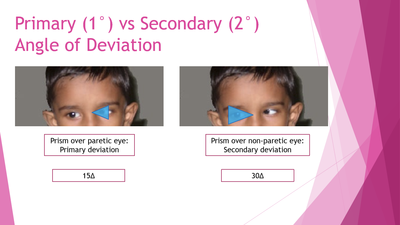
What is the difference between the primary and secondary angle of deviation?
Primary deviation (1°): Angle of deviation when the normal (non-paretic) eye is fixing.
Secondary deviation (2°): Angle of deviation when the paretic eye is fixing.

Why is the secondary deviation larger than the primary deviation in a paretic muscle palsy?
According to Hering's Law of Equal Innervation, equal neural input is sent to yoked muscles in both eyes.
When the paretic eye must fixate, extra innervation is required to move the weak muscle. That increased innervation is also sent to the fellow eye's yoke muscle, causing a larger deviation.
How are primary and secondary deviations measured clinically?
Primary deviation
Normal eye fixes
Prism placed over the paretic/deviating eye
Measure the angle of deviation
Secondary deviation
Paretic eye fixes
Prism placed over the non-paretic eye
Measure the angle of deviation
What common errors can lead to inaccurate Alternate Cover Test (ACT/PACT) measurements?
Inappropriate target or poor instructions
Poor dissociation (cover moved too quickly)
Inappropriate fixation (e.g., deep amblyopia)
Poor fixation stability
Insufficient reversal/bracketing
Examiner observation errors
What is Hering's Law, and why is it important in strabismus evaluation?
Hering's Law of Equal Innervation states that yoked muscles in both eyes receive equal neural input.
Clinical significance:
Explains why secondary deviation is larger than primary deviation in paretic strabismus.
Helps identify cranial nerve palsies and incomitant deviations.
What special situations can make the Alternate Cover Test (ACT) more difficult to interpret?
Common challenges include:
Latent nystagmus
Rebound saccades
Diplopia during cover testing
Eccentric fixation
Poor fixation stability
How should ACT be modified in a patient with latent nystagmus?
Covering an eye may induce or worsen latent nystagmus, making fixation unstable.
Strategies:
Use a frosted occluder
Use a high-plus lens instead of complete occlusion
What is a rebound saccade, and how can it affect ACT measurements?
A rebound saccade is an additional corrective eye movement that may occur after refixation.
Problem: It can make the examiner misjudge the true direction or size of the deviation.
Solution: Repeat the ACT several times and look for a consistent endpoint.
How does eccentric fixation (EF) affect cover test results?
Eccentric fixation can contaminate both UCT and ACT findings.
Effects on UCT:
Alters the apparent direction of movement
Alters the apparent size of movement
Effects on ACT:
Produces inaccurate measurements of the angle of deviation
Why can eccentric fixation lead to inaccurate estimates of strabismus magnitude?
Because the patient fixates with an eccentric retinal locus rather than the fovea.
As a result:
Refixation movements may appear larger or smaller than the actual deviation.
The measured angle may not represent the true ocular misalignment.
What level of deviation can most trained examiners detect during ACT?
Most examiners can reliably detect approximately 2Δ of deviation.
What are the major causes of inaccurate ACT/PACT measurements?
Poor fixation target
Inadequate dissociation
Covering too quickly
Deep amblyopia
Eccentric fixation
Nystagmus
Rebound saccades
Insufficient bracketing/reversals
Examiner sensitivity limitations
How does eccentric fixation affect the angle measured by ACT?
Eccentric fixation contaminates ACT measurements because fixation occurs with a non-foveal retinal locus.
∠Dt=∠Dm+EF
Nasal EF = positive (+)
Temporal EF = negative (−)
Therefore:
Nasal EF → ACT underestimates the true angle
Temporal EF → ACT overestimates the true angle
Why should you avoid stacking multiple prisms of the same orientation during strabismus measurements?
The effective prism power of stacked prisms is greater than the simple arithmetic sum of the individual prism powers.
Stacking introduces measurement error.
Error increases as prism power increases.
Large deviations may be overestimated if prism stacking is not accounted for.

How should large-angle deviations be measured when high prism powers are required?
Instead of stacking multiple loose prisms in front of one eye, split the prism between both eyes.
When is prism stacking acceptable during strabismus testing?
Stacking prisms in different meridians is acceptable.
Examples:
Horizontal prism + Vertical prism
Base-out prism + Vertical prism