The Temporomandibular Joint & Muscles of Mastication
 Call Kai
 Learn
 Practice Test
 Spaced Repetition
 Match
 Flashcards
 Knowt Play
1/75

There's no tags or description
Looks like no tags are added yet.
76 Terms
the articulation of the two condyles of the mandible and the two temporal bones
TMJ
TMJ consists of three articulating parts per side:
• Mandibular condyle
• Articular fossa & eminence of the temporal bone
• Articular disc

what are the names of the pointed structured in the red boxes?


what are the terms in the red boxes pointed to the skull?

TMJ movement: Ginglymoid=
hinging movement (rotation)
TMJ movement: arthrodial=
gliding movement (translation)
elevate vs depress mandible
closes vs opens the mouth
retrude vs protrude the mandible
retracts/pulls back vs protract/move anteriorly
lateral excursions:
move mandible sideways
mandibular condyles are wider _______ than _______
mesiolaterally than anteroposteriorly
the functioning regions of the condyles are covered with what?
avascular, fibrous CT
the portion of the mandibular fossa anterior to the petrotympanic fissure
Articular/glenoid fossa
located anterior & inferior to articular fossa
Articular eminence
which portion is the non-functioning portion of the joint, articular fossa or eminence?
articular fossa

what are all the arrows pointing to? (ignore letters-don’t help w/ answer)
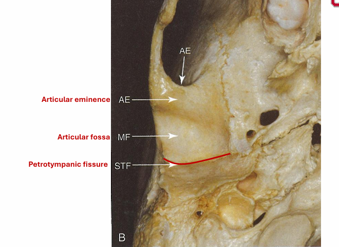
Tough oval pad of dense fibrous CT between the condyle & the articular fossa/eminence
articular disc
how is the articular disc shaped for the superior surface?
concave anteriorly & convex posteriorly
how is the articular disc shaped for the inferior surface?
concave
Center of the disc is ___ and ____
what are the articular disc functions?
1. acts as a shock absorber
2. moves w/ the mandible
3. creates upper and lower synovial cavities between the condyle and the fossa
4. proprioceptive nerve fibers contained in anterior and posterior parts of the disc
improper movement of the disc results in….
popping/crepitus

what would normally be here?
articular disc

what is the general function of the ligaments?
Function to support the joint and limit movement of the mandible
do the ligaments move the joint?
NO
list the 4 major ligaments of the TMJ?
• Fibrous capsule or capsular ligament
• Lateral ligament
• Stylomandibular ligament
• Sphenomandibular ligament
Fibrous tube of tissue that encloses the joint
Fibrous Capsule/Capsular Ligament
list the action of the Fibrous Capsule/Capsular Ligament:
limits protrusion of the mandible
what are the attachments of the Fibrous Capsule/Capsular Ligament?
temporal bone around articular fossa & eminence, around neck of condyle, & the anterior portion of the articular disc

Reinforces the capsular ligament
lateral ligament
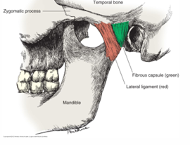
list the action of the lateral ligament?
keeps condyle close to fossa & prevents excessive lateral & posterior displacement of the mandible
what are the attachments of the lateral ligament?
zygomatic arch, & posterior to the lateral & posterior neck of the condyle

what ligament is the red one?
Stylomandibular Ligament
list the action of the stylomandibular ligament
prevents excessive protrusion of the mandible
what are the attachments for the stylomandibular ligament?
styloid process & posterior border of the angle of the mandible

what is the yellow ligament?
sphenomandibular ligament
what is the action of the sphenomandibular ligament?
prevents excessive opening of the mandible
what are the attachments for the spehnomandibular ligament?
angular/sphenoidal spine & the lingula of the mandible (near the mandibular foramen)
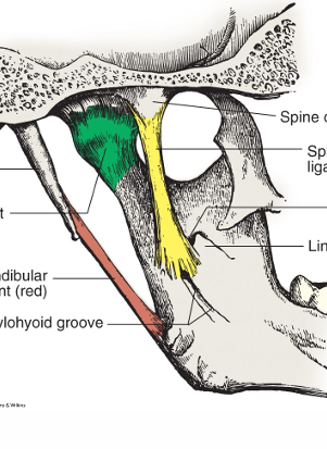
what is the green one?
capsular ligament
what are the 4 muscles that come in pairs?
• Masseter
• Temporalis
• Medial pterygoid
• Lateral pterygoid
MOM function to do what?
move the mandible

what are the 2 red boxes pointing to?


what is the blue/red muscle?
temporalis and masseter
function of masseter m.
elevates
Fan-shaped, large & flat muscle with both vertical anterior & middle fibers & more horizontal posterior fiber
temporalis muscle
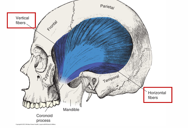
anterior and middle fibers of temporalis m. action:
elevation
posterior fibers of temporalis m. action?
retrude the mandible
origin of temporalis m:
temporal fossa

insertion of temporalis m:
coronoid process, anterior border of the ramus, & temporal crest

origin of medial pterygoid m:
medial surface of lateral pterygoid plate, pterygoid fossa & tuberosity

insertion of medial pterygoid muscle:
medial surface at angle of mandible

action of medial pterygoid m:
elevation

medial pterygoid
The prime mover of the mandible (except elevation); has two heads
lateral pterygoid muscle

origin of lateral pterygoid m:
greater wing of sphenoid (superior head) & lateral surface of lateral pterygoid plate (inferior head)

insertion of lateral pterygoid m:
Pterygoid fovea, anterior portion of articular disc & capsular ligament

when the lateral pterygoid heads are working together, what action do they do?
protrude & depress mandible
when the lateral pterygoid m. is working alone what action does it do?
lateral excursion to contralateral side
suprahyoid muscles:
extend from hyoid bone to mandible
infrahyoid muscles:
extend from hyoid bone to clavicle

what are the 4 suprahyoid m?
• Stylohyoid
• Mylohyoid
• Geniohyoid
• Digastric


what are the 4 infrahyoid m?
• Omohyoid
• Sternohyoid
• Sternothyroid
• Thyrohyoid

when the suprahyoid and infrahyoid muscles act together what action do they perform for the mandible?
depress and retrude mandible
what muscles perform elevation?
• Masseters • Medial pterygoids • Temporalis (anterior & middle fibers)
what muscles depress the mandible?
• Suprahyoids & infrahyoids together • Lateral pterygoids working together
what muscles perform retrusion?
• Temporalis (posterior horizontal fibers) • Suprahyoids (with help from infrahyoids)
what muscles perform protrusion?
Both lateral pterygoids simultaneously
what muscles perform lateral excursion?
• R lateral pterygoid moves mandible to L • L lateral pterygoid moves mandible to R
Orbicularis Oris action:
closes/purses the lips
buccinator action:
aids in chewing by pulling cheeks inward
Muscles that raise upper lip (smiling):
• Zygomaticus major & minor
• Levator labii superioris
• Levator anguli oris
• Risorius: retracts angle of the mouth
Muscles that lower the lower lip (frowning):
• Depressor labii inferioris
• Depressor anguli oris
• Mentalis
what other action does the mentalis perform?
raises or protrudes chin (pouting)
muscle of the neck that contracts while grimacing:
platysma

what are the muscles the red arrows are pointing to?

nerve innervation for MOMs:
: Mandibular branch of trigeminal nerve • CN V, branch III
nerve innervation of muscles of facial expression:
Facial nerve • CN VII