PHS 3300 : Cardiovascular II (still Part 1)
 Call Kai
 Learn
 Practice Test
 Spaced Repetition
 Match
 Flashcards
 Knowt Play
1/79

There's no tags or description
Looks like no tags are added yet.
80 Terms
How is heart failure defined?
A clinical syndrome with symptoms and/or signs caused by a structural and/or functional cardiac abnormality, corroborated by elevated natriuretic peptide levels and/or objective evidence of pulmonary or systemic congestion.
When does heart failure occur
heart is unable to generate adequate CO→ inadequate perfusion of tissues
there is increased diastolic filling pressure of left ventricle or both→ pulmonary capillary pressures increased
Why is heart failure often called congestive heart failure (CHF)?
Because heart failure commonly leads to fluid overload and congestion of blood in the venous circulation.
What is a common symptom of left-sided heart failure?
Dyspnea (shortness of breath), especially during physical activity.
Why does SOB occur with left-sided heart failure
The left ventricle cannot effectively pump blood forward, causing blood to back up into the lungs
Pulmonary edema→ SOB
Consequence of blood backing up into lungs
Pulmonary venous pressure increases, leading to pulmonary congestion
How can cardiac disease affect the lungs?
Left-sided heart failure increases pressures in the pulmonary circulation, causing pulmonary congestion and edema, which leads to dyspnea.

Where does blood back up if the right heart fails?
Into the systemic venous circulation (systemic veins).
Where does the blood in the right heart come from?
The systemic circulation (body tissues) via the superior and inferior vena cava.
What is the main consequence of right-sided heart failure?
Systemic venous congestion
What abdominal signs can occur in right-sided heart failure?
Hepatomegaly (enlarged liver) and ascites (fluid in the abdomen).
Why does hepatomegaly occur in right-sided heart failure?
Congestion of blood in the hepatic veins leads to liver enlargement and tenderness.
What is ascites?
Accumulation of fluid in the peritoneal (abdominal) cavity due to venous congestion.
What is the overall pattern of blood flow in right-sided heart failure?
Right ventricle fails → blood backs up into systemic veins → increased venous pressure → fluid leakage into tissues.
How do kidneys interpret heart failure
↓ Cardiac output → ↓ kidney perfusion ("effective circulating volume" appears low)
Kidneys interpret this as a low blood volume problem
How do kidneys respond to heart failure
RAAS activated → ↑ Angiotensin II → vasoconstriction
Aldosterone released → ↑ Na⁺ and water retention
ADH released → ↑ water reabsorption
What is the result of the kidneys responding to heart failure
increased BP
increased BV
If the heart is already having trouble handling the existing blood volume, are the kidneys and ADH helping by increasing plasma volume in heart failure?
No, just creates extra workload for the heart
What is the general approach to diagnosing heart failure?
based on a constellation of symptoms, physical exam findings, and supportive tests.
not determined with just a SINGLE test
What is the Framingham Criteria used for?
A set of clinical criteria used to help diagnose heart failure
Heart failure is diagnosed when 2 major criteria OR 1 major +1 minor criteria are met
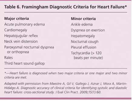
Commonly used criteria for diagnosing heart failure
presence of dyspnea
Pulmonary rales (crackles), indicating fluid in the lungs.
pulmonary edema
S3 hear sounds (ventricular gallop)
tachycardia

What does a chest X-ray show in heart failure?
Cardiomegaly
pleural effusion.
What is the main use of echocardiography in heart failure?
assess chamber size
valvular flow dynamics (EF)
What is ejection fraction (EF)?
The percentage of blood ejected from the left ventricle with each heartbeat
AKA % of end diastolic volume
What is a normal ejection fraction?
Approximately 60%-80%.
Why is ejection fraction important in heart failure?
It helps categorize heart failure as left-sided or right-sided
whether EF is preserved or reduced
What is Preserved EF Heart failure ?
signs and symptoms of HF with LEFT ventricular EF of ≥ 50% with signs and symptoms of HF.
Impaired ventricular filling (diastolic dysfunction)
2 common causes of HF Preserved EF?
Coronary artery disease (CAD) and hypertension.
What is HF Reduced EF?
SYSTOLIC Heart failure with reduced ejection fraction (EF < 40%)→ decreased CO
reduced contractility due to loss of cardiomyocytes showed by low EF during ventricular systole
What is a common cause of HF Reduced EF?
Myocardial infarction (MI).
Are compensatory mechanisms in heart failure initially beneficial or harmful?
Initially beneficial (short-term), but harmful over the long term.
What is myocardial hypertrophy?
Results from: chronic elevation of myocardial wall tension (takes longer)
High SP in the ventricle needed to overcome a high afterload leading to hypertrophy → increased pumping force
Eventually remodeled LV undergoes fibrous changes and stiffens
What are the two common problems caused by heart failure
Decreased forward flow
Backup of flow

How to improve forward flow?
Increasing the force of ventricular contraction (digoxin/digitalis)
Decrease the rate of contraction to increase filling time (beta-blockers)

How to decrease backup of flow?
Decrease preload
decrease afterload
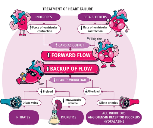
How does reducing afterload improve cardiac output?
The ventricle expends less effort ejecting blood, increasing forward flow.
What may a chest X-ray show in left-sided heart failure?
Cardiomegaly, pulmonary edema, and pleural effusions.
What is Coronary Artery Disease (CAD)?
also called ischemic heart disease, myocardial ischemia or coronary heart disease (CHD) forms a pathophysiologic continuum
characterized by insufficient delivery of oxygenated blood to the myocardium (ischemia) because of atherosclerotic coronary arteries (CAD)

Sequelae of CAD
Angina pectoris
Myocardial Infarction
Dysrhythmias
Heart failure
Sudden cardiac death
What is the primary cause of Coronary Artery Disease?
Atherosclerosis of the coronary arteries.
How does atherosclerosis cause CAD
causes narrowing of the arterial lumen that can lead to cardiac ischemia through:
thrombus formation
coronary vasospasm
endothelial cell dysfunction
tends to develop in large- and medium-size arteries
most frequently the coronary, cerebral, carotid, femoral arteries and the aorta
Uncommon causes of Ischemia
abnormalities of blood O2 content (i.e. respiratory failure)
poor perfusion pressure through coronary arteries
abnormalities in microcirculation via small coronary blood vessels
What are non-modifiable risk factors for coronary artery disease?
Age, sex, family history, and ethnicity.
Why do men have earlier onset of CAD compared to women?
Hormonal protection(i.e. Estrogen) in premenopausal women delays atherosclerosis development.
What are the major modifiable risk factors for CAD?
Dyslipidemia
non-lipid risk factors
hypertension
smoking
diabetes/insulin resistance
obesity
sedentary lifestyle
poor diet.
Pathogenesis of Atherosclerosis
Chronic endothelial ‘injury” leads to Lipids (especially LDL) enter the arterial wall and become oxidized.
endothelial dysfunction, monocyte adhesion, and emigration
Macrophage activation and smooth muscle recruitment
Macrophages and smooth muscle cells engulf lipid and become foam cells
smooth muscle proliferation, collagen and other extracellular matrix deposition, extracellular lipid
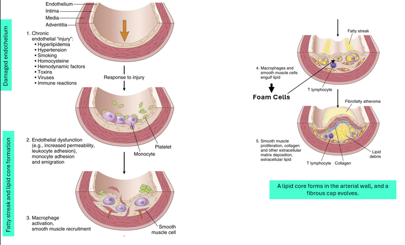
What forms after foam cell accumulation?
A lipid core develops within the arterial wall.
What is the lipid core made of?
Lipids, foam cells, and cellular debris.
What is a fibrous cap?
A layer of smooth muscle cells and collagen that covers the lipid core.
What is the function of the fibrous cap?
It stabilizes the atherosclerotic plaque and separates it from blood flow.
What happens if an atherosclerotic plaque ruptures?
It can trigger thrombus formation, leading to acute coronary syndromes (e.g., MI).
At what level of arterial narrowing does significant reduction in blood flow usually occur?
When a plaque occupies more than ~75% of the vessel lumen.
Where can atherosclerotic plaques form in the coronary circulation?
In any major coronary artery or their major secondary branches.
Alternative/collateral pathways may develop

Factors affecting plaque vulnerability?
Active inflammation within the plaque
Large lipid core with a thin cap
Endothelial denudation (erosion) with superficial platelet adherence
Fissured or ruptured cap
Severe stenosis predisposing to high shear stress
2 critical factors affecting cellular demand for O2
Rate of coronary perfusion
Myocardial workload
Rate of coronary perfusion
large, stable atherosclerotic plaque
acute platelet aggregation & thrombosis
vasospasm
failure of autoregulation by the microcirculation
poor perfusion pressure

Myocardial Workload
HR
preload
Afterload
Contractility

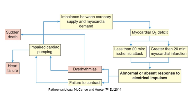
Cycle of Myocardial Ischemic Events
Dysfunction can range from
Rapid and full recovery of myocyte function (e.g. after a brief episode of stable angina)
Prolonged contractile dysfunction without myocyte necrosis with potential recovery of normal function (i.e. stunned myocardium, hibernating myocardium)
Irreversible myocardial necrosis (i.e. myocardial infarction).
Angina/Angina Pectoris
the hallmark of coronary artery disease
In economic terms, the vessels cannot supply sufficient oxygen for the heart’s demand in an atherosclerotic heart
Precipitated by increased O2 demand (exercise, stress, SNS activation, increased preload, afterload & HR)
Characteristics of Angina/Angina Pectoris
Intermittent chest pain (caused by myocardial ischemia)→ MI
Burning, crushing, squeezing, choking or referred pain
Often describes as having a clenched fist over the left sternal border
Pallor, diaphoresis, and dyspnea may be present
May result in inefficient cardiac pumping with resultant pulmonary congestion and dyspnea (shortness of breath)
3 patterns of angina pectoris
Stable or typical angina
Vasospastic or Prinzmetal angina
Unstable or crescendo angina
Stable or typical angina
most common form
Fixed coronary obstruction (usu. stenotic atherosclerotic coronary vessel) cannot respond to increased O2 demand
Predictable: onset of anginal pain is predictable and elicited by similar stimuli (physical or emotional exertion) each time
no change in cause, duration or pain over time
relieved with rest and nitrates (e.g. nitroglycerine causes vessel dilation & reduced preload)
Prinzmetal (vasospastic) or variant angina
Unpredictable attacks of angina
Onset of symptoms is unrelated to physical or emotional exertion, HR, or other obvious causes of increased myocardial oxygen demand
Characterized by vasospasms, atherosclerosis-induced hypercontractility, abnormal secretion of vasospastic chemicals by local mast cells, and abnormal calcium flux across vascular smooth muscle
vasospasm proposed as possible mechanism
may intensify or worsen but does not carry the same concern as unstable angina
relieved by calcium channel blockers which inhibit muscle contraction
Unstable angina and MI
Difficult to distinguish based on clinical manifestations.
Common characteristics
chest pain usually more severe and lasts longer than typical angina
may occur in previously asymptomatic individuals
Both life threatening conditions with a common initiating pathological mechanism

Unstable angina vs MI
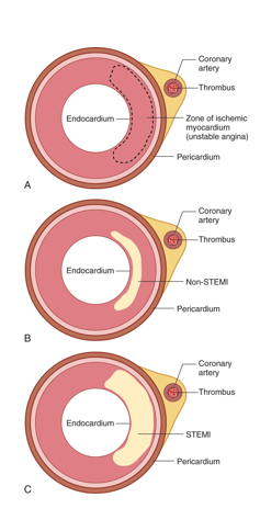
Unstable angina: partial occlusion/clot dissolved before cell death
MI: complete occlusion/thrombus persists→ irreversible cell death
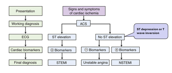
3 presentations of acute coronary syndrome
unstable angina
NSTEMI: Non-ST elevation MI
STEMI: ST-elevation MI
3 primary indicators of acute coronary syndromes
Signs & symptoms
ECG changes
Serum biomarkers
cardiac catheterization, echocardiography, radionuclide scintigraphy may provide additional information
Signs and symptoms of acute coronary syndromes
Pain accompanies by nausea, diaphoresis and vomiting
last longer than 15 min / not relieved by rest and nitrates
some MIs are asymptomatic – silent MI
others present atypically: nausea, back pain and abdominal discomfort
ECG changes of acute coronary syndromes
ST- segment changes indicate injury and ischemia
ST elevation - acute cellular injury and ischemia/ injury ongoing
ST depression or T wave changes patients may benefit from timely PCI.

Serum Biomarkers of acute coronary syndromes
Cardiomyocytes contain specific isoforms of: CK-MB, troponin I, & troponin T
elevated level of serum CK-MB is a highly specific indicator of MI and considered to be diagnostic.
Unstable angina and MI indicators
Traditionally: distinguished by the presence or absence of serum biomarker
Reperfusion therapy effective only if administered early so treatment for unstable angina and MI combined
chest pain + evidence of acute ischemia on ECG (ST-elevation): acute reperfusion therapy + fibrinolytics
symptoms of unstable angina with no ST elevation: percutaneous coronary intervention (PCI)
Differentiation made using serum biomarkers
Acute occlusion
causes a range of cellular events, depending on
Availability and adequacy of collateral blood flow
Relative workload
Length of time that flow is
interrupted Infraction area not homogenous: multiple zones with cells in various stages of ischemia & death / almost always located in the left ventricular wall
Ultimate size of the infarcted tissue depends on the extent, duration, and severity of ischemia
Compensatory mechanisms for MI
MI leads to drop in CO, triggering compensatory responses
Compensatory responses → increase SV and maintain CO
SNS activation leads to increased myocardial workload by increasing: Heart rate Contractility
Potential complications of MI

Treatment goals of MI
- minimizing the duration of exposure of myocardium to ischemia
preventing recurrent ischemia and re-occlusion
How does minimizing the duration of exposure of myocardium to ischemia help treat MI
Decreasing myocardial workload
preload & afterload reduction, HR control, pain relief & activity restriction
SNS antagonists, nitrates, morphine
Increasing myocardial O2 supply
Reestablish perfusion: O2 administration, antiplatelet therapy (aspirins, thrombolytic drugs), anticoagulant drugs, angioplasty, stent placement, coronary bypass grafting
How does preventing recurrent ischemia and re-occlusion help treat MI
managing arrythmic + mechanical complications
modifying underlying atherosclerosis
How to monitor and mange complications of MI
Early detection and management of dysrhythmias and conduction disorders; continuous
ECG monitoring
Chronic Ischemia Cardiomyopathy
Heart failure that develops insidiously due to progressive ischemic myocardial damage.
affected individuals have a history of angina or MI, often many years before the onset of heart failure.
failure due to slow, progressive death of myocytes from chronic ischemia
usually found in elderly individuals.
Sudden Cardiac death
AKA sudden cardiac arrest
Unexpected death from cardiac causes within 1 hr of symptom onset
Most cases associated with coronary atherosclerosis
Lethal dysrhythmia (e.g., ventricular fibrillation) is usually the primary cause
Use of external defibrillators and CPR has increased survival