Advanced Medical-Surgical Exam 2
 Call Kai
 Learn
 Practice Test
 Spaced Repetition
 Match
 Flashcards
 Knowt Play
1/61

There's no tags or description
Looks like no tags are added yet.
62 Terms

Sinus Rhythm
Rhythm: regular
HR: 60-100 bpm
P wave: rounded, upright, before each QRS interval
PR interval: 0.12 - 0.20 seconds
QRS interval: less than 0.10 seconds
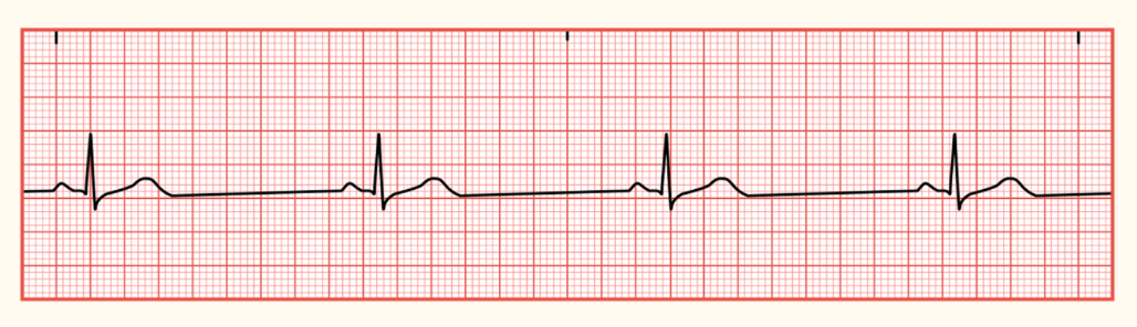
Sinus Bradycardia Characteristics
Occurs when the SA node sends an electrical impulse lower than 60
Rhythm: regular
HR: less than 60 bpm
P wave: rounded, upright, before each QRS complex
PR interval: 0.12 - 0.20 seconds
QRS interval: less than 0.10 seconds
Sinus Bradycardia
Causes: genetics, MI, sleep apnea, ICP, eating disorders, increased exercise tolerance, hypothyroidism, vagus nerve stimulation (frequent coughing, excessive gagging, and straining for BM)
Associated with: lyme disease, typhoid fever, malaria, Rocky Mountain Spotted Fever, meds, normal aging, and prolonged hypoxia
Medication cause: acetylcholine (parasympathomimetics), beta-blockers (lol), digitalis glycosides (digoxin), calcium channel blockers (diltiazem), antiarrhythmics (amiodarone), chemotherapy agents (thalidomide), and lithium

Sinus Tachycardia Characteristics
Occurs when the SA node sends a faster electrical impulse, greater than 100 bpm; prolonged tachycardia could lead to decrease in cardiac output (affecting the amount of oxygenated blood being pumped thru the body)
Rhythm: regular
HR: 101-180 bpm
P wave: rounded, upright, before each QRS complex
PR interval: 0.12 - 0.20 seconds
QRS interval: less than 0.10 seconds
Sinus Tachycardia
Causes: fluid volume loss or excess, pain, fever, MI, shock, hyperthyroidism, and stress
Medication cause: atropine, catecholamines, theophylline, illicit drugs (cocaine or amphetamines), caffeine, and nicotine

First-Degree Atrioventricular (AV) Block
When the cardiac conduction system is delayed in transmitting an electrical signal through the right atrium to the AV node
Causes:
Advanced age; usually males over 60
History of cardiac disease (MI)
Electrolyte imbalance (hypokalemia + hypomagnesemia)
Antiarrhythmic medications
Infection
Athletes; usually caused by a higher resting vagal tone that allows for tolerance of slower HRs

Second-Degree Atrioventricular (AV) Block Type I (Mobitz Type I or Wenckebach)
An abnormality in the heart’s electrical conduction system where signals from the atria are delayed more and more with each beat until a heartbeat is “dropped”
Causes:
High vagal tone: common; normal variant of healthy individuals or well-conditioned athletes
Medications: beta-blockers, calcium channel blockers, digoxin, or certain antiarrhythmics
Heart conditions: ischemia or MI (especially involving the inferior wall), myocarditis, or age-related changes in the AV node
Second-Degree Atrioventricular (AV) Block Type I (Mobitz Type I or Wenckebach) treatment
Asymptomatic: does not require treatment or a permanent pacemaker
Symptomatic: if it causes dizziness, fainting, or severe slow HR, can be treated with atropine, or a temporary pacemaker; permanent pacemaker is rare
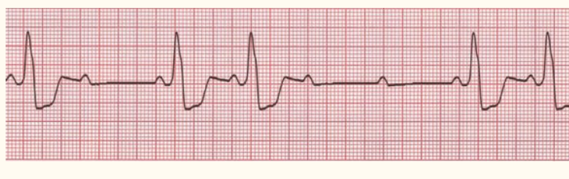
Second-Degree Atrioventricular (AV) Block Type II (Mobitz Type II or Hay Block)
Identified through an ECG and is caused by an irregular block of atrioventricular conduction below the AV node; unstable warning rhythm that can lead to complete heart block or ventricular asystole
Second-Degree Atrioventricular (AV) Block Type II (Mobitz Type II or Hay Block) Characteristics
Constant PR interval
Infranodal Conduction defect (happens below the AV node within the His-Purkinje; results in wide QRS complex
Unpredictable dropped beats; failure to trigger a QRS complex
Second-Degree Atrioventricular (AV) Block Type II (Mobitz Type II or Hay Block) Causes
Structural damage:
Past or current heart attacks
Chronic fibrosis or degeneration of the conduction system (aging)
Cardiac surgery or inflammatory/infectious heart conditions
Second-Degree Atrioventricular (AV) Block Type II (Mobitz Type II or Hay Block) Treatment
Permanent pacemaker, even without symptoms but confirmed diagnosis

Third-Degree Atrioventricular Block (Complete Block) Characteristics
Rhythm: regular, atria/ventricle independent
HR: atrial 60-100 bpm
20-40 bpm (ventricular focus)
40-60 bpm (junctional focus)
P waves: normal; independent QRS complex
No PR interval
QRS interval: greater than 0.10 (ventricular), less than 0.10 (junctional)
More P waves than QRS
Third-Degree Atrioventricular Block (Complete Block) Immediate and Acute Treatment
Temporary pacing: for unstable pt with severe symptoms; primary, most reliable treatment to stabilize the HR
Medication: atropine is first line, dopamine or epinephrine used if atropine fails and pacing isn’t available
Reversing causes: treat the underlying trigger or stop the offending drug
Long-term:
Permanent pacemaker; to maintain a safe, coordinated heartbeat
Third-Degree Atrioventricular Block (Complete Block) Causes + Symptoms
Causes:
Coronary heart disease
MI
Digoxin
Aging
Symptoms:
Dizziness
Syncope
Fatigue
SOB
Chest pain
Hypotension
Diminished or absent peripheral pulses
Electrolytes Causing Cardiac Heart Block
First-Degree AV Block (slower AV node conduction): hypokalemia + hypomagnesemia
Medications to Decrease HR
Digoxin: slows AV conduction; check apical pulse 1 full min before administration
Beta-blockers: metoprolol, atenolol, propranolol
Calcium channel blockers: diltiazem, verapamil
Amiodarone; monitor ECG, QT interval
Clonidine
Medications to Increase HR
Atropine: blocks vagal stimulation (first-line for symptomatic bradycardia)
Epinephrine: stimulates beta-1 receptors
Dopamine
Dobutamine
Norepinephrine
Cardiac Arrest (Cause + Etiology)
Leading etiologies:
Heart disease
Arrhythmias
Sepsis
Trauma
Other etiologies:
Hypovolemia
Cardiac tamponade
Valvular heart disease
Pulmonary embolism
Airway obstruction
Greatest risk:
Higher baseline C-reactive protein levels
Cocaine use
Genetics
Cardiac Arrest Presentation
Fatigue/weakness
Dizziness
Syncope
Chest pain
SOB
Vomiting
Palpitations
Backpain
Late:
No pulse
No respirations
Shockable Rhythms
Ventricular fibrillation: when the lower chambers of the heart (ventricles) contract at an irregular rapid rate; heart will not pump blood to the rest of the body
Ventricular tachycardia: when the heart is beating at a rapid rate which prevents the chambers of the heart to fill with blood; not enough blood is pumped to the body
Non-sustained: intermittent (a few seconds)
Sustained: lasts longer than 30 seconds
Unstable polymorphic VT
Non-shockable Rhythm
Asystole (flat-line): absence of electrical and mechanical activity; heart stops pumping
Pulseless electrical activity (PEAA): no ventricular contraction, or the electrical activity is not strong enough for the heart to contract
Cardiac Arrest Nurses Roles + Immediate Actions
First responder
Ensure IV access
Medication administration
Assist with oxygen delivery and ventilation
Provide CPR
Record time, rhythms, and actions during code
Leader
Family support
Cardiac Arrest: First Line Medications
Epinephrine
Amiodarone
Lidocaine
Atropine
Sodium Bicarbonate
Calcium
Magensium
Dextrose
Shock Stage 1: Initial
Too little oxygen in the blood to feed organs; body switches from aerobic to anaerobic metabolism
Increased lactic acid
Shock Stage 2: Compensatory
Aldosterone released to maintain BP
Vasoconstriction to shunt blood to vital organs
Increased HR
SNS releases catecholamines to compensate for low oxygen
Shock Stage 3: Progressive
Oliguria
Altered LOC
Pallor, cool clammy skin
Electrolyte imbalance
Hypotension
Shock Stage 4: Refractory
Irreversible cellular and organ failure
Impending doom
Distributive Shock
Pathophysiology:
Septic: inflammatory cytokines cause damage to internal vessel layers and initiate clotting mechanisms
Anaphylactic: severe Ige-mediated hypersensitivity reaction increased vascular permeability and vasodilation, decreasing SVR
Neurogenic: autonomic dysregulation from spinal cord injury above T6
Toxic: Staphylococcus aureus infection causing excessive activation of cytokines and inflammatory cells
Etiology: sepsis/infection, allergens (food, meds, bee stings), spinal cord trauma (GBS, cerebral hemorrhage), soft tissue/post-surgical infections, pancreatitis + burns
Clinical Presentation
Sepsis: fever, tachycardia, hypotension, oliguria, flushed/dry/warm skin
Anaphylactic: tachycardia, hypotension, bronchospasm, laryngeal edema, wheezing, stridor, itchy skin, hives, swelling of tongue
Neurogenic: bradycardia, hypotension, orthostatic hypotension, flushed/dry/warm skin
Toxic: fever, tachycardia, hypotension, flushed/dry/warm skin
Treatment
IV fluids (NS or LR)
Vasopressors (norepinephrine)
Antibiotics
Epinephrine IM (first-line treatment) for anaphylactic shock
Oxygen
Hypovolemic Shock
Pathophysiology: intravascular volume is decreased by 25% to 30%, resulting in poor cardiac output; hemorrhagic shock leads to imbalance in oxygen supply and demand
Etiology:
Hemorrhagic — traumatic blood loss, upper/lower GI bleed, ruptured aneurysm, postpartum bleed, anticoagulants
Non-hemorrhagic — excessive diuresis, burns, GI fluid loss
Clinical Presentation
CV: tachycardia, hypotension
Resp: tachypnea
Integumentary: cold, clammy skin
Digestive: possible hematemesis, bloody stool
Treatment
IV fluid (NS or LR)
Blood transfusions
Vasopressors
Oxygen
Cardiogenic Shock
Pathophysiology: circulatory failure and systemic hypoperfusion caused by cardiac dysfunction, leading to myocardial ischemia
Etiologies: acute MI, aortic dissection, arrhythmias, medication overdose, RV failure, metabolic acidosis, electrolyte imbalances, or pulmonary embolism
Clinical Presentation
Neuro: anxiety, restlessness, deteriorating consciousness
CV: tachycardia, hypotension, arrhythmias, narrow pulse pressure
Resp: adventitious lung sounds, hemoptysis
Integumentary: cold, clammy skin
Treatment
Vasopressors (increase BP)
Dobutamine (improve cardiac output)
Diuretics
Obstructive Shock
Pathophysiology: physical blockage of blood flow that disrupts circulation to major organs and arteries
Etiology: tension pneumothorax, mechanical ventilation with high PEEP, cardiac tamponade, pulmonary embolism, cardiac mass, aortic dissection
Clinical Presentation
CV: tachycardia, hypotension, jugular vein distention, cyanosis
Resp: asymmetrical breathing pattern, absent breath sounds in 1 lung field, subcutaneous emphysema
Integumentary: cold, clammy skin
Digestive: abdominal distention
Treatment
Pericardiocentesis: removes fluid/blood surrounding the heart)
Cardiac Tamponade: Pulsus Paradoxus
Can indicate that a pericardial effusion is the cause of the cardiac tamponade
A fall of systolic BP greater than 10 mmHg during inspiration
Korotkoff sounds disappear with inspiration and return with expiration
Cardiac Tamponade: Diagnostic Testing
An echocardiogram is the gold standard
Can confirm:
Pericardial effusion
Size of pericardial effusion
Will show:
Tachycardia
Chest X-Ray is another option; will show enlarged cardiac silhouette with a large amount of fluid volume
Computerized Tomography (CT)
Electrocardiogram (ECG)
Magnetic Resonance Imaging (MRI)
Coronary Angiography
Right Heart Catheterization
Labs:
Creatinine kinase levels
Renal (BUN)
Coagulation studies
Septic Shock: Vasopressors
Action: cause narrowing of the blood vessels to increase systemic vascular resistance and blood pressure
Therapeutic effect: increase MAP (goal: greater than or equal to 65 mmHg) to improve blood flow and oxygen delivery to vital organs
Complications:
Tachycardia or bradycardia
Cardiac dysrhythmias
Hypertension
Peripheral ischemia
Decreased cardiac output
Tissue necrosis from IV extravasation
First line: Norepinephrine
Vasopressin added when norepinephrine alone is insufficient
Code Sepsis
Alert & mobilize
Trigger: a healthcare professional catches warning signs of sepsis
Team huddle: emergency or attending, nurses, pharmacist, lab tech/phlebotomist, and respiratory or radiology staff
Immediate testing (within first 30 min of symptom recognition)
Blood cultures
Lactate level: high levels signal poor organ perfusion
Vital signs
Rapid treatment
IV fluids: BP too low or lactate is high — large bolus of IV crystalloid fluids
Broad-spectrum antibiotics — administer within 1 hour of the code to stop spreading infection
Source control: search and treat the root cause of infection
Reassessment & transfer
Huddle and monitoring: check back within a few hours to review lab results and vitals
Vasopressors: fluids not working to bring up blood pressure? use vasopressors to tighten blood vessels
Level of care: patient transfer to ICU or high-observation step-down unit
CABG: Indications
Chronic conditions contribute to CAD and may result in the need for CABG surgery:
Diabetes
Hypertension
Hypercholesterolemia
Obesity
Unhealthy diet
Smoking
CABG: Post-Operative Care + Rehabilitation
Hemodynamic monitoring: ABGs, bleeding, pain, anxiety, readiness to wean, hypovolemia
Closely monitor VS, frequent assessments, minimize risk of site infections (redness, fever, pain, warmth, swelling), encourage early mobilization and deep breathing, enteral nutrition within 24 hr post-op
Valve Replacement: Infection Prevention
Prophylactic antibiotic use before any invasive dental or oral procedure; reduces risk of endocarditis
Perform good oral hygiene
Valve Replacement: Vascular/Transcatheter Aortic Valve Implant (TARV)
Minimally invasive option to remove the damaged aortic valve
Used to treat severe, highly symptomatic aortic stenosis
Valve Replacement: Open Replacement Complications
Death
Stroke
Paralysis
Pericardial effusion
Pulmonary edema
Kidney failure
Aortic Aneurysm: Risk Factors
Atherosclerosis
Smoking
Sex (males more likely)
Family history
Hypertension
Coronary Artery DIsease
Peripheral Artery Disease
Renal Insufficiency
COPD
CHF
Aortic Aneurysm: Intervention
AA is a dilation of the aorta greater than 30 mm in diameter.
If the diameter is greater than 55 mm, it is considered large and requires urgent treatment.
Aortic Aneurysm: Physical Assessment Signs
Cullen’s sign: umbilical bruising
Turner’s sign: flank bruising
Sudden onset back and abdominal pain
Noticeable abdominal distention
Signs and symptoms of hemorrhagic shock
Transplant: Ethical Principles
Autonomy
Patients should be involved in decisions about transplantation, including understanding the procedure, risks, benefits, and lifelong immunosuppressant therapy
Beneficence
Transplantation is intended to improve survival and quality of life compared with remaining on dialysis
Nonmaleficence
Providers must minimize harm by evaluating donor and recipient risks, monitoring complications, and preventing infection and rejection
Justice
Organ allocation involves fairly distributing a limited supply of donor kidneys
Transplant: Post-Op Emergencies
Hemorrhage
New-onset flank plan
Palpable mass or bulging near incision
Hypertension
Renal vein or arterial thrombosis (rare, but high risk graft loss)
New-onset hematuria
Sudden decrease in urine output (oliguria, less than 30 ml/hr)
Anuria
Infection
First 3-6 months, high immunosuppression
Opportunistic infections: CMV, EBV, BK polyomavirus
May receive prophylactic medications (Bactrim, Valcyte, antifungal coverage)
Lymphocele — lymphatic vessels are disrupted during surgery
Pain near transplant
Bulging over site
Infection or compression of graft
Decreased kidney function
Urinoma (urine leakage)
Usually occurs within the first week
Pain/swelling near incision
Decrease graft function
Possible infection
Confirmation: elevated creatinine in fluid aspirate
Treatment: foley catheter, surgery
Arterial stenosis
Later complication
May be asymptomatic
Identified through ultrasound when graft function decreased, creatinine rises
Diagnosis: angiography
Transplant: Post-Op Hypertension
Blood pressure goal: less than 130/80
First line medications:
Dihydropyridine calcium channel blockers
Angiotensin receptor blockers (ARBs) — preferred when proteinuria is present; early on avoided because it can affect the GFR
Do not use ARBs in pregnant women, trying to conceive, or breastfeeding
Transplant: Medication Non-Adherence
Transplant recipients require lifelong immunosuppressive medications.
Missing or stopping medications can cause acute or chronic rejection.
Rejection can lead to graft dysfunction, graft failure, and potentially the need for another transplant or dialysis.
Even occasional missed doses can put the transplanted organ at risk.
Post Transplant Cancer Risk
Immunosuppression increases cancer risk
Skin cancer: annual clinical skin examinations
Renal cell carcinoma: monitor the native kidneys (every 1-3 years)
Post-transplant lymphoproliferative disorder (PTLD): associated with Epstein-Barr, particularly in those non-immune
Signs — low-grade fever, dyspnea, headaches, abdominal discomfort
Post Transplant Pregnancy
Fertility increase: uremia-related hormonal disruptions improve
Maternal complications: increased risk of preeclampsia, hypertension, acute rejection, and worsening kidney graft function
Fetal complications: increased risk of small-for-gestational-age, prematurity, and low birth weight
Mycophenolate mofetil (MMF): increased risk of fetal structural malformations
Contraception: most effective when started before MMF, throughout treatment, and for 6 weeks after discontinuation
Can become pregnant at least 1 year post-transplant
Stable kidney function
Creatinine less than 1.5 mg/dL
No or minimal proteinuria
No recent rejection episodes
Stable maintenance immunosuppression
Need to switch to azathioprine for less teratogenic effect from the immunosuppressant
Transplant Patients: Bone Health
Pt experiences decreased bone strength and a higher fracture risk due to advanced CKD
CKD causes mineral and hormonal imbalances involving calcium, phosphorus, vitamin D, parathyroid hormone (PTH) and FGF-23
Highest risk period: 6 months after transplant (trabecular bone fractures)
Bone density loss: rapid bone density loss and increased fracture risk (first 12-18 months post-transplant)
Organ Donation: When to Call Procurement
Procurement cannot begin until the heart has stopped beating
An independent physician pronounces the patient following terminal extubation
Brain death
Deep coma with no recovery chance
Family decides to end life support
Do not wait until death is declared to make a required referral
Post Transplant Diabetes: Basal Insulin
Helps protect pancreatic beta cells and can prevent further damage; reduces the incidence of PTDM
For post-transplant diabetes: tacrolimus can be toxic to pancreatic islet cells and corticosteroids can contribute hyperglycemia
Transplant: Infection Prevention and Immunizations
Immunosuppression risk due to medications used to prevent rejection
First 3-6 months high immunosuppression and infection risk
Live vaccines can be given 4 weeks before transplantation
Inactivated vaccines: most can be given 1 month post-op
Bactrim: PJP prophylaxis
Valganciclovir: CMV prophylaxis
BK virus: can cause nephropathy and damage the transplanted kidney
Active infection: contraindication to transplantation