Week 11 Respiratory care modalities: care of the artificially ventilated patient
 Call Kai
 Learn
 Practice Test
 Spaced Repetition
 Match
 Flashcards
 Knowt Play
1/106

Earn XP
Description and Tags
ventilator modes will be on exam i think. alarms too
107 Terms
Do you need an order to administer oxygen?
yes, it is a medication
Oxygen is administered at what concentrations?
21% (higher than room air)
How long does it take for brain damage to occur d/t no/low oxygen?
3 minutes
Goal with oxygen therapy
decrease workload of heart and lungs
increase tissue perfusion
adequate transport of oxygen
adjuvant for pain relief
How much oxygen does nasal cannula deliver low flow?
1-2 L/min= 24-28%
3-5 L/min= 32-40%
6L/min= 44%
Which form of oxygen therapy is better tolerated by children?
nasal cannula high flow
10L/min= 65%
15L/min= 90%
How much oxygen does Simple Mask deliver?
low flow 5-8L/min= 40-60%

Nursing interventions for Simple Mask
may need nasal cannula at meals
vents for exhaled CO2 claustrophobia
How much oxygen does Partial Rebreather mask deliver?
low flow 8-11L/min= 50%-75%

Nursing interventions for Partial Rebreather mask
watch for kinks in bags, tubing, skin checks
helps conserve oxygen- exhaled air trapped in bag and mixes with oxygen
How much oxygen does Nonrebreather mask deliver?
low flow 10-15L/min= 80%-95%

Nursing interventions for Nonrebreather mask
delivers the highest O2 concentration, do not rebreathe exhaled air
How much oxygen does Venturi Mask deliver?
high flow 2-15L/min= 24-60%
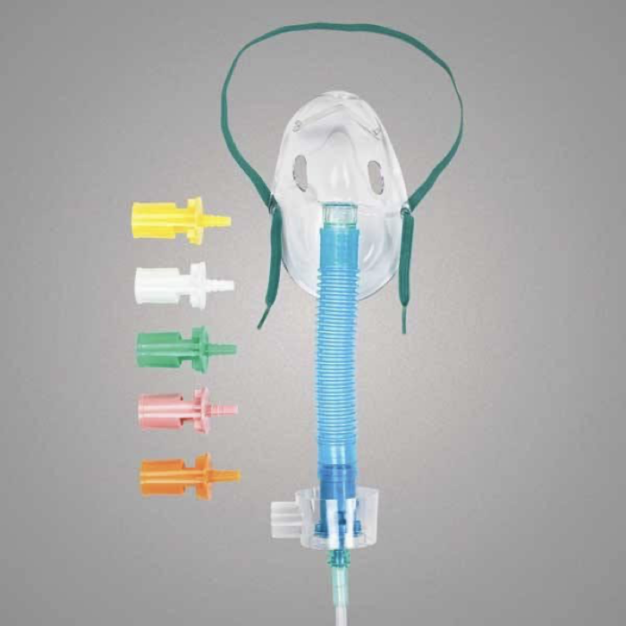
Nursing interventions for Venturi mask
most precise delivery of O2
exception for COPD patients
Other oxygen therapy include
nebulizer
intermittent positive pressure breathing
continuous positive airway pressure (C-pap)
Bi level positive airway pressure (Bi-pap)
ventilator
Noninvasive Positive Pressure Ventilation (NPPV) is good for patients who
cannot undergo intubation or invasive ventilation with the goals of decreasing the work of the respiratory muscles and relieve dyspnea
NPPA refers to
BiPAP and CPAP
Continuous Positive Airway Pressure (CPAP)
method of keeping airways open by providing constant milk air pressure
helps to hold the airway open, mobilize secretions, treat atelectasis, and ease the work of breathing
Bilevel Positive Airway Pressure (BiPAP) is also called
pressure support ventilation
Pressure delivery during inspiration vs expiration in BiPAP
BiPAP delivers higher pressure during inspiration and a lower pressure during expiration
Key mechanical feature of BiPAP
2 distinct pressure settings that changes depending on whether the patient is in inspiration or expiration
Basis for adjusting inspiratory and expiratory pressure in BiPAP
independently adjusted based on patient’s condition and target inspiratory-to-expiratory ratio (I:E)
Inspiratory Positive Airway Pressure (IPAP) is set higher to
aid airflow in
Expiratory Positive Airway Pressure (EPAP) is set lower to
make exhalation easier
When to use supplemental oxygen?
hypoxemia
hypoxia
dyspnea
other S/S respiratory distress
dyspnea on exertion (DOE)
changes in respiratory pattern
abnormal arterial blood gas (ABG)
fatigue
level of consciousness (LOC) changes
trauma
pain
Hypoxemia is
decrease arterial oxygen in blood
Hypoxia is
decrease in oxygen supply to cells and tissues (cellular level condition/systemic)
Dyspnea is
shortness of breath from hypoxia
Cautions of supplemental oxygen
hypercapnia
oxygen toxicity
Hypercapnia is
high amounts of CO2 in the blood (seen in COPD patients)
What is the primary drive to breathe in a healthy individual vs. a patient with chronic hypercapnia?
Healthy individual: High CO2 levels (hypercapnic drive).
Chronic hypercapnia patient: Low O2 levels (hypoxic drive).
Why do patients with severe chronic hypercapnia shift to a hypoxic drive to breathe?
Because their central chemoreceptors become desensitized to chronically high PaCO2 levels, forcing the body to rely on peripheral oxygen sensors instead.
What condition commonly causes patients to adapt to a hypoxic drive to breathe?
Chronic Obstructive Pulmonary Disease (COPD) and other severe chronic lung diseases.
What is the clinical danger of severely elevated PaCO2 in chronic lung disease?
It can cause CO2 narcosis/toxicity and lead to respiratory arrest
Oxygen toxicity is held
in the lungs
Oxygen toxicity occurs when patient is given
long term percentages of O2, causing lung damage (50-60%)
Oxygen toxicity destroys
lung surfactant
Why is lung surfactant important?
helps to maintain alveoli and keep open
kills pathogens in respiratory tract
aid in immune response
S/S Oxygen Toxicity
fibrotic changes
increased capillary congestion
interstitial space thickening
paresthesia
dyspnea
restlessness
pulmonary edema
Treatment for oxygen toxicity
Positive End Expiratory Pressure (PEEP)
Continuous Positive Airway Pressure (CPAP)
Treatments that help increase oxygenation
incentive spirometer
chest physiotherapy (PT)
How does incentive spirometer increase oxygenation?
encourages deep inspiration
How does Chest Physiotherapy (PT) increase oxygenation?
postural drainage
position that uses gravity to drain secretions
mobilizes secretions from bottom of lungs
chest percussion
hand clapping to chest wall loosen secretions
vibration
device used to mobilize secretions
Artificial Airways include
Endotracheal tube and Tracheostomy tube
What is the primary method of choice for establishing a patent airway in emergency care?
Endotracheal Tube
How is an Endotracheal Tube (ETT) inserted?
Inserted through the mouth or nose
How long should you keep Endotracheal Tube (ETT) in for?
no longer than 3 weeks
What is the difference in aspiration protection between a cuffed vs. uncuffed Endotracheal Tube (ETT)?
Cuffed provides better secretion clearance and some aspiration protection
Uncuffed provides airway clearance but no aspiration protection
What are five main reasons to use a Tracheostomy Tube?
by pass upper airway obstruction
permit long-term mechanical vent
permit oral intake and speech
replace an endotracheal tube
remove tracheobronchial secretions
How is the Tracheostomy Tube inserted?
into the trachea
Duration for Tracheostomy Tube
can be temporary or permanent
Who can place an Endotracheal Tube (ETT)?
need special training
anesthesia provider
paramedic
respiratory therapist
provider with ACLS training
Characteristics of Endotracheal Tube (ETT)
flexible tube that is placed with laryngoscope into trachea
cuff inflated and taped/secured with collar to assure placement
multiple sizes
How is Endotracheal Tube (ETT) placement checked?
with lung sounds, end-tidial carbon dioxide levels, and with chest x-ray
Characteristics of Tracheostomy Tube
semi-flexible, rigid, or metal tube that is surgically inserted that is placed into an artificial opening made into the trachea at the 2nd or 3rd tracheal ring
tube has a plate that is secured with sutures and/or with trach ties
multiple sizes
What do we call the artificial opening that is made to place Tracheostomy Tube?
tracheostomy
Advantages of Endotracheal Intubation
keeps airway open
able to administer O2 directly to lungs
ability to perform tracheal suctioning
short-term therapy (usually less than 3 weeks)
can attach to ventilator
Advantages of Tracheostomy Tube
keeps airways open
able to administer O2 directly to lungs
ability to perform tracheal suctioning
can be long term
can attach to ventilator, humidified oxygen, or room air
can permit oral intake and speech (if not on vent)
Disadvantages of Endotracheal Intubation
discomfort
patient unable to speak
cough reflex depressed
closure of glottis inhibited
thicker secretion- requires suctioning
swallowing reflexes depressed
risk of aspiration and ventilator-associated pneumonia (VAP)
unintentional displacement of the tube
Disadvantages of Tracheostomy Tube
discomfort
patient unable to speak (if on vent)
cough reflex depressed
secretions tend to be thicker
risk of aspiration and ventilator-associated pneumonia (VAP)
unintentional displacement of the tube
can be long term
Disadvantages of Endotracheal Tube
requires patient cooperation or restraint use
requires special training of nursing staff
oral assessment and care needed frequent
Disadvantages of Tracheostomy tube
requires special training of nursing staff
oral assessment and care needed frequently
Complications with Endotracheal Tube
Tube dislodgement
accidental removal
high cuff pressure
low cuff pressure
trauma to tracheal lining
vocal cord paralysis
Accidental removal of Endotracheal Tube can lead to
laryngeal swelling, hypoxemia, bradycardia, hypotension, death
High cuff pressure d/t Endotracheal Tube can lead to
tracheal bleeding, ischemia, or necrosis
Low cuff pressure d/t Endotracheal Tube can lead to
risk of aspiration and hypoxia
Early Complications with Tracheostomy Tube
tube dislodgement
accidental decannulation
bleeding
pneumothorax
air embolism
aspiration
subcutaneous emphysema
laryngeal nerve damage
posterior tracheal wall penetration
Later Complications with Tracheostomy Tube
airway obstruction from secretions
infection
dysphagia
tracheoesophageal fistula
tracheal dilation, ishcemia, or necrosis
Nursing care of pt with ET or Trach
monitor O2
cuff management
maintain patent airway (tube)
oral care
skin care
safety and comfort
Monitoring oxygenation
assess for signs and symptoms of hypoxemia
change in mentation, anxiety, dusky skin, dysrhythmias
ABGs
continuous SpO2
Gee, Clap, I Can See Nasty Respirations, Too stand for?
Grunting
Change in LOC
Intercostal spaces evident
Color (Cyanosis, Circumoral Pallor)
Seesaw chest movement
Nasal flaring
Retractions/dysRhythmias
Tachypnea
Managing the cuff
needs to be inflated if pt requires mechanical ventilation or high risk for aspiration
cuff pressure should be maintained at 20-25 mmHg
check every 6-8 hours
Promoting effective airway clearance
chest physiotherapy
frequent position changes
increased mobility
suctioning
Indications for suctioning
visible secretions in the tube
sudden onset of respiratory distress
suspected aspiration of secretions
auscultation of adventitious breath sounds over bronchi or trachea
increased respiratory rate & sustained coughing
sudden or gradual decrease in SpO2
changes in LOC, restlessness, tachycardia
cyanosis or pallor
increased peak airway pressure
Incline/closed suctioning
allow suctioning without disconnection from vent
decreases hypoxemia
sustains PEEP
decrease patient anxiety
protection of staff from secretions
Open suctioning
sterile procedure needing sterile gloves
staff needs PPE
higher risk for hypoxia
Preventing Complications for a patient with an ET or Trach Tube
administer warmed humidity- should see mist in tubing
maintain appropriate cuff pressure
suction as needed
maintain skin integrity
auscultate lung sounds
monitor for signs and symptoms of infection (temp, secretions, WBC)
administer prescribed O2; monitor O2 sat
monitor for cyanosis
maintain adequate hydration
sterile technique for suctioning and trach and ET tube care
Nursing consideration with Endotracheal Tube
monitor tube for correct placement at least every 2-4 hours
confirm the exit mark on the tube
symmetrical chest expansion- respiratory assessment
need suction set up at bedside
head of bed 30-45
ambu bag at bedside
if dislodged, may need to manually ventilate- call for necessary help
Nursing consideration with Tracheostomy Tube
Involve patient or family in teaching for long-term tracheostomy placement
Need spare tracheostomy (smaller size) at bedside
Need inner cannulas bedside
Need suction set up at bedside
Head of Bed 30 – 45
Ambu bag at bedside
If dislodged, may need to manually ventilate – call for necessary help
Mechanical ventilation are
breathing device which maintains ventilation and oxygenation
Mechanical ventilation indications
compromised airway
severe decrease in oxygenation
altered breathing patterns
drug overdose
inhalation injury
shock
multi-system failure
COPD
Thoracic or abdominal surgery
Trauma or lung injury
Acute respiratory distress syndrome (ARDS)
Neuromuscular disorders
Brain injury or damage
Rest the respiratory muscles
Coma
Types of mechanical ventilation
negative pressure ventilation
positive pressure ventilation
Negative Pressure Ventilation exerts
negative pressure on external chest
Negative Pressure Ventilation decreases
the intrathoracic pressure during inspiration and allows air to flow into lung
Negative Pressure Ventilation is similar to
spontaneous respiration
When is negative pressure ventilation used?
chronic respiratory failure
Neuromuscular conditions (poliomyelitis, MD, ALS, myasthenia gravis)
What is positive about negative pressure ventilation?
it’s simple to use, does not require intubation, adaptable for home use
Positive Pressure ventilation is used in
acute care, operating room, or at home but generally used with an ET tube or tracheostomy
4 types of Positive Pressure ventilation
volume cycled
pressure-cycled
high frequency oscillatory
noninvasive positive-pressure
How does positive pressure ventilation work?
the ventilator pushes air into the lungs under + pressure during inspiration
intrathoracic pressure is raised during lung inflation (unlike normal inspiration)
patient exhales passively
High pressure alarm is caused by
increased airway secretions, bronchospasm, displaced or obstructed tube, water in tubes, stiff airway (ARDS), kinked tubing
Low pressure alarm is caused by
disconnection or leak in the system
Apnea alarm occurs when
patient is off ventilator, tubing disconnected from patient
Temperature alarm occur when
more water is needed for humidification
Risks Associated with Mechanical Ventilation
Infection – Ventilator Acquired Pneumonia
Pneumothorax
Lung damage
Side effects of sedation/paralytic meds
Maintenance of life when not wanted
Hypotension
sodium and water imbalance
stress ulcers, GI bleeding, peptic ulcer prophylaxis
decreased peristalsis
Why does mechanical ventilation cause hypotension?
increased positive pressure increases intrathoracic pressure, which inhibits blood return to the heart
Nutritional Issues while on a Ventilator
Hypermetabolism associated with critical illness
Elimination of normal route for eating
Inadequate nutrition puts person at risk for poor oxygen transport secondary to anemia and poor exercise
Decreased total protein, albumin
Delays weaning
Decreased response to infections
Intervention – enteral feeding tube – what are the complications?
↑ risk for stress ulcer
Mechanical ventilator weaning takes place in 3 stages
Gradual removal from the ventilator
Removal of ET or tracheostomy tube
Removal from oxygen
When can you start weaning patient off mechanical ventilator?
earliest time possible when safe for patient
Hemodynamically and physiologically stable
Stable VS, Stable ABGs
Spontaneous breathing
Recovered from acute-stage medical or surgical issues
Cause of respiratory failure is reversed
Weaning process
Do not abruptly remove patient from ventilator
May decrease respiratory rate
May give short times off vent or with 0 rate
May place on T-piece or Briggs
Patient may develop psychological dependence on ventilator - anxiety
Assess for respiratory distress or cardiac compromise
Not done at night
Explain that they may feel SOB
Monitor oxygen status, pulse ox, EKG, and respiratory pattern