Week 10 & 11 - Respiratory System
 Call Kai
 Learn
 Practice Test
 Spaced Repetition
 Match
 Flashcards
 Knowt Play
1/41

There's no tags or description
Looks like no tags are added yet.
Name | Mastery | Learn | Test | Matching | Spaced | Call with Kai | Chat |
|---|
No analytics yet
Send a link to your students to track their progress
42 Terms
Functions of respiratory system
Exchanges of gases (O2 & CO2) between atmosphere and blood
Maintain blood pH homeostasis (via CO2)
Protection from airborne pathogens
Vocalisation/speech
External respiration
4 parts
Ventilation: Exchange of air b/w atmosphere and lungs
Inspiration/inhalation: Movement of air into the body
Expiration/exhalation: Movement of air out of the body
1st Gaseous exchange: CO2 and O2 exchange at lung site
Gas transport: Movement of O2 and CO2 through the blood around the body
2nd Gaseous exchange: CO2 and O2 exchange at body tissue site
Segregation of respiratory system
Upper respiratory system: Anything above trachea
Lower respiratory system: Including trachea and anything below
Thoracic cavity
Where the lungs are, ribs on the side and diaphragm on the bottom.
Inside bordered by a pleural membrane cushion (2 sides) - contains ~ 25ml of pleural fluid
Moist, slippery surface for when lungs move
Holds the walls of the lungs open like a “suction”
Thus, lungs are usually partially open at all times
Cartilage in lower respiratory system
Cartilage: is in trachea to bronchi - conducting system
Smooth muscle: is mainly in bronchioles and alveoli - exchange surface
Alveoli structure
Wrapped with capillaries for gas exchange.
Wrapped with collagen & elastin for elastic recoil while breathing
Type 1 Alveolar cells - ~90% of cells in alveoli, where gas exchange occurs
Type 1 Alveolar cells/ surfactant cells - not used in gas exchange, keeps alveoli inflated
Alveolar macrophages - immune cells that eliminate pathogens
“Conditioning” air before it enters the lungs
Warming the air in the nasal cavity via ridges making turbulent flow
Making air 100% humid in the nasal cavity via ridges making turbulent flow
Filtering out foreign material (e.g dust)
Filtering out foreign material (e.g dust)
Occurs in the conducting zone (a.k.a trachea & bronchi) where there are ciliated epithelial cells. Goblet cells sandwiched b/w cilia produce mucus.
Between the sticky mucus & cilia is a barrier of saline lubricant, preventing adhesion.
Bronchioles and alveoli contain no mucus
Cilia move in waves to shunt mucus up and out of the trachea — mucociliary escalator.
Once pharynx is reached, swallowed or coughed out

How its made?
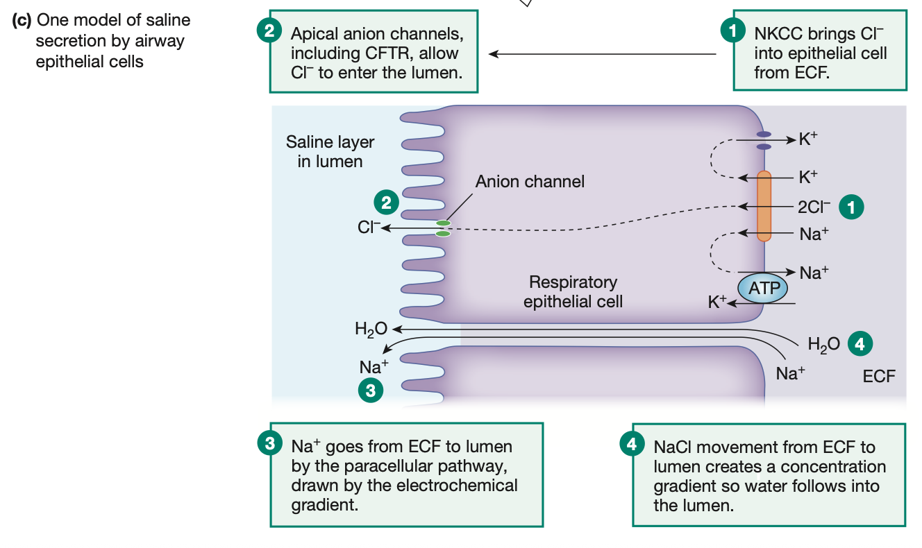
Saline lubricant
NKCC transporter = 1 Na+, 1 K+, 2 Cl- ions moved
CFTR transporter = Moves Cl- ions into the lumen/cilia side
Na+ ions move across the cells to balance out Cl- ion movement
Now NaCl (salt) formed to make saline as water follows the movement of ions
Ppl with cystic fibrosis = no CFTR transporter, no Cl- ion movement, no saline produced
Atmospheric pressure
At sea level, 760 mmHg.
At HIGHER altitudes, atmospheric pressure DECREASES
Boyle’s law
p1v1 = p2v2
Pressure is INVERSELY proportional to volume
volume up, pressure down
volume down, pressure up
Quiet breathing
Inspiration is an ACTIVE process - muscles are put into action
Expiration is a PASSIVE process - muscles relax
Diaphragm
External intercostals
Elastic recoil from alveoli also play a part in increasing the pressure of lungs above atm pressure during exhalation
Quiet breathing: Pressure of pleural fluid, alvoli, and volume of air
Pleural fluid is subatmospheric - slightly below atmospheric pressure
During inspiration, pleural fluid decreases pressure even further as the ribs pull on the pleura which pulls on the lungs.
But lungs have some resistance to being pulled and this decreases pleural pressure even further

Active expiration
During exercise, when we force more air out of our lungs each breath.
Actively use inner intercostals and abdominal muscles

Physical factors effecting ventilation:
Lung compliance
Surface tension
Pulmonary resistance
Physical ability for the lungs to stretch.
High compliance = High volume change for little pressure change = easier to inflate
Elastic recoil: Ability for lungs to resist being deformed/stretch.
Elastic recoil = 1/compliance
High compliance = Low elastic recoil
e.g Alveoli have elastic, this helps for shape to recoil
If not present, easy to inflate (high compliance), but hard to deflate to original size (exhalation is hard = low elastic recoil)
High compliance = Low elastic recoil → EXHALE is HARD
Low compliance = High elastic recoil → INHALE is HARD

Physical factors effecting ventilation:
Lung compliance
Surface tension
Pulmonary resistance
At any air-water interface the surface of the fluid is under tension due to the presence of hydrogen bonds.
Surfactants = Surface active agents = secreted by type 2 Alveolar cells
Protein and phospholipid combo
Disturbs surface tension of watery surface on alveoli
Decreased surface tension = Easier to expand alveoli = increased compliance
Physical factors effecting ventilation:
Lung compliance
Surface tension
Pulmonary resistance
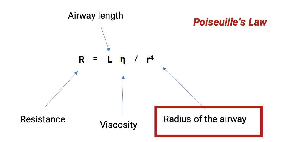
Resistance of the respiratory system to air flow during inspiration and expiration.
From Poiseuille’s Law, only radius of airway can be changed.
From trachea → bronchi = Most resistance as little 90% of cross-sectional area
However, cartilage rings keep airways open
From bronchioles → alveoli = Little resistance as HUGE cross-sectional area

Lung diseases:
Restrictive lung disease – ventilation is restricted
Obstructive lung disease – obstruction to ventilation
Due to decreased compliance of the lungs
decreased ability to stretch, decreased ability to inhale
Causes:
increased alveolar surface tension - decreased surfactant production
Formation of inelastic tissue in the lung - Pulmonary fibrosis
Fibrous masses toughening up the lungs, likely due to occupational hazards like asbestos, coal mines, silica etc.

Lung diseases:
Restrictive lung disease – ventilation is restricted
Obstructive lung disease – obstruction to ventilation
Increased airway resistance. (i/e smaller airways)
During exhalation, smaller airways can possibly collapse
Decreased exhalation efficiency, decreased elastic recoil
CO2 remains trapped in lungs, poor O2 exchange
Asthma

Volume types in the lung
Tidal volume = Volume of air moved in and out for each quiet breath = 500 ml (REMEMBER)
Expiratory reserve volume (ERV) = Volume left remaining in lungs that can be exhaled after a quiet breath
Inspiratory reserve volume (IRV) = Extra volume that can be breathed into lungs after a quiet inhale
Residual volume = volume left after a full active exhale

Capacity types in the lung
Sum of 2 or more volumes
Inspiratory capacity = TV + IRV = Total amount of air inhaled
Functional Residual capacity = ERV + RV = Amount of residual air in lungs after quiet exhale
Total lung capacity = TV + IRV + ERV + RV = Total max lung capacity
Vital capacity = TV + IRV + ERV = Total amount of exchangeable air in lungs
Forced vital capacity (FVC) and FEV1 = How much can you breath out FAST
IF lower than normal indicative of asthma
Pulmonary ventilation/ minute ventilation
TOTAL volume of air moved into and out of the lungs each minute
However, NOT all of this air is going to commit gas exchange, as not all reaches alveoli
Air in conducting zone = anatomical dead space = 150 ml (REMEMBER)
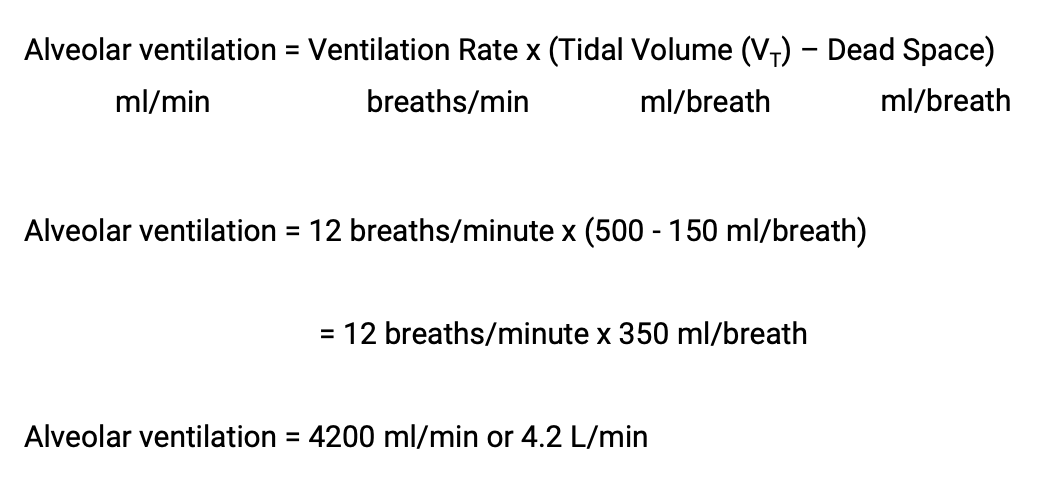
REMEMBER FORMULA

Alveolar ventilation
Volume of air that reaches the alveoli in 1 minute and undergoes gas exchange.
REMEMBER THE FORMULA
Dalton’s Law
Total pressure of a gas mixture is the sum of the partial pressure of the gases that the mixture
Each gas in a mixture exerts its own pressure called its partial pressure (Pgas)
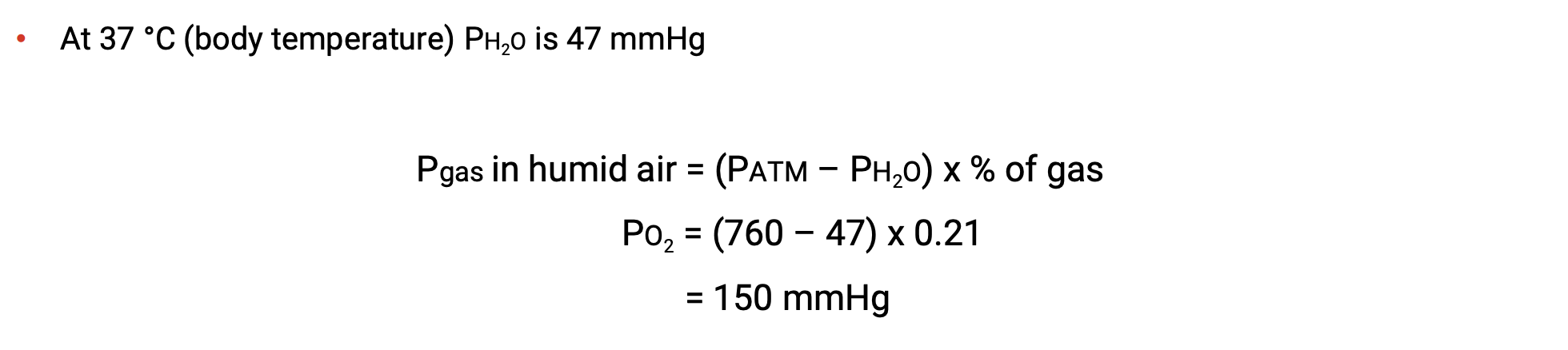
DALTON’s LAW DOES NOT ACCOUNT FOR WATER VAPOUR


Partial pressure of O2 in dry air vs humid air
REMEMBER FORMULAS
Humid air present inside lungs

Partial pressures of O2 & CO2 in blood
High CO2, low pH (acidic)
vice versa

Factors effecting alveolar ventilation
Enough O2 reaching alveoli
Enough gas exchange of CO2 & O2
Enough bloodflow to alveoli - “perfusion” of alveoli

Perfusion of lungs and alveoli
Usually 0.5L of blood in lungs at anytime.
BUT flow rate is high
Pulmonary arterial pressure is 25/8 mmHg vs 120/80 mmHg in systemic arteries
Capillaries in the lungs are collapsible
Enables only sections of the lungs to be actively perfused and used at rest, prevents waste of energy
if pCO2 increase, bronchioles dilate (more CO2 exhaled)
if pCO2 decreases, bronchioles constrict (less CO2 exhaled)

Hypoxia – decreased O2 levels in the blood
Hypercapnia – increased CO2 in the blood
Can happen NOT at the same time
Emphysema = COPD


O2 consumption:
Mass Balance (any gain must be offset by equal loss)
Mass flow (how much O2 is transported around the body in 1 minutes)
QO2 = Oxygen consumption
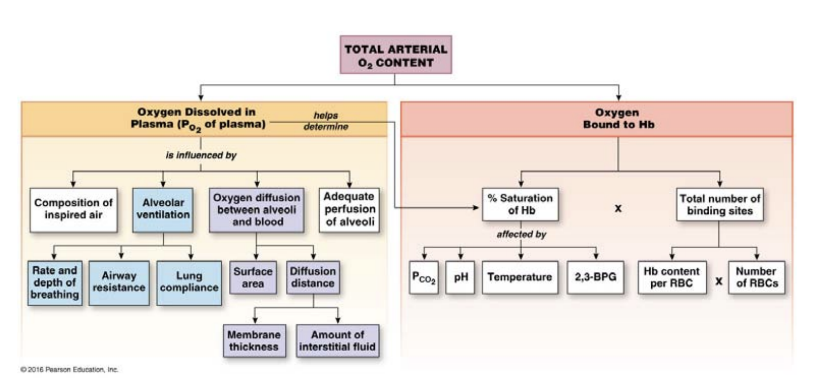
O2 is poorly soluble in blood, much more soluble with haemoglobin
Haemoglobin can help blood carry 200ml/L of oxygen (4x what we need at rest)
Reserves left for sharp spikes in oxygen demand
Total blood O2 = dissolved O2 + O2 bound to Hb
1 haemoglobin molecule carries 4 iron that binds to 4 O2 molecules
Can be fully or partially saturated

Law of mass action
When a reaction is at equilibrium, the ratio of products to the substrates is always the same
If the ratio is disturbed by either adding or removing one of the participants of the reaction, the equation with shift to restore the equilibrium

Haemoglobin to pO2 graph
Decreased pH, increased temperature or increased PCO2 shift the curve to the right
(decreased affinity for O2 - more O2 given to body tissues)
Mimics exercise conditions (more acidic, more temp, more CO2)
CO2 transport
7% dissolves in plasma
93% diffuses into red blood cells (23% bind to haemoglobin — diff site from O2, 70% become HCO3- ions (acid))
HCO3- ions diffuse out of red blood cells in exchange for Cl- ions entering RBCs (chloride shift)


Neural control of breathing
Phrenic nerve involved somehow??
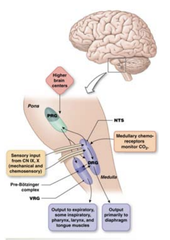
Motor neurons are ALWAYS firing — even when exhaling/relaxing in quiet breathing
Pre-Botzinger complex — pacemaker neurons that maintain quiet breathing pace
Ventilation rhythm — what keeps it in check?
Peripheral chemoreceptors
Central chemoreceptors
Mainly CO2 surprisingly, not O2 or pH
O2 still plays a part, but not as important.
Where? on Carotid arteries
Can INCREASE ventilation
Detect a decrease in PO2 or pH (more H+) or an increase in PCO2
Send info to DRG
Does not play a major role in maintaining ventialtion, major changes have to occur for anything notable
Glomus cells trigger Ca2+ release after stimuli by Peripheral chemoreceptors, sends stimuli to DRG
Ventilation rhythm — what keeps it in check?
Peripheral chemoreceptors
Central chemoreceptors
Mainly CO2 surprisingly, not O2 or pH
O2 still plays a part, but not as important.
Where? on medulla
Can INCREASE or DECREASE ventilation
Detect ONLY changes in pCO2
Senses pH changes in cerebrospinal fluid
If changes to pH are not rectified, Central chemoreceptors can become used to the pH change
In chronic conditions (like COPD), oxygen becomes the main regulator of ventilation
Voluntary control via the limbic system cannot override involuntary chemoreceptor control of ventilation

Regulating pH
Chemical buffers - weak acids/bases (think carbonic acid HCO3-)
Respiratory responce - Peripheral & central chemoreceptors
Renal response -
Proximal tubule: Overall↑HCO3 reabsorbed & ↓H+ secreted
Distal tubule: I-cells 2 types
Type A I-cells: ↑HCO3 reabsorbed & ↓H+ secreted — acidosis
Type B I-cells: ↓HCO3 secreted & ↑H+ reabsorbed — alkalosis
Respiratory acidosis:
Hypoventilation
Hypoventilation → CO2 buildup → acidosis → causes like COPD (emphysema), bronchitis, improper muscle movement
How to fix? Renal Type A I-cells: ↑HCO3 reabsorbed & ↓H+ secreted — acidosis
Respiratory alkalosis:
Hyperventilation
Hyperventilation → Little CO2 → alkalosis → causes like panic attack, asthma, hypoxia from high altitude
How to fix? Renal Type B I-cells: ↓HCO3 secreted & ↑H+ reabsorbed — alkalosis
Metabilic acidosis
Think too much exercise, renal dysfunction (H+ improperly filtered out, buildup in body)
Compensated by respiratory or kidneys
Metabilic alkalosis
Causes like excessive vomiting (stomach acid lost too much), too much alkaline drugs
Compensated by respiratory or kidneys