Anatomy 1
 Call Kai
 Learn
Learn Practice Test
 Spaced Repetition
 Match
Match Flashcards
 Knowt Play
1/140

There's no tags or description
Looks like no tags are added yet.
Name | Mastery | Learn | Test | Matching | Spaced | Call with Kai | Chat |
|---|
No analytics yet
Send a link to your students to track their progress
141 Terms
Name for humerus region of the thoracic limb (upper arm)
Brachium
Name for radius/ulna region of the thoracic limb
Antebrachium
Most proximal part of manus
Carpal
Middle part of manus
Metacarpal
Most distal part of manus
Digits/phalanges
How are mammaries classified in dogs
Cranial Thoractic
Caudal Thoractic
Cranial Abdominal
Caudal Abdominal
Inguinal
Overall: 2 sets of thoractic, 2 sets of abdominal, and 1 set of inguinal mammaries
How are mammaries classified in cats
Thoracic
Cranial Abdominal
Caudal Abdominal
Inguinal
Overall: 1 set of thoraccic, 2 sets of abdominal, and 1 set of inguinal

Name of major arterial trunk in shoulder/scalpular region
Axillary Artery
Note the axillary vein next to it

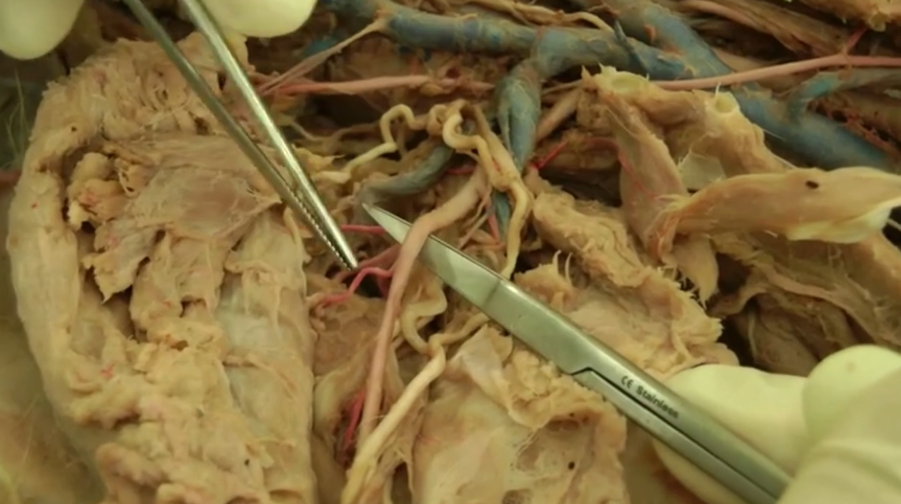
Name of major arterial trunk in brachium
Brachial Artery
After subscalpular branch of axillary. Name change from axillary

Most proximal branch of the axillary artery
Subscalpular artery
Connects to subscalpularis
Supplies scalpular region

More distal branch of the axillary artery (dog) / branch of subscalpular artery (cat)
Cranial circumflex humoral artery
thinner than subscalpular and brachial
Supplies the upper front leg
Name of major arterial trunk in antebrachium
Median artery
Vein commonly used for venipuncture in forelimb
cephalic vein
cMost distal split of arterial trunk in front leg
Radial artery
Splits off of brachium
Runs medial along radius bone
Muscle in skin
Cutaneous trunci m.
Name for scar on stomach
umbilicus
Name for connective tissue under skin and around muscle
Fascia
Superficial is less dense and deep is more dense
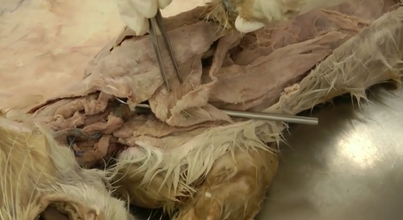
Muscle being pointed to by tweezers
Superficial pectoralis - Transverse
Will be cut and reflected on demo

Muscle being pointed to
Superficial pectoralis - Descending
Will be cut and reflected on demo
Where in the body are pectorals located
Proximal medial side of forelimb.
Medially connects to sternum

Muscle
Deep pectoral
Located just deep to the superficial pectoral

Muscle
brachiocephalicus m.
Located just laterally to pectoralis

Bone here
Clavicular bone (cats only)
Located in brachiocephalicus muscle

Region of brachiocephalicus muscle between clavicle and forelimb
cleidobrachialis m.

Region of brachiocephalicus muscle between clavicle and neck
cleidocephalicus m.
Has 2 heads

Muscle, specific muscle part, and species here
brachiocephalicus
White bit is clavicular intersection, separates it into cleidocephalicus and cleidobrachialis
Clavicular intersection is in dog only

Directly deep to cleidocephalicus
omotransversarius m.
Connects shoulder region to head
Lymph nodes in neck
superficial cervical lnn.
Lymph node in upper arm/armpit
axillary ln.

muscle
trapezius m.
cervical part (cranial) and thoracis part (caudal) - named for the vertebrae they attach to
Triangle-shaped muscle very superficial to shoulder

muscle
rhomboideus m.
along spine directly deep to trapezius
Three parts of rhomboideus m.
capitis, cervicis, and thoracis parts

Muscle
latissimus dorsi m.
long, flat muscle directly caudal to forelimb

Muscle
serratus ventralis m.
Large, fan shaped muscle deep to latissimus dorsi and caudal to forelimb
2 sections of serratus ventralis m.
cervicis & thoracis

Muscle
deltoideus m.
distal to trap
“checkmark” shape

Muscle
infraspinatus m.
Large muscle on lateral side of proximal forelimb deep to deltoideous
Below the spine of the scapula

structure
subtendinous synovial bursa
deep to the distal end of infraspinatus m.
Shiny and has fluid in it

muscle
supraspinatus m.
Large muscle on craniolateral side of proximal forelimb deep to deltoideous and omotransversarious
BUT: above scapula spine

muscle
subscapularis m.
Large muscle on medial side of proximal forelimb.

muscle
Teres major m.
Ventral to subscalpularis
Runs from bottom edge of shoulder blade to the armpit

muscle
tensor fasciae antebrachii m.
Located on medial side of antebrachium. Attach to origin of cutaneous trunci and latissimus dorsi
long, flat muscle

muscle
Triceps brachii m. long head.
caudal side of antebrachium, deep to tensor fasciae antebrachii m.
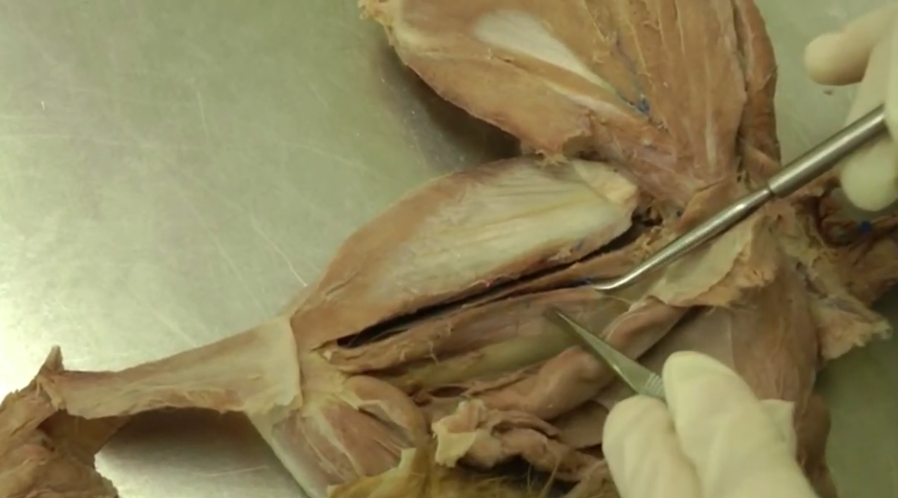
muscle
Triceps brachii m. medial head.

muscle
Triceps brachii m. accessory head.
Located deep to lateral head, or wedged between medial and long head when looking at it from medial plane.

muscle
Triceps brachii m. lateral head.
on lateral side of antebrachium

muscle
anconeus m.
located on lateral side of elbow joint.

muscle
brachialis m.
Located on lateral side of antebrachium, ventral to triceps lateral head

muscle
biceps brachii m.
medial side of antebrachium
deep to pectoralis

structure
transverse humeral retinaculum
bit of connective tissue on proximal side of biceps brachii and distal side of subscalpularis

nerve
suprascapular n.
runs cranially around scalpula
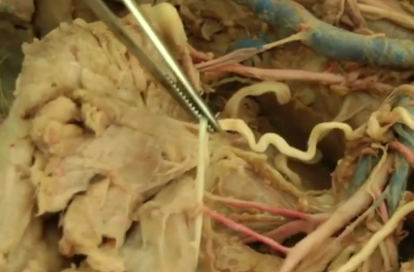
nerve
musculocutaneous n.
runs along caudal aspect of biceps

nerve
medial n.
runs along to brachial artery

nerve
ulnar n.
connects to elbow
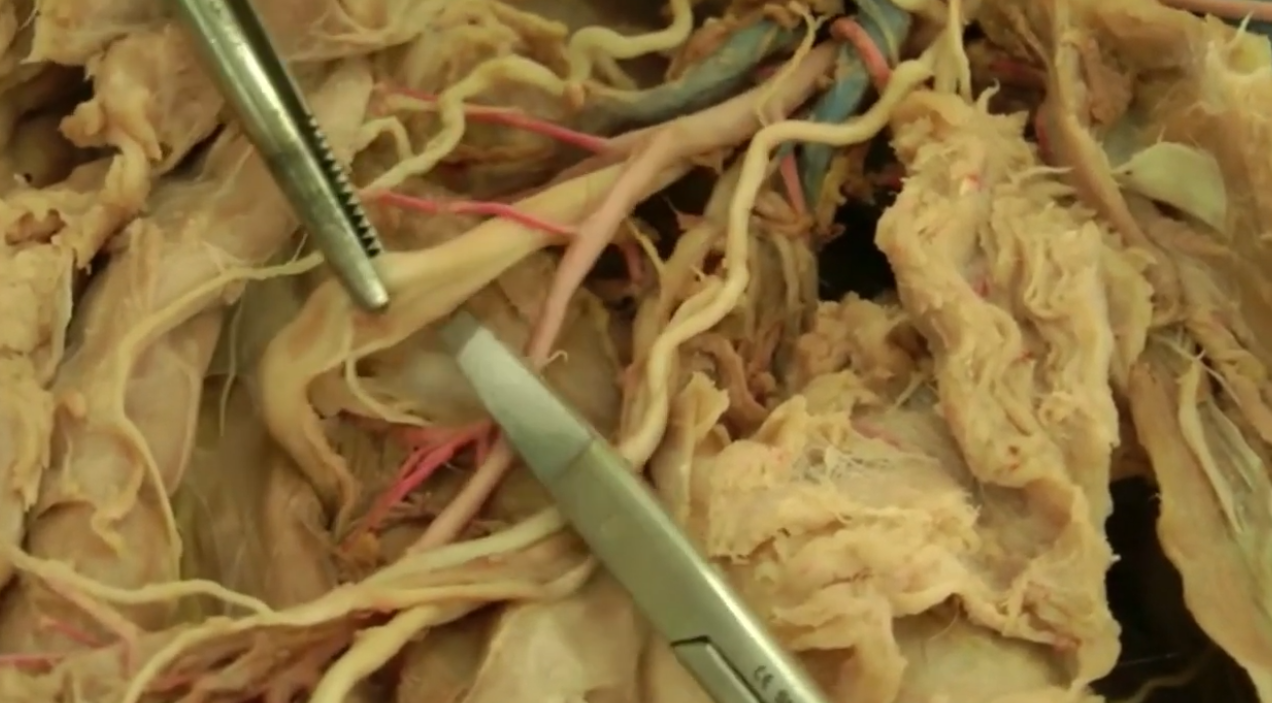
nerve
radial n.
huge nerve that goes into triceps
you can see it if you cut the lateral head of triceps
splits into 2 branches under triceps

nerve
axillary n.
splits from radial nerve really close to trunk
Muscles that cross the shoulder joint
deltoideus
infraspinatus
supraspinatus
subscapularis
teres major
long head of triceps brachii
Which side is the extensor surface of limb
craniolaterally (top)
which side is the flexor surface of limb
caudomedially (bottom)
hip bone name and region
os coxae / rump
thigh bone
femur
middle leg region name / bones
crus / tibia (longer, front) & fibula (smaller, back)
hind paw lowest region and bones
pes / tarsus (hock), metatarsal bones, and phalanges (digits)
Muscles that cross the front elbow joint
Brachialis
Biceps Brachiii
Triceps brachii
ONLY extrinsic muscle in hind limb
iliopsoas m. Everything else is intrinsic
Major muscles attaching to fascia lata
tensor fasciae latae and the biceps femoris
Clinical significance of fascia lata
Helps stabilize hips and knees, attach muscles, and helps with propulsion
Tissue can be used to repair hernia
veins commonly used for venipuncture in the hind limb
lateral saphenous vein in dog and medial saphenous vein in cat
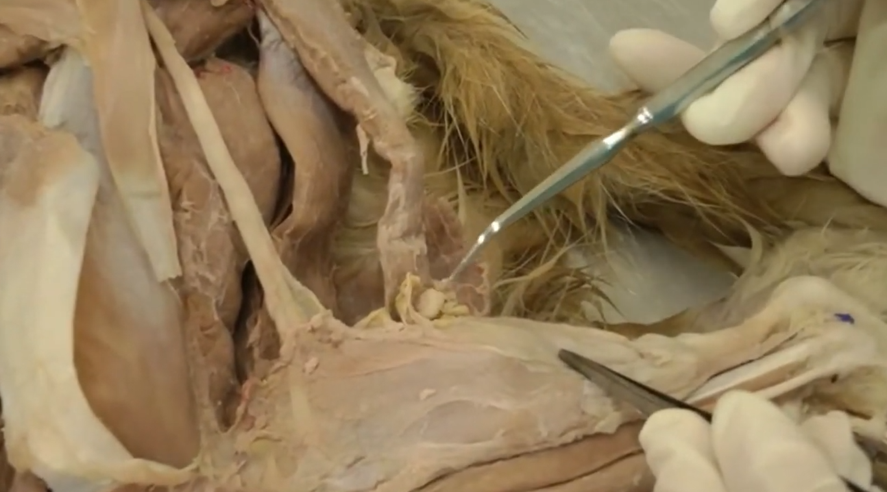
Name for lymph node of back leg and general location
popliteal lymph node
located behind the stifle (knee)
Summarize how hind limb attachment to the trunk differs from the forelimb.
Back limbs are connected via bone attachments, which give more strength and less flexibility than front limbs, which are connected via muscle attachments.
major muscle groups contributing to the common calcanean tendon
Gastrocnemius, Superficial digital flexor
soleus in cat
front shoulder region nerves for extensors and flexors
flexor: Axillary nerve
extensor: Suprascapular
front brachium region nerves for extensors and flexors
Flexor: Musculocutaneous nerve
Extensor: Radial nerve
front antebrachium region nerve for extensors and flexors
flexors: Median nerve and ulnar nerve
extensors: radial nerve
front manus region nerve for extensors and flexors
flexor: Median nerve
extensor: Radial nerve
Main functional difference between Extrinsic Muscles and Intrinsic Muscles
extrinsic: Gross motor movements, force production, large joint actions
Note: when under weight, need muscle activation and tissue stiffness to prevent the joint from collapsing inward. The extrinsic muscles transition from simple "movers" to primary load-bearing
Intrinsic: Fine motor control, precise stability, micro-adjustments

muscle
extensors of the digits
in the middle of the dorsal plane of front leg
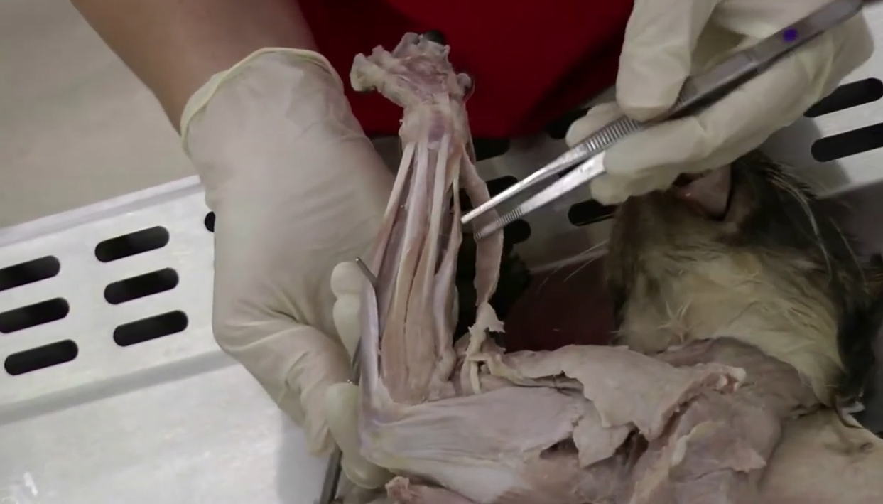

muscle
extensors of the carpus
on craniolateral side of front manus, on outside of the digit extensors

structure
extensor retinaculum
band of connective tissue that holds down extensor tendons as they pass into the manus

muscle
flexors of the digits
on bottom side of paw in middle

muscle
flexors of the carpus
on bottom side of paw on outside

structure
flexor retinaculum
where flexor tendons pass to manus

structure
carpal canal
area in manus where nerves, blood vessels, and tendons cross the manus
on bottom side of paw, under flexor retinaculum
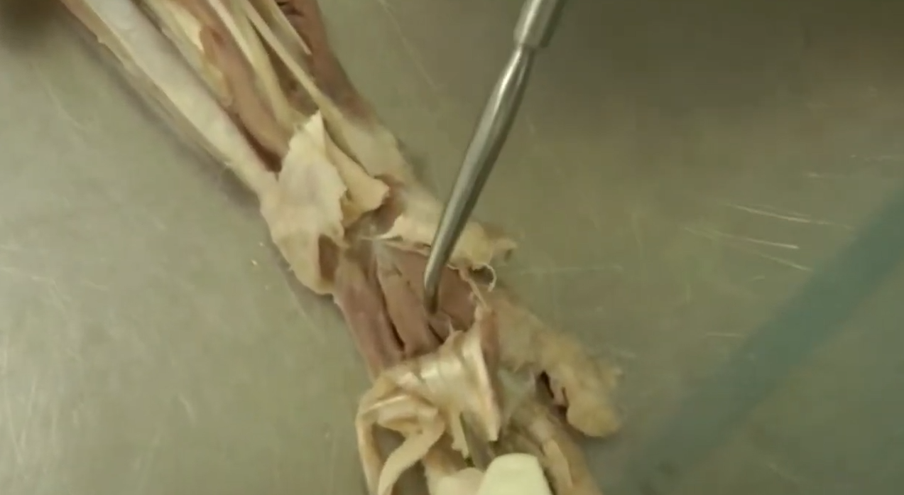
muscle
interossei muscles
located below flexor tendons on bottom side of paw.

ligament
lateral elastic ligament
on cats only
lateral side of toes
used for claw retraction

ligament
dorsal elastic ligament
dorsal side of toe
2 in dog, 1 in cat usually

ligament
palmar annular ligament
goes across base of toe on bottom side of paw
holds tendons in place

ligament
digital annular ligaments
horizontal on toe in about the middle of the toe on bottom side of foot
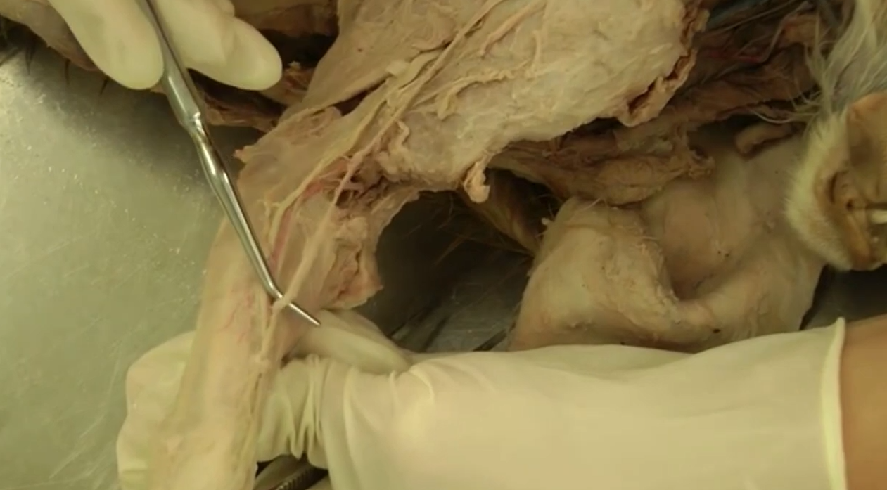
vein
cephalic

vein
accessory cephalic
branch of cephalic that comes off in antebrachium

vein
median cubital v.
branches off cephalic in brachium (further up than accessory cephalic)
describe attachment of common calcanean tendon to tuber calcanei (heel bone)
Gastrocnemius Tendon: proximal (top side) of tuber calcanei
Superficial Digital Flexor Tendon: runs over gastroc. tendon and broadens to form a cap over the tuber calcanei. calcaneal bursa sits underneath this tendon to cushion it from the underlying gastrocnemius tendon.
biceps femoris (lateral tract), gracilis, and semitendinosus (medial tract) muscles: insert on the sides/plantar aspects of the calcanei
main arterial trunk in thigh (femoral) region
femoral artery
main arterial trunk in crus (tibial/fibular) region
Cranial Tibial Artery
main arterial trunk in pes (foot) region
dorsal pedal a. (p)

borders of femoral triangle
cranial: Sartorius muscle
caudal: pectineus and adductor muscles
proximal: inguinal ligament / groin crease

clinical importance of femoral triangle
easily palpable region with strong pulse
landmark for blood vessels and nerve
main nerve of the hip area
femoral n.
main nerves of thigh area
cranially: femoral
caudally: sciatic

main nerve of crus (lower leg/tibia/fibula)
lateral: fibular nerve (left, side of leg)
caudal: tibial n. (right behind tibia, dives into muscle on lateral side)
note: both are branches of sciatic nerve
