Cell Structure
 Call Kai
 Learn
 Practice Test
 Spaced Repetition
 Match
 Flashcards
 Knowt Play
1/25

There's no tags or description
Looks like no tags are added yet.
26 Terms
what is cell fractionation?
a technique that allows scientist to study the several different functions of organelles (etc mitochondira/ ribosomes)
describe the homogenization process
take a sample of tissue containing the cells were interested in ( etc heart muscle tissue)
homogenize the tissue (break up the tissue/break open the cells) can do this using a blender or homogeniser (glass tube containing plunger)
place tissue sample into the glass tube and cover the tissue with buffer solution (buffer keeps PH constant so theres no change in PH which may cause enzymes in the cells organelles to denature ) water potential of buffer should be the same as as w.p inside cell to prevent water moving into cell by osmosis which may cause them to burst
place the homogenizer on ice as cooling the sample means that enzymes work more slowly, preventing any destructive enzymes from damaging the organelles
then push the plunger up and down to disrupt the tissue and break open the cells , producing a cell homogenate that contains all the organelles we find in a cell.
filter- filtering removes large cell debris and whole cells before centrifugation
what do we need to find organelles function
we need to seperate the organelles by looking at realitive sizes of different organelles
largest to smallest =
nucleus, mitochondria, lysozomes, ribosomes
(endoplasmic reticulum is a very large organelle but tends to get broken up during cell homogenisation )
2nd stage of cell fractionation is ultracentrifugations , describe the steps
place tube containing cell homogenate into the sample holder, the centrifuge spins the sample n the organelles are flung towards the bottom of the tube by forces generated (larger organelles experience a greater force therefor move towards the bottom of the tube faster- at lower speeds)
first start w a low speed, as centrifuge spins, larger organelles such as nuclei are flung towards the bottom of the tube forming a nuclei pellet, remaining organelles stay suspended in the liquid(supernatant)
transfer supernatant into a new test tube and centrifuge at a higher speed, now the pellet will contain mitochondria
transfer the new supernatant into new test tube n centrifuge at even higher speed , -now pellet contains lysosomes
finally, take supernatant one more time n transfer to new test tube w a very high speed, now pellet contains ribosomes
(now organelles are seperated we can test each fraction)
limitations of cell fractionation (2)
we need to keep pellets on ice till we use them to slow enzymes down which might damage organelles
its very difficult to seperate organelles fully (etc mitochondira fraction may contain a little nuclei)
unit conversions
m- mm - um - nm
x1000
adv of light microscopes
can use them on living cells so we can use them on processes such as cell division/ cell movement
2 disadvs to light microscope
sometimes we need to use a stain which can kill cells
low mag/ low resolution-limit of 200nm
definition of resolution
the minimum distance between 2 objects where they can still be seen as 2 seperate objects
why do light m have low resolution -200nm
due to the nature of light , the wavelength of visible light is around 400-700 nm so if 2 objeccts are closer than 200nm then we cant see them as 2 seperate objects using a light m , (can use electron microscope)
mag triangle
always convert to um
watch vid n do q’s on scale bars
use same units

adv of electron microscope
uses electrons- have a very short wave length so the resolution is 200x better than light m
describe an electron microscope
we start w an electron gun which is producing a beam of electrons , where the electrons pass down the microscope (inside of an electron m contains a vacuum so electrons can pass through w/out bouncing off the molecules in air
e are (-) so we can focus the electron beam by using electromagnetic lenses
the specimen is plaed in the path of the electron beam
electrons can pass through some parts of specimen more easily than others
the final image is produced on a fluroscent screen
3 disadv of electron microscopes
the interior of an e.m is a vacuum to it cannot be used toview living organisms
e.microscopy requires very careful staining and the specimen must be very thin
e.m can lead to artefacts
one type of em is transmission em (3)
the electron beam passes through the specimen
produces flat 2D images, only works if specimen is very thin
has very high resolution
scanning e.m
the electron beam doesnt pass through the specimen
instead electrons are scattered from the surface of the specimen and detected
produces 3D images
doesnt require specimen to be thinly sliced
lower resolution than t.e.m
requires specimen to be coated w a metal e.g gold (could lead to artefacts)
key features of eukaryotic cells (animals/plants/fungi) (7)
dna is in a memb bound nucleus
dna is tightly wrapped around proteins called histones (DNA+ histones= chromosomes)
by tightly coiling DNA into chromosomes they can pack alot of dna into nucleus
their dna is a linear molecule (ends arent joined tg to form a loop)
contain other memb bound organelles(mitochondria, golgi a, e.r)
also contain non memb bound organelles etc 80s ribosomes (larger)
surrounded by a cell memb
similairty betwee plant/ fungi
both have cell wall which helps maintain structure
plantcw= made from cellulose
fungi cw= made from polysaccharide chitin
prokaryotic cell key features
musch smaller than e.c
no memb bound organelles- dna is found loose om cytoplasm (not nucleus)
dna is arranged in a circular chromosome w no free ends
dna isnt bound to histone proteins)
70s ribosomes (smaller)
bacterial cell wall is made from peptidoglycan (murein)-polymer formed between peptides/ polysacharides that help maintain strucutre
what do all prokaryotes have ?
cytoplasm
circular chromosome
ribosomes
cell wall
what do only some prokaryotes have?
sometimes contain loops of dna ‘plasmids’(contain a little bit of genes but enough to make bacteria resistant to a.b
some bacetria have a slime capsule on outside of cell wall which can help protect bacteria from phagocytosis by wbc
flagellum to help move
fine protein strands on surface called pilli - help baccteria to attach to other bacteria/surfaces
contain lipid droplets/ glycogen granules - act as nutrient stores for bacterial cell
mesosomes
when using a e.m we sometimes see infoldings in the cell memb of p.c =’mesozomes’
initally, scientists thought mesozomes play a role in respiration but now they beleieve theyre artefacts that are created when baceria cells are prepared for e.microscopy
viruses
20nm- 30nm
they cant reproduce independently- can only reproduce inside a host cell
1) first a virus particle attaches to the structure of a host cell and enters
2) viruses use the host cells enzymes to produce copies of itself
3) virus particles leave the host cell and can go onto infecting new hostcells and continue reproducing
structure of a virus
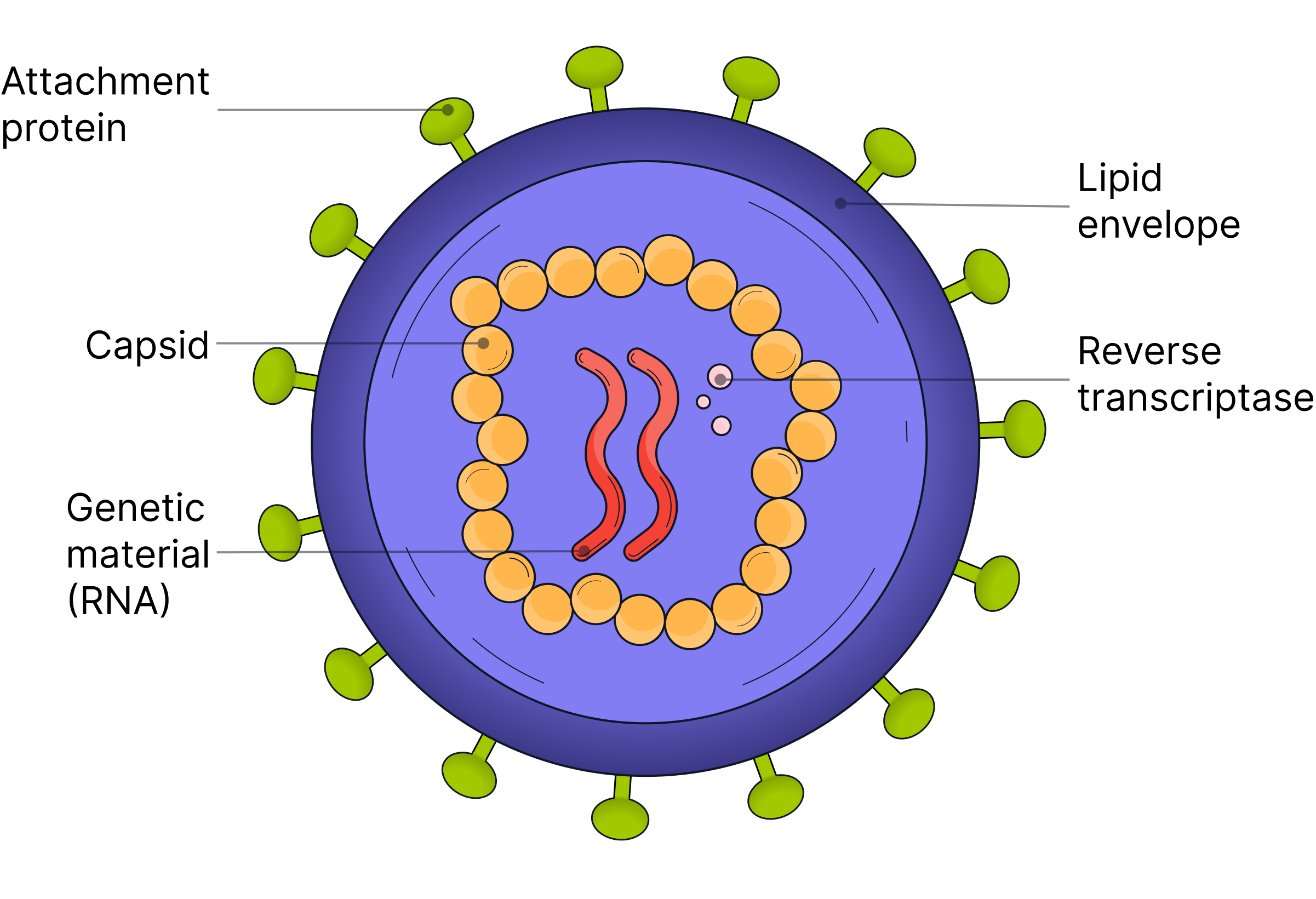
all contain genetic material- dna/rna
genetic material is inside a protein structure called a capsid
surfaces have attachment proteins whichallow viruses to attach/ enter host cells
some are surrounded by a lipid envelope, formed from host cell memb
Key ideas of viruses
viruses arent cells theyre ‘ acellular’ ,not based on cells
cant reproduce independently - must invade host cells / use its enzymes to make copies of itself , therefore arent counted as living organisms