Haemodynamic disorders
 Call Kai
 Learn
Learn Practice Test
 Spaced Repetition
 Match
Match Flashcards
 Knowt Play
1/68

Earn XP
Description and Tags
Formation of atheromatous plaque & consequences
Name | Mastery | Learn | Test | Matching | Spaced | Call with Kai | Chat |
|---|
No analytics yet
Send a link to your students to track their progress
69 Terms
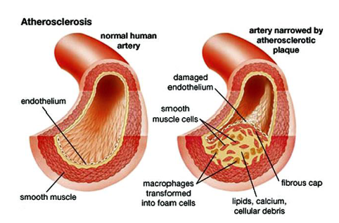
What is atherosclerosis?
Chronic inflammatory disease, caused by the formation of ‘fibro-fatty’ lesions in the arterial wall
Which arteries does atherosclerosis occur in?
All arteries - including aorta, carotid, coronary, iliac, femoral …
What is atherosclerosis a major cause of?
Heart attacks
ischemic stroke
PAD
What two conditions are major risk factors for atherosclerosis?
hypercholesterolaemia
inflammation
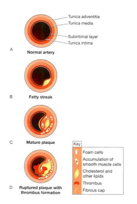
What are the 3 stages of atheroma?
Initiation (fatty streak)
Plaque progression (fibrous cap atheroma)
Plaque rupture (atherothrombosis)

What layer of artery structure does atherosclerosis occur in?
Intimal layer - specifically the tunica intima

Initiation (fatty streak)
Endothelial dysfunction - lipids accumulate and modify in this area forming fatty streak
Leukocyte recruitment - includes macrophages from tissue and monocytes from blood
These cells phagocytise the lipids, before becoming foam cells
How are lipids modified by macrophages?
Macrophages oxidise LDL
How are foam cells formed?
Recruited monocytes differentiate into macrophages and engulf lipids trapped in subendothelial space = foam cells
Plaque progression (fibrous cap atheroma)
Has a lipid rich necrotic core comprised of dead foam cells, macrophages, smooth muscle cells, lymphocytes and ECM.
What separates the necrotic core of the atheroma from the vessel lumen?
Fibrous cap
What is atherothrombosis
Rupture of the fragile atheroma into the blood
What can atherosclerosis cause?
acute narrowing of vessel lumen
chronic occlusion of vessels (due to growing plaque)
Thrombosis and embolism (rupture of plaque)
What is PAD?
Peripheral arterial disease - it is the presence of systemic atherosclerosis distal to the arch of the aorta
What is the primary symptom of PAD?
Intermittent claudication
Intermittent claudication
pain with walking
What are other symptoms of PAD?
cold feet
weak/absence of popliteal and pedal pulses
blanching of limb colour with elevation
Non-modifiable risk factors of PAD
Increasing age
male gender
Genetic disorders of lipid metabolism
Family history of premature CAD
Modifiable risk factors of PAD
Smoking
Obesity
Hypertension
Hyperlipidaemia?
Diabetes mellitus
What is ischemia?
Restriction in blood flow/ supply to tissues = shortage of oxygen and glucose needed for cellular metabolism.
Also the reduced ability to remove metabolites
Result of ischemia?
Tissue damage - by build up of metabolic waste, inability to maintain cell membranes, mitochondrial damage, and eventual leakage of autolyzing proteolytic enzymes into the cell and surrounding tissues.
What characterises hyperaemia & congestion of haemodynamic disorders?
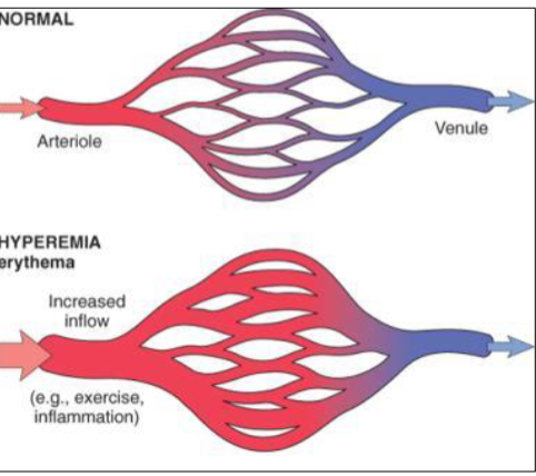
local increase in tissue blood volume
focused on microcirculation (arterioles, capillaries, venules)
What is hyperaemia?
It is an adaptive local increase in blood volume in tissues due to a change in environment
What are 2 kinds of hyperaemia?
reactive
active
Reactive hyperaemia
Local vasodilation due to oxygen debt or accumulation of metabolic waste
eg: raynaud syndrome
Active hyperaemia
Increased blood flow/vasodilation due to period of activation (increase in blood in skeletal muscle during exercise)
What is congestion?
Its is a passive process caused by impaired venous return from affected area

Hyperaemia vs congestion - process
Hyperaemia = adaptive process
congestion = passive process
Hyperaemia vs congestion - MOA
Hyperaemia = increase in blood volume in tissues in response to change in environment
Congestion = due to impaired venous return from affected area
What causes congestion?
failure of heart to pump blood away from affected area
Physical obstruction in veins
What impact does congestion have on venous pressure?
increases VP
Types of congestion?
Local
Systemic
Cause of local congestion
Blood vessel compression due to venous obstruction (eg: tumour, VT)
Cause of systemic congestion
Often associated with heart failure = widespread oedema
Examples of congestion
Chronic pulmonary congestion
Centrilobular hepatic congestion
What is oedema?
Abnormally increased fluid in interstitial tissue spaces
Causes of oedema?
Increased hydrostatic pressure
Decreased colloidal osmotic pressure
Lymphatic obstruction
Sodium retention
Inflammation
Causes of increased hydrostatic pressure
Can be generalised (termed anasarca)
e.g. in heart failureCan be localised as in limbs
e.g. deep vein thrombosis (DVT)
Causes of decreased colloidal osmotic pressure
Reduction in the generation of plasma proteins
e.g. liver cirrhosis, malnutritionLoss of protein
e.g. via skin (burns), urine (nephrotic syndrome),
faeces (bowel disease)
How can lymphatic obstruction cause oedema?
Lymph no longer circulates and drains into tissues
Obstruction by malignancy, radiation therapy, surgery, parasites
How can sodium retention cause oedema?
As in excessive salt intake in renal insufficiency, or increased RAAS activation
Increases hydrostatic pressure
How can inflammation cause oedema?
Blood vessel dilation increases blood flow to the area = redness
increased blood vessel permeability = increased movement of fluid, proteins and leukocytes from blood into interstitial space
Haemorrhage
Extravasation of blood due to vessel rupture
What % of blood volume loss can cause hypovolemic shock?
>15%

Petechiae
Minute haemorrhage (1-2mm diameter) of skin and
mucosa.
• Due to thrombocytopenia, clotting factor deficiency, or
increased pressure in capillaries

Purpura
Small haemorrhage (3-10mm diameter)
• Due to trauma or vasculitis

Ecchymosis - haematoma/ bruise
Subcutaneous haematoma (>1cm)
• Trauma, platelet or clotting factor deficiencies
• Colour change (red–blue green–yellow brown) due to
metabolism of haemoglobin to bilirubin and hemosiderin
What is a thrombus?
Blood clot that has formed within the blood vessel AND which remains attached to the vessel wall
resulting from inappropriate activation of the haemostatic system
What 3 factors are the main cause of thrombi?
endothelial injury
abnormal blood flow
hypercoagulability
Thromboembolism
Thrombi that has detached from the blood vessel wall
Composition of thrombi?
platelets
RBCs
Neutrophils
Lymphocytes
All held together by fibrin mesh
2 types of arterial thrombi
mural thrombi - doesn’t occlude vessel
occlusive thrombi - occludes vessel
Are arterial thrombi platelet of fibrin driven?
platelet driven
Are venous thrombi platelet or fibrin driven?
Fibrin driven - due to hypercoagulation
Embolism
Solid, liquid or gaseous mass carried in blood to site distant from its point of origin - lodging in smaller vessels = occlusion and infarction
Pulmonary embolism
Blockage of an artery in the lungs, may cause sudden death
90% result from DVT
Infarct
An area of ischemic necrosis caused by occlusion of either arterial supply or venous drainage in a tissue
99% result from thrombosis or embolism
What is the most common kind of occlusion?
Arterial occlusion
List common clinical conditions caused by infarctions
MI
Cerebral infarction (ischemic stroke)
PI
Intestinal infarction
Ischemic necrosis of extremities (gangrene)

Aneurysm
Blood vessel enlargement caused by vessel weakness
Main causes of aneurysm?
Hereditary conditions or birth defect
Acquired disease - high BP, atherosclerosis, trauma
Aneurysm classification by morphology?
Fusiform: bulge all sides of the vessel
Saccular: bulge on one side

What is a true aneurysm?
It involves all three layers of the wall of an artery
(intima, media and adventitia)
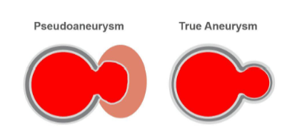
What is a false/pseudoaneurysm?
Does not contain any layer of the vessel wall.
• Result of trauma that punctures the artery (needle, knife, bullets), percutaneous surgical procedures (coronary
angiography, arterial grafting)

Shock
Clinical state characterised by systemic hypoperfusion (lack of blood flow) leading to reduced delivery of oxygen and nutrients
What does shock ultimately lead to?
Cellular injury, inadequate tissue function, multi-organ dysfunction syndrome