Bacterial infection
 Call Kai
 Learn
 Practice Test
 Spaced Repetition
 Match
 Flashcards
 Knowt Play
1/22

There's no tags or description
Looks like no tags are added yet.
23 Terms

Gingivits
▪ Dental plaque induces gingival disease.
▪ It is an inflammation that affects the gums surrounding the teeth.
▪ Gingival disease which is initiated by dental plaque is common.
▪ The studies have shown a predominance of facultative anaerobic Gram-positive bacteria and strict anaerobic bacteria.
▪ Enlarged gingival contour.
▪ Redness.
▪ Spontaneous bleeding or bleeding on probing.
▪ The inflammation only affects the gums.
▪ Reversible with dental plaque removal.
▪ It is common that gingivitis converts to periodontal disease.
▪ Dental plaque, enlarged gingival contour, redness.
▪ Spontaneous bleeding or bleeding on probing.
▪ The inflammation only affects the gums.

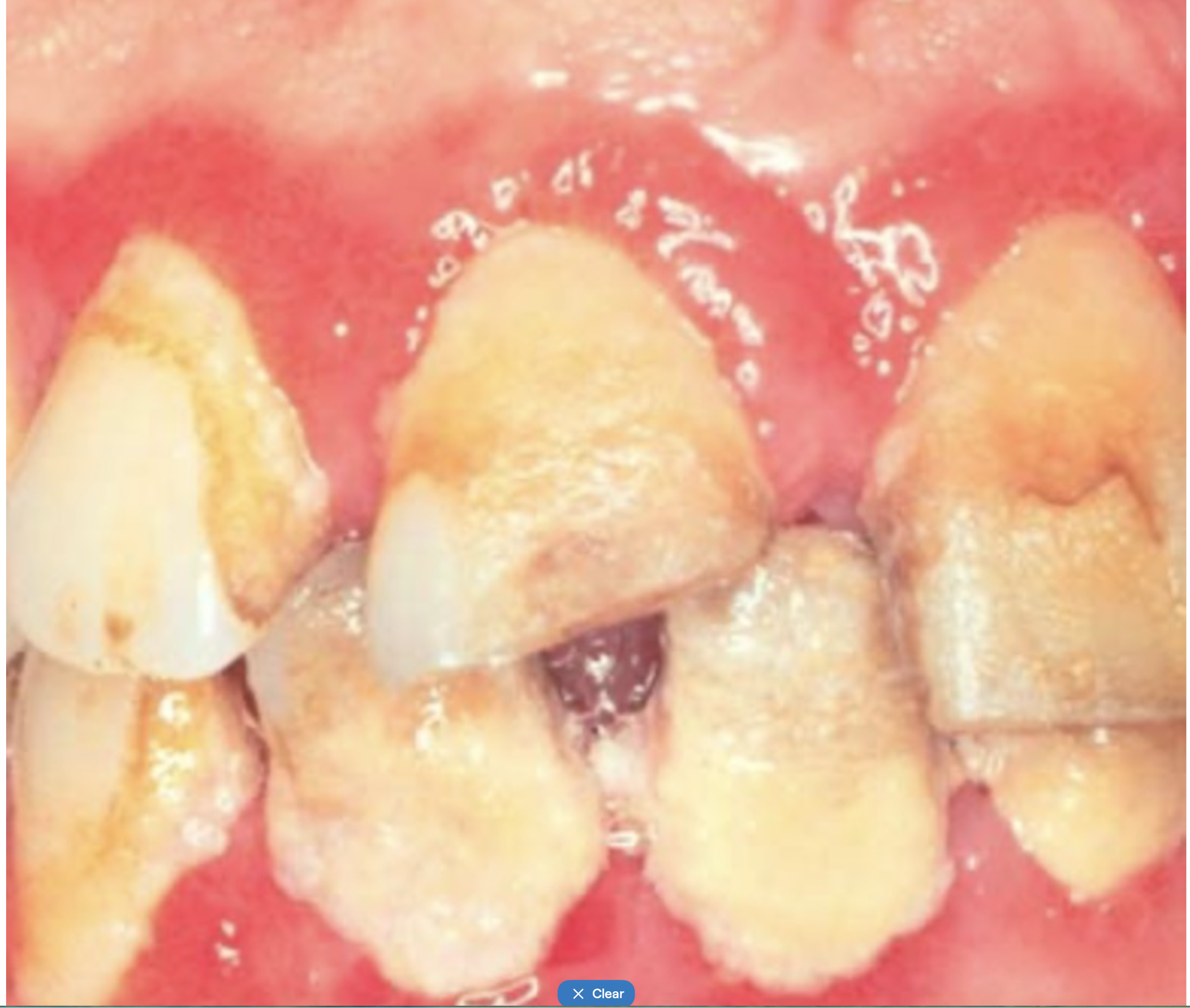
Gingivitis 2
▪ Pathology diagnosis is based on:
▪The combination of gingivits and dental plaque.
▪ Hygienic measures applied confirm the diagnosis.
▪ There are predisposing host factors that can modify the response to bacterial infection:
▪Local factors: crowding, overcontorned fillings...
▪Systemic factors: pregnancy, diabetes...
▪ Dental plaque removal.
▪ Oral hygiene.
▪ Control of predisposing factors.
Treatment
▪ Dental plaque removal.
▪ Oral hygiene.
▪ Control of predisposing factors.
NECROTIZING ULCERATIVE GINGIVITIS
▪ It is an opportunistic infection caused by germs of dental plaque.
▪ It is caused by a bacterial infection that includes Selenomonas spp., Prevotella Intermedia, Porphyromonas gingivalis and Treponema spp.
There are predisposing host factors that help in the development:
▪ Local factors: poor oral hygiene, gingivitis, smoking...
▪ General factors: emotional stress, malnutrition, immunosuppressive therapy, systemic diseases (HIV, diabetes, leukemia or agranulocytosis).
Malaise,
▪ Fever,
▪ Painful necrotic ulcerative gingival lesions that begin in the interdental papilla and may spread to the rest of the gingiva (necrotizing ulcerative stomatitis),
▪ Spontaneous gingival bleeding,
▪ Halitosis.

NECROTIZING ULCERATIVE GINGIVITIS 2
▪ Dental plaque control.
▪ Minor painkillers (ibuprofen 400-600mg every 8 hours, paracetamol 500-1000 mg every 8 hours).
▪ Antibiotic therapy:
▪Metronidazole (250 mg every 8 hours for 8-10 days).
▪In severe cases, it can be prescribed along with amoxicillin clavulanic acid (500-875/125 mg every 8 hours).
▪Surgical debridement of soft tissue.
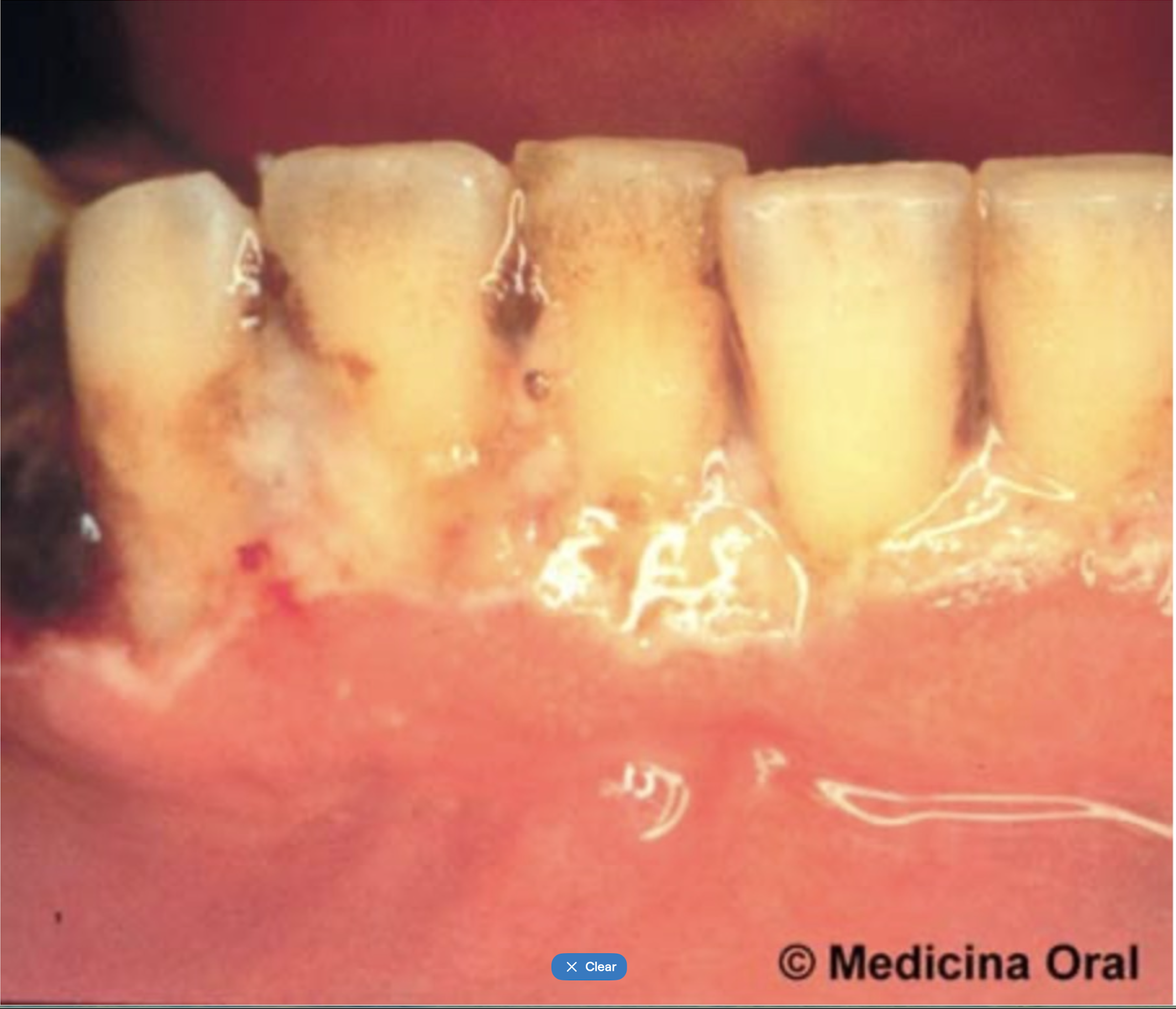
Oral tuberculosis
▪ Bacterial infection secondary to a pulmonary tuberculosis or primary oral tuberculosis.
▪ Oral tuberculosis - 1% of cases of extra pulmonary tuberculosis (20%).
▪ Transmission through blood or sputum.
▪ Primary oral tuberculosis is uncommon.
Etiology- ▪Mycobacterium tuberculosis or Koch's bacillus.
▪Tubercle bacilli are strict, obligate aerobes.

ORAL TUBERCULOSIS SECONDARY TO PULMONARY TUBERCULOSIS
▪ A typical oral lesion consists of a stellate ulcer, with irregular border, soft consistency and peripheric inflammation.
▪ Pain.
▪ Several months of evolution.
▪ General symtoms: odynophagia (20%), fever or weight loss.
▪ The differential diagnosis of a tuberculous ulcer includes oral squamous cell carcinoma.

PRIMARY ORAL TUBERCULOSIS
▪ A typical oral lesion consists of a stellate and irregular ulcer, soft consistency and it’s painless.
▪ The surface of the ulcer is usually covered by a grey- yellowish exudate.
▪ Regional lymphadenopathy usually accompanies the oral lesions. They are calcified lymph nodes with a tendency to form fistulas.
▪ Tuberculosis of cervical lymph nodes is the most common extra pulmonary tuberculosis type.
▪ It is more commonly found in children and adolescents.
TUBERCULOSIS INFECTION TESTING 1
1. Tuberculin skin testing or Mantoux skin testing:
▪ Intradermal injection of tuberculin purified protein derivative (PPD) into the inner surface of the forearm.
▪ It is the most common diagnostic tool for tuberculosis infection.
▪ The skin test reaction should be read between 48 and 72 hours after administration.
▪ The reaction is an induration area (5-10 millimeters).

TUBERCULOSIS DISEASE TESTING 2
2. Chest radiograph
• A person whose tuberculin skin test result is positive should be given a chest radiograph to rule out active tuberculosis disease.
Dense, homogeneous parenchymal consolidation in left lung.

TUBERCULOSIS DISEASE TESTING 3
3. Histology
• Granulomas composed of:
• caseous necrosis,
• epithelioid cells
• Langhans-type giant cells.
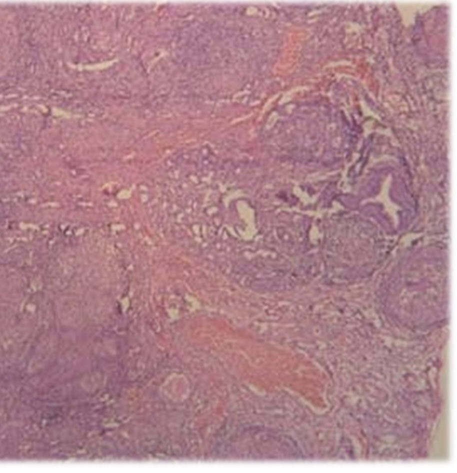
TUBERCULOSIS DISEASE TESTING 4
Ziehl-Neelsen staining
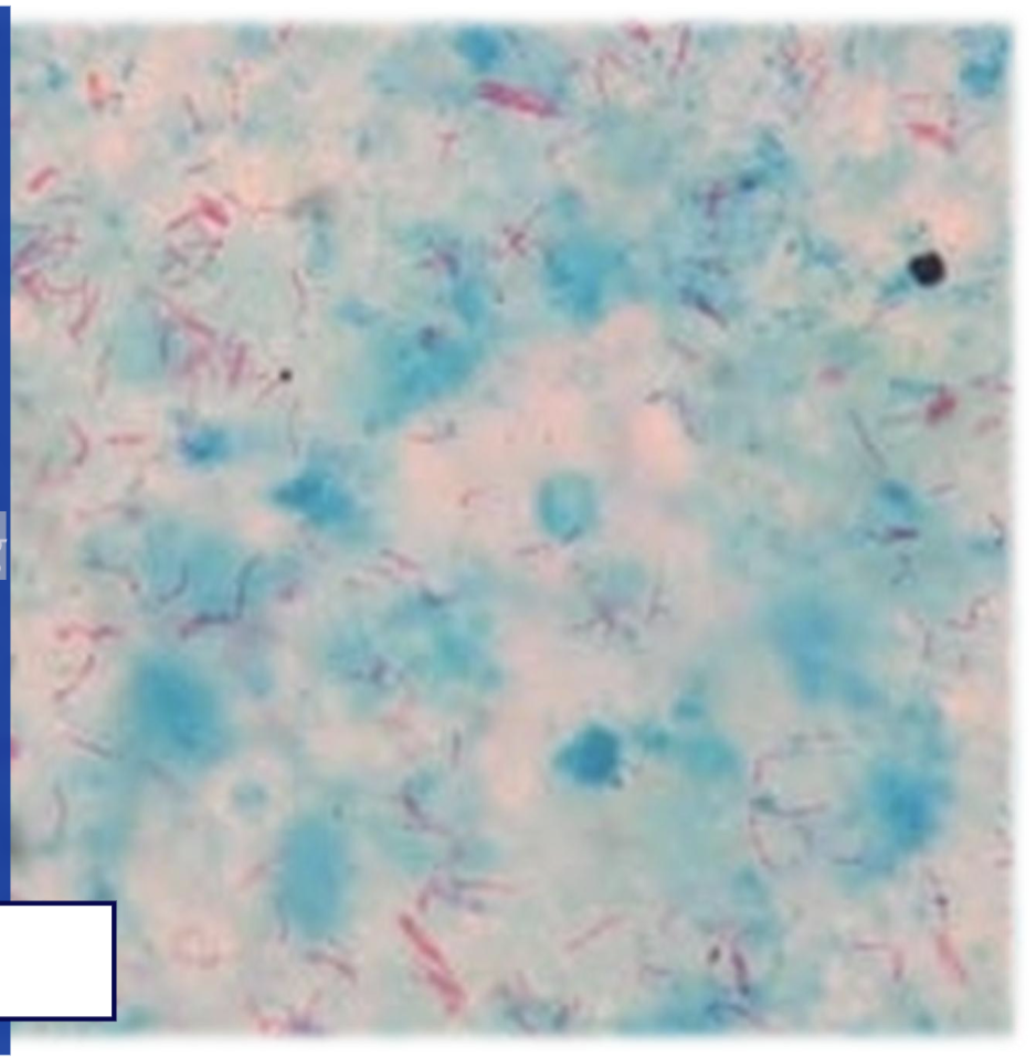
TUBERCULOSIS DISEASE TESTING 5
Cultures in specific medium (Lowenstein-Jensen, M-GIT 90): Tubercle bacilli grow slowly in culture and require 3 or more weeks to develop colonies.
Polymerase chain reaction (PCR): detects the DNA of the bacteria.
Treatment of tuberculosis
▪ Prolonged treatment (6 months therapy).
▪ First-line drugs are:
▪ Isoniazid + rifampin + pyrazinamide: during the first two months of treatment.
▪ Isoniazid + rifampin: for a further four months.
▪ Other combining drugs:
▪ Ethambutol+streptomycin+fluoroquinolones
Syphillis
▪ Chronic infection caused by Treponema pallidum.
▪ Sexually transmitted or from mother to fetus.
▪ There are three stages: primary, secondary and tertiary.
PRIMARY SYPHILIS
Chancre
▪ An oral chancre appears 3-4 weeks after infection.
▪ Most common on the lip (60% extragenital syphilis).
▪ Other oral sites are the tongue and gums.
▪ Usually solitary, firm and painless.
▪ The surface breaks down after a few days, leaving an ulceration with a clean base and sharp borders.
▪ Heals in 1-5 weeks.
▪ Regional lymphadenopathy can appear, and persist after healing of the chancre.
▪ Highly infectious.
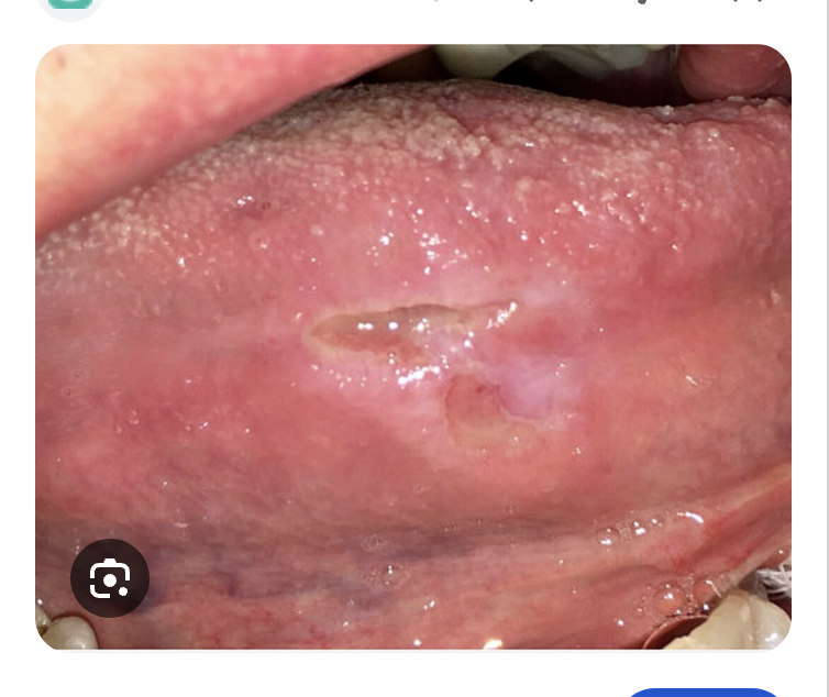
Primary syphilis clinical features
▪ Solitary,
▪ Firm
▪ Painless,
▪ The surface breaks down, leaving an ulceration with a clean base and sharp borders.

Secondary syphillis clinical features
▪ The secondary stage develops 1-3 months after healing of the chancre.
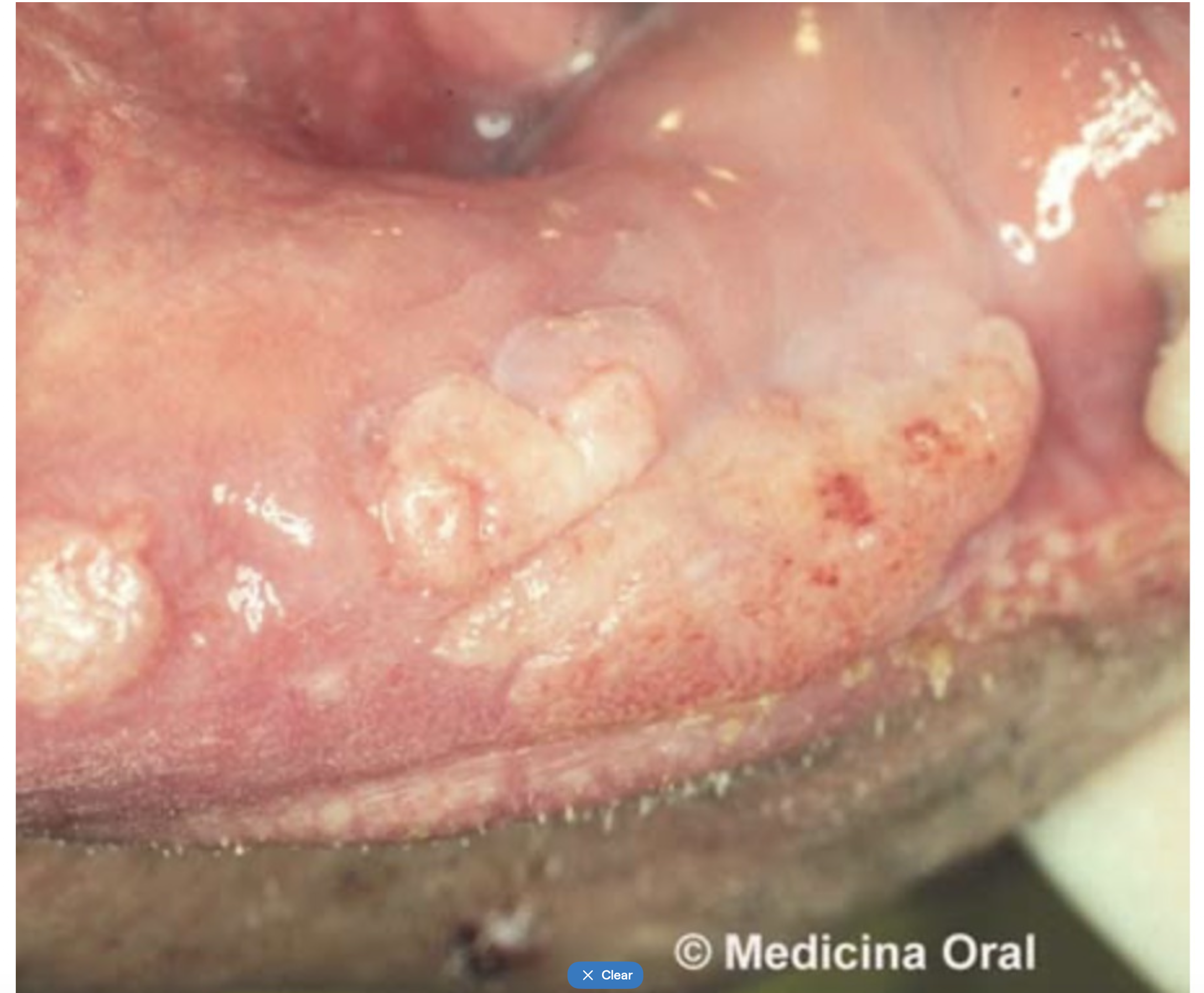
▪ Characteristic lesions are mucous patches:
▪ White raised maculopapular lesions.
▪ Multiple lesions.
▪ They may coalesce to give rise to serpiginous lesions.
▪ Highly infectious.
▪ Spontaneous resolution or can persist much longer.
Tertiary syphillus clinical features
▪ Develops in many patients around three or more years after infection.
▪ A characteristic lesion is the gumma:
▪ It may affect the palate, tongue or lips.
▪ It begins as a swelling, sometimes with a yellowish centre which undergoes necrosis, leaving a painless deep ulcer.
▪ It eventually heals with severe scarring which may produce bone destruction, palatal perforation and oro- nasal fistula formation.
▪ Little infectious.
▪ Intertital glossitis:
▪ Atrophy of dorsum of the tongue.
▪ It favours the development of oral cancer (precancerous lesion).

Diagnosis of syphillis
In primary and secondary stages, diagnosis depends on finding Treponema pallidum by dark ground illumination of a smear from the chancre or adenopathy.
Serological confirmation of the infection is essential:
1. Serologic screening tests – nonspecific:
▪ VDRL (Venereal Disease Research Laboratory) and RPR (Rapid Plasma Reagin): only positive in the first two stages. VDRL becomes positive 15-30 days after infection.
2. Serologic tests-specific: they use treponemic antigens. Positive in all three stages.
▪ FTA-Abs (Fluorescent Treponemal Antibody- ABsortion): becomes positive 4 days after infection.
▪ TPHA (Treponema pallidum Hemo-Agglutination).
▪ TPI (Treponema pallidum Inmobilization).
Treatment of syphilis
PRIMARY AND SECONDARY SYPHILIS
▪ Penicillin G benzathine: 2,4 million units intramuscularly (IM) in a single dose.
▪ Penicillin allergy: Erythromycin or tetracycline.
TERTIARY SYPHILIS
▪ Penicillin G benzathine: 3 doses of 2.4 million units IM each at 1-week intervals.
Dental abscess
- Periapical abscess
- Result of decay and infection extending into pulp of tooth
- Pain is severe, persistent,& throbbing
- Tooth is tender to touch
- If not treated pus tracks to surface inside or outside the mouth
1. Pharmacological treatment
- Minor painkillers (ibuprofen 400-600mg every
8 hours, paracetamol 500-1000 mg every 8 hours).
- Antibiotic therapy:
- Amoxicillin 500-750 mg every 8 hours
- Amoxicillin clavulanic acid (500-875/125 mg every 8 hours).
2. Debridement
3. Dental treatment:
- Root canal treatment
- Extraction

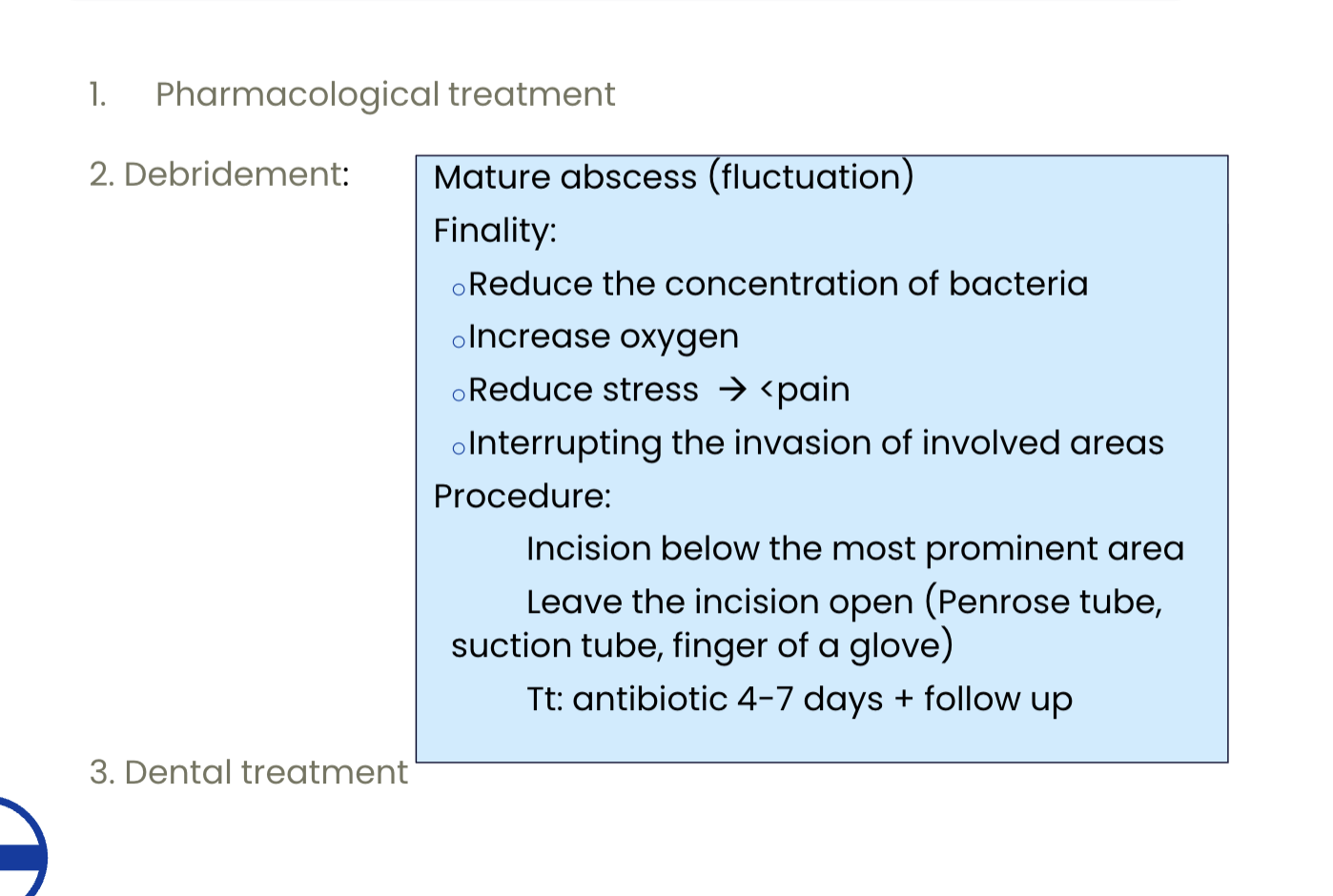
Treatment of dental abscess
1. Pharmacological treatment
2. Debridement
3. Dental treatment
------------------------------------------------
Granuloma Treatment: root canal treatment /re treatment
Cyst Treatment: extraction / apical resection

Periocoronitis
- Pericoronitis is an inflammation that develops around the crown of partially erupted tooth.
- Third molars
- Estreptococus, actynomices and Propionibacterium.
- Acute irradiate pain
- Halitosis
Periocoronitis treatment
▪ Minor painkillers (ibuprofen 400-600mg every 8 hours, paracetamol 500-1000 mg every 8 hours).
▪ Antibiotic therapy:
▪amoxicilin+clavulanic acid (500-875/125 mg every 8 hours).
▪ Follow up or extraction; depends on the position