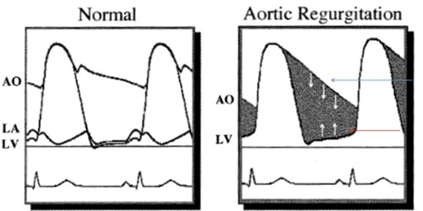
Aortic Insufficiency
 Call Kai
 Learn
 Practice Test
 Spaced Repetition
 Match
 Flashcards
 Knowt Play
1/21

There's no tags or description
Looks like no tags are added yet.
22 Terms
When is AI considered pathologic?
ANY amount - even trace
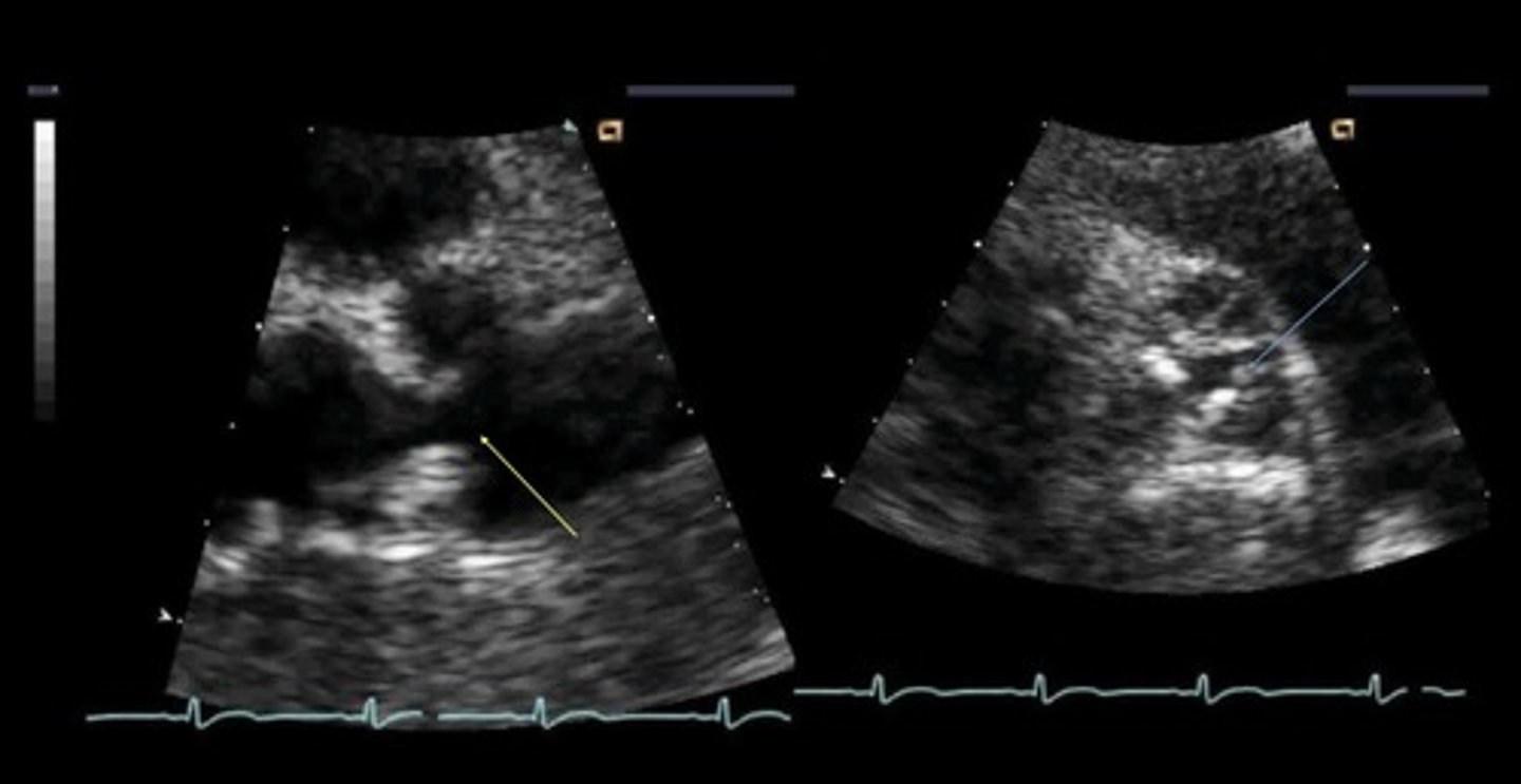
2D Features of AI
LV overload/enlargement
Thickened & stenotic leaflets
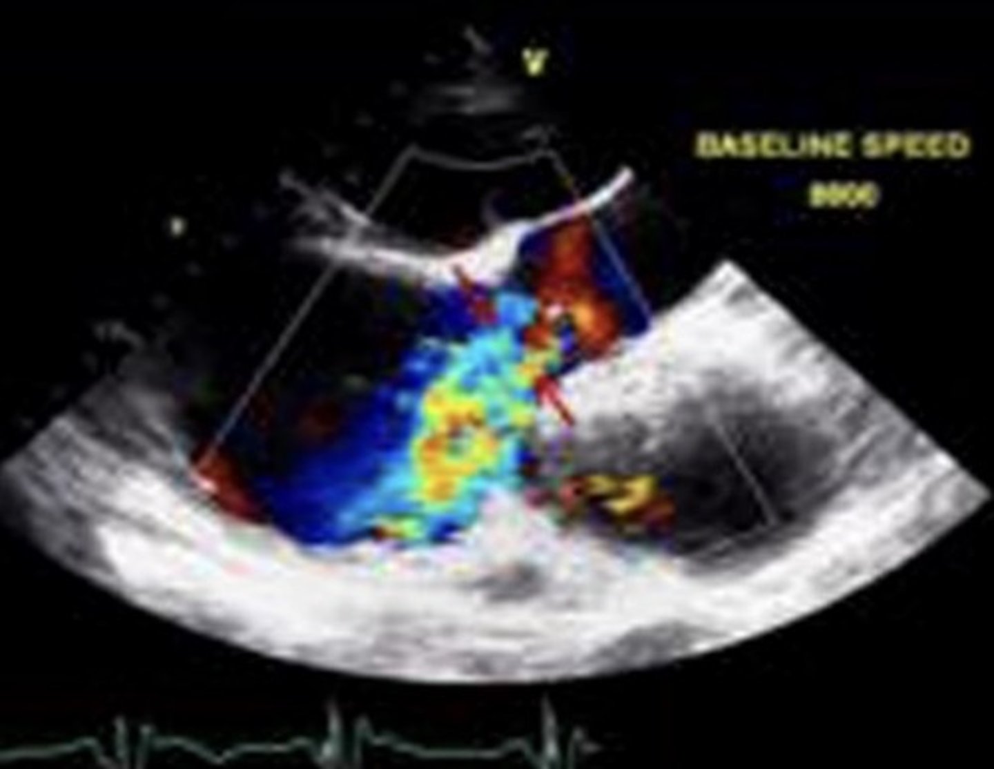
AI Jet Diameter/LVOT Diameter Ratio
Zoomed PLAX view
Measure LVOT diameter and compare it to the AI jet width diameter
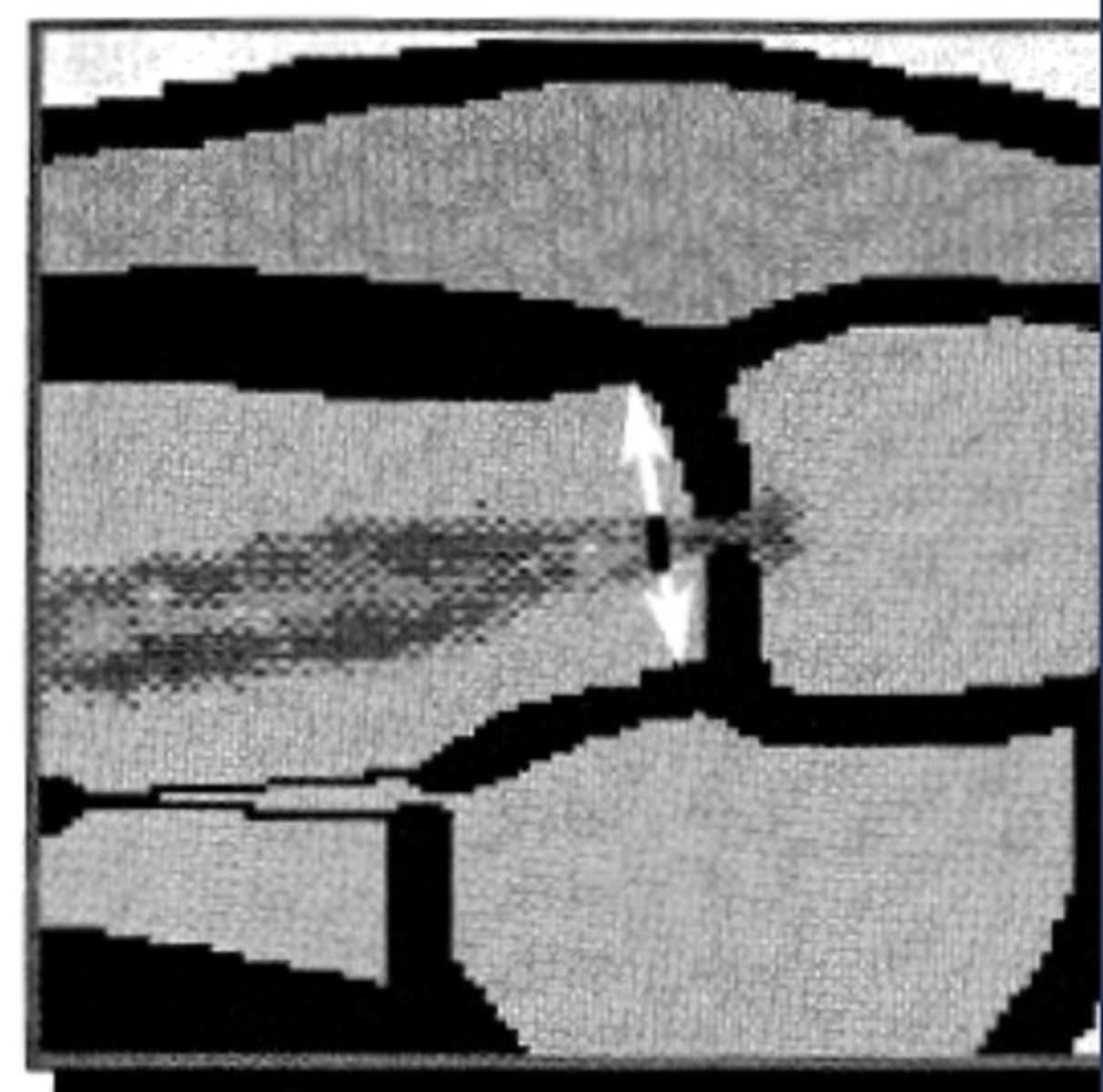
Mild AI Jet Width
< 25% of LVOT
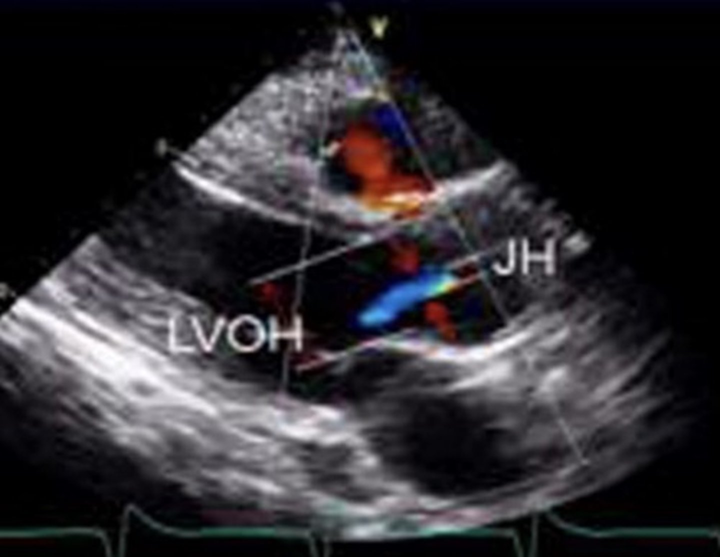
Severe AI Jet Width
≥ 65% of LVOT
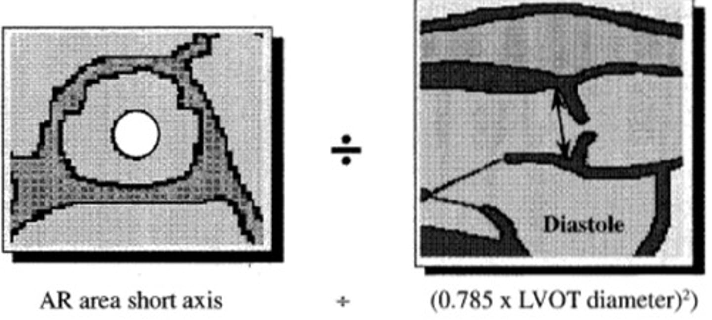
AI Jet Area/LVOT Jet Area Ratio
Compares PLAX LVOT area to PSAX AI jet area
Mild AI VCW
< 0.3 cm
Severe AI VCW
> 0.7 cm
Mild AI Doppler Waveform
Dimmer signal
Flatter slope
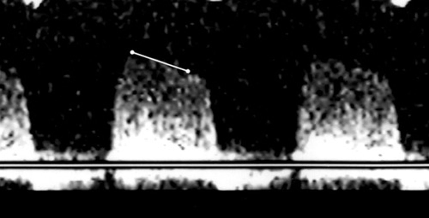
Severe AI Doppler Waveform
Brighter signal
Steeper slope
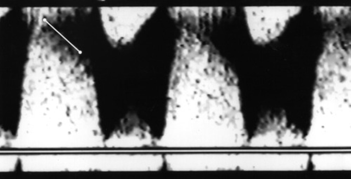
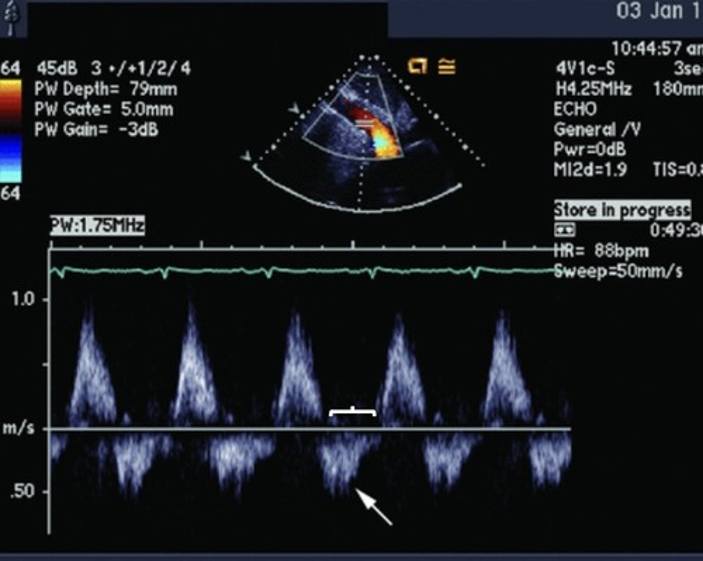
Tace Flow Reversal in Descending Aorta
Normal in early diastole
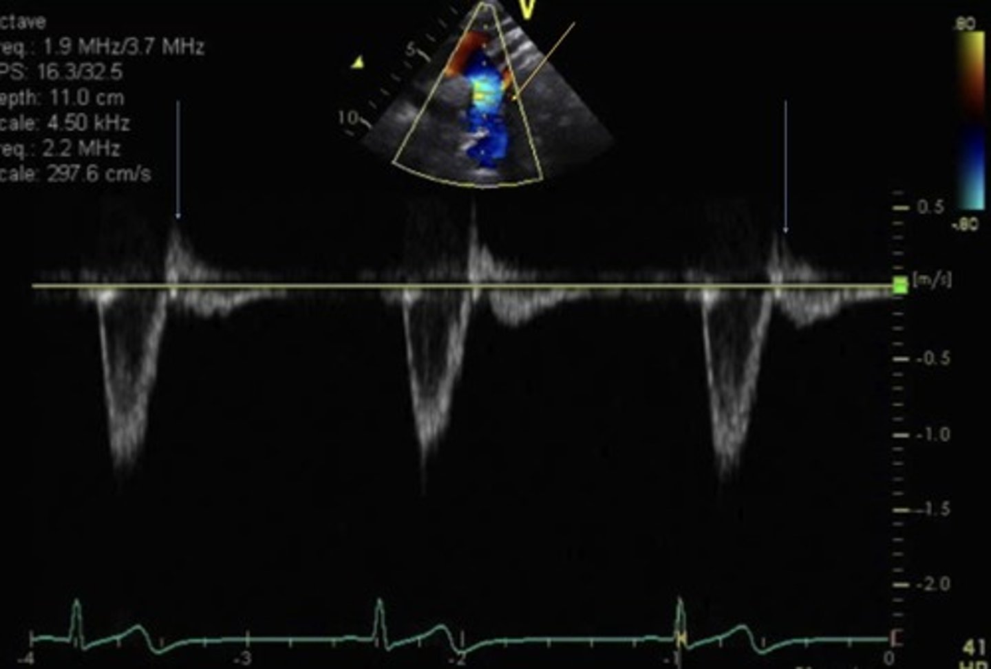
Mild Flow Reversal In Descending Aorta
Seen with mild AI
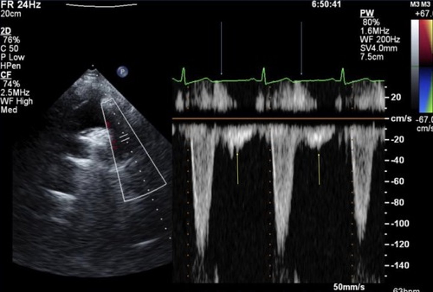
Holodiastolic Flow Reversal in Descending Aorta
Seen with at least moderate AI
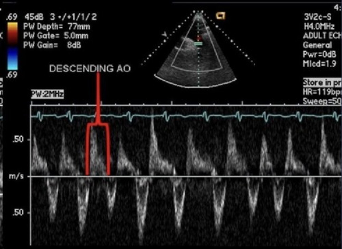
Normal Flow in Abdominal Aorta
Triphasic - normal to have some early flow reversal
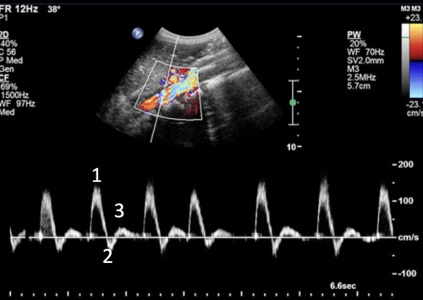
Holodiastolic Flow in Abdominal Aorta
Seen with severe AI
Pressure Half Time (PHT)
Time it takes for peak LV pressure to drop to half the original pressure value
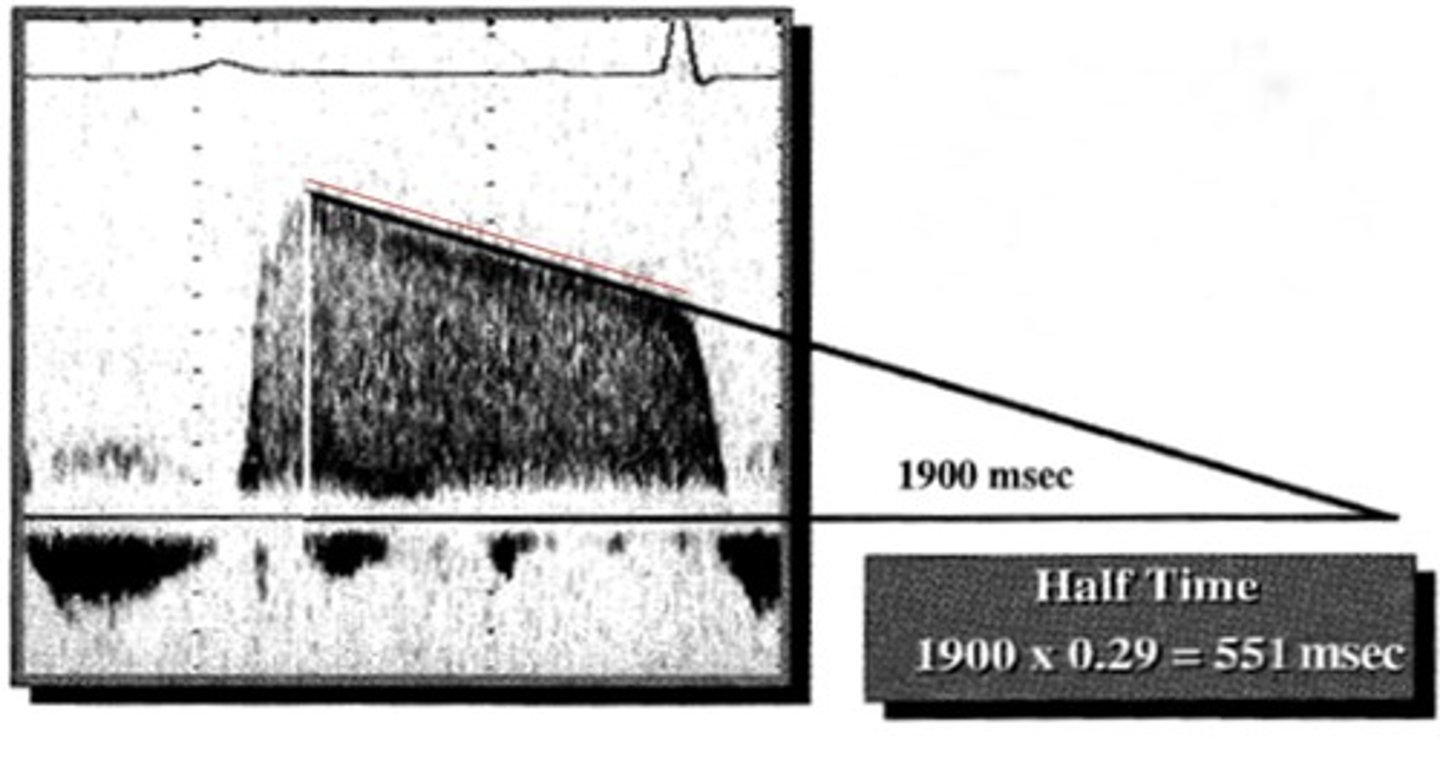
A shorter PHT value is seen more with...
Severe AI
Mild PHT
> 500 msec
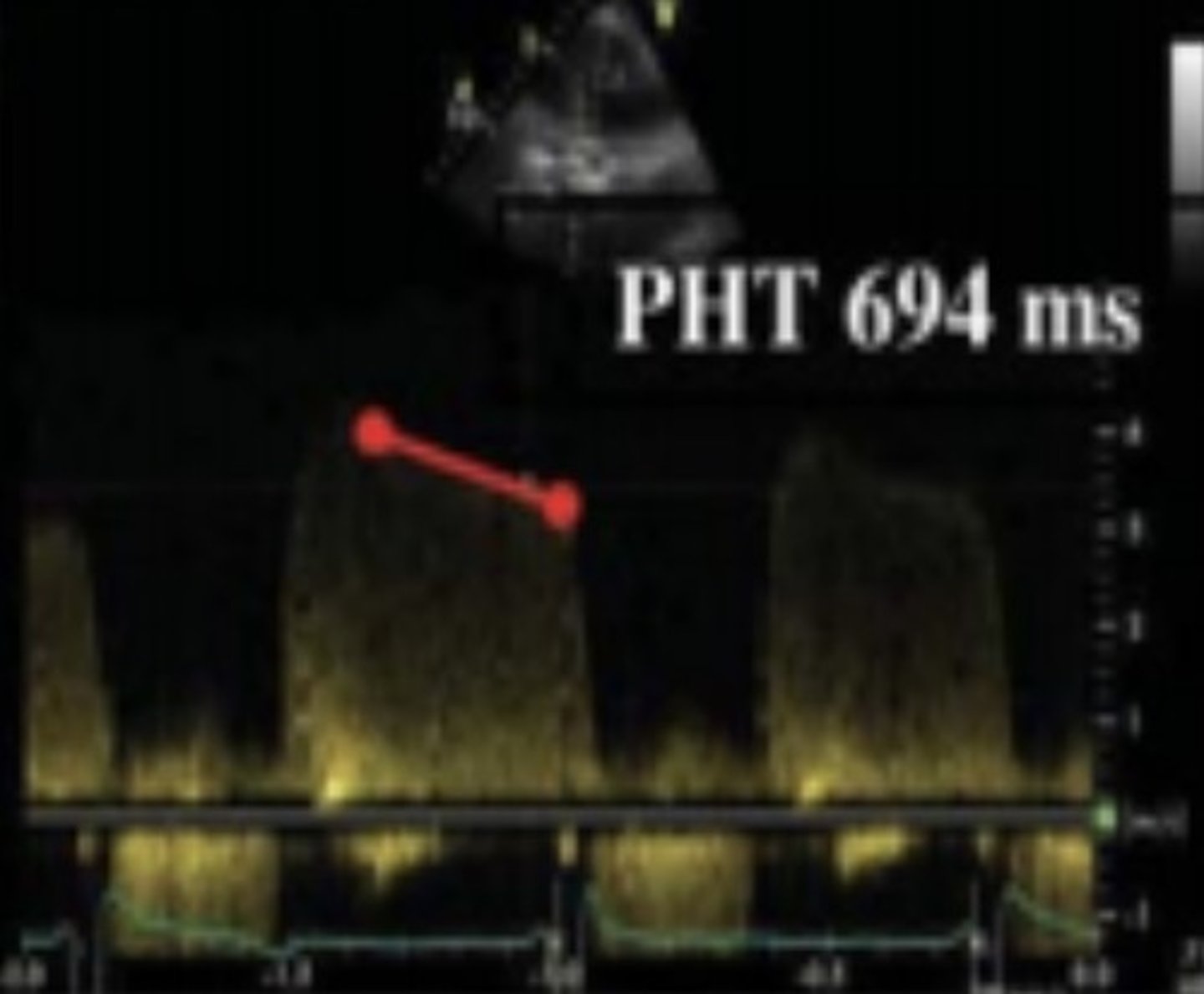
Severe PHT
< 200 msec
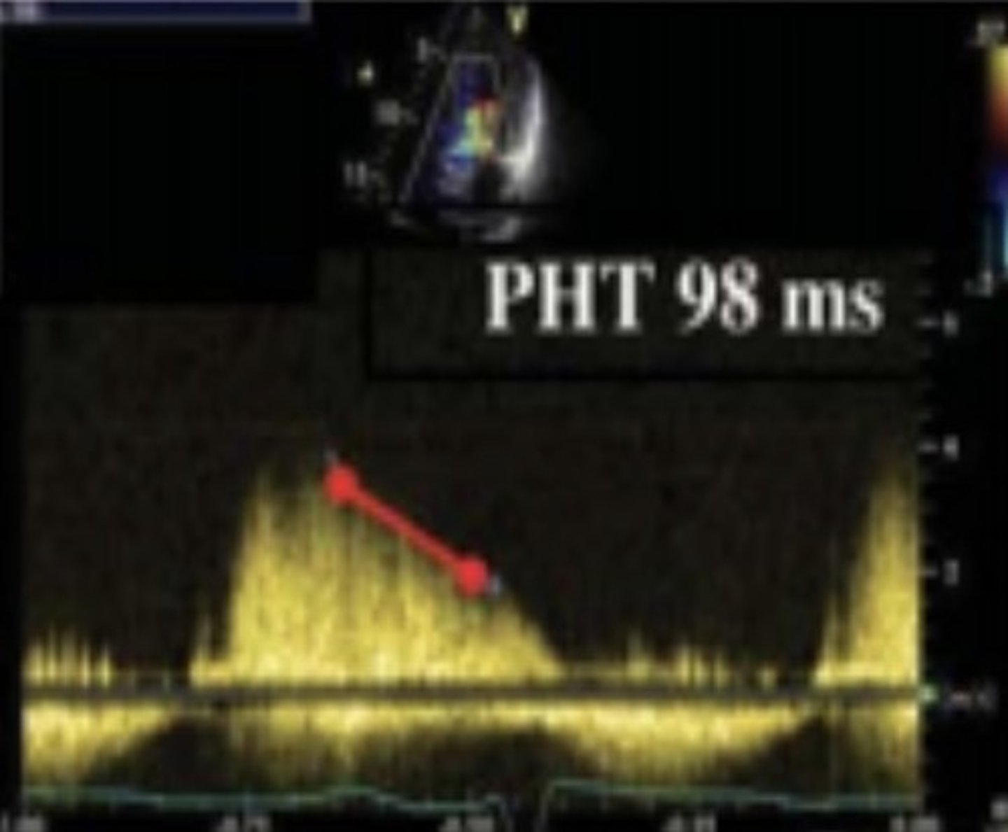
PISA for AI
Zoomed in apical 3 chamber or PLAX view
Measure convergence
Measure AR VTI & peak velocity
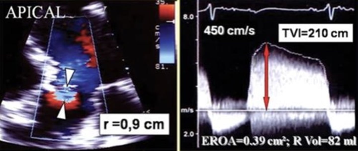
Regurgitant Fraction for AI
Only use if PISA can't determine severity
Compares stroke volumes of the aortic and mitral valves
Pressure Changes with AI
Increased LV pressure
Decreased AO pressure