Week 2: Muscular system, action, Wk 1 skeletal list, case study
 Call Kai
 Learn
 Practice Test
 Spaced Repetition
 Match
 Flashcards
 Knowt Play
1/71

There's no tags or description
Looks like no tags are added yet.
72 Terms
General Functions of the Muscular System: What are the 4 main functions of the muscular system?
Movement: Moves your bone, blood, food, and body parts.
Posture: keeps your body sitting or standing upright against gravity.
Joint stability: holds your bones securely together at joints so they don’t slip.
Heat Generates: Generates body heat whenever muscles contract to keep you warm.
Skeletal Muscle Connective tissue Coverings and Fascicle organization: what are the 3 connective tissue coverings of a muscle and the 5 ways fascicles are arranged?
3 Coverings:
Epimysium: Tough outer wrap around the entire muscle.
Perimysium: middle wrap around bundles of muscle fibers called fascicles.
Endomysium: Delicate inner wrap around each individual muscle fiber.
5 fascicle Arrangements:
Circular: rings around openings (ex: mouth muscles).
Convergent: Broad fan shape coming together at one tendon (ex: chest muscles).
Parallel: Running straight along strap-like muscles ( ex: thigh muscles).
Fusiform: spindle-shaped with a thick middle belly (ex:biceps)
Pennate: Feather-like diagonal arrangement on a central tendon.
Neuromuscular Junction: What is the Neuromuscular junction?
The neuromuscular Junction is the exact spot where a Nerve cell (motor neuron) connects to a muscle fiber. It passes the electrical nerve signal to tell the muscle when to pull or contract.
Functional Groups of Skeletal Muscles: what are the 4 functional groups (roles) muscles play during a movement?
Prime Mover (agonist): the main muscle responsible for making the movement happen.
Antagonist: the muscle that pulls in the opposite direction or reverses the movement.
Synergist: A helper muscle that adds extra power or stops unwanted extra movement.
Fixator: a special synergist that locks a bone in place so another muscle has a steady base.
Muscle attachments to the skeleton: How do muscles attach to bones ( direct vs, indirect attachments)?
Direct attachment: the muscle’s outer skin (epimysium) fuses straight onto bone skin (periosteum) or cartilage.
Indirect Attachment: Connective tissue extends a past the muscle as a ropelike tendon or a flat sheetlike ribbon called an aponeurosis that attaches to bone.
Rules of Naming muscles: what are the 7 ways skeletal muscles get their names?
Location: dot region or nearby bone ( ex: temporalis, temporal bone).
Shape: Geometric shape ( example: deltoid= triangle).
Relative size: Maximus ( largest), Minimus (smallest), Longus (long).
Direction of Fibers: rectus (straight), Transversus( across), oblique(angled).
Number of origins: Biceps (2 heads), triceps (3 heads)
Attachment Points: Named By origin and insertion.
Action: Named by the movement it creates (ex: flexor, extensor).
Identifying Muscles and their actions: what does it mean to identify a muscle and describe its action?
It means lookin at a muscle on a model or diagram, naming that specific muscle, and explaining the specific movement (action) it causes when it pulls on a bone.
Embryologic Difference Between Extrinsic and intrinsic Back Muscles: What is the embryologic difference between extrinsic and intrinsic back muscles?
Extrinsic Back Muscles(hypaxial): Formed from Hypaxial muscle blocks and controlled by front nerve branches ( ventral rami).They are superficial back muscles that move your arms and ribs.
Intrinsic back Muscles (epaxial): formed from epaxial muscle blocks and controlled by back nerve branches (dorsal rami). They are deep “true” back muscles muscles that hold your spine straight and maintain posture.
Abdominal muscle aponeuroses and rectus sheath: Why are abdominal muscle aponeuroses and the rectus sheath important?
The flat sheet tendons (aponeuroses) of the 3 side belly muscles (external oblique, internal oblique, and transversus abdominis wrap around the front “six-pack” muscle (rectus abdominis). This forms protective sleeve called the rectus sheath that holds your belly muscles tight and meets down the center seam called the linear alba.
Microscopic Characteristics of the 3 types of muscle: How do skeletal, caridoa and smooth muscle compare under a microscope?
Skeletal Muscle: voluntary ( you control it), long cylinder-shaped cells, striped (striated), with many nuclei along the outer edges.
Cardiac Muscle: Involuntary (heart controls itself), found only in the heart wall, striped (striated), branched cells with special connecting lines called intercalated discs.
Smooth Muscle: involuntary (automatic control), spindle-shaped cells (thick middle, pointed ends), smooth with no stripes ( non-striated), found in walls of hollow organs and blood vessels.
Muscle Movements
Frontalis (part of Occipitofrontalis)
Lifts up your eyebrows, wrinkles your forehead and pulls your scalp forward.

Occipitals (part of occipitofrontalis)
Pulls your scalp backward

Corrugato Supercilli
Pulls your eyebrows down and inward toward the middle ( makes you frown/squint)

Orbicularis Oculi (Palpebral and Orbital Parts)
Closes your eyelids.
Palpebral part: Gently closes your eyes ( blinking)
Orbital Part: tightly squeezes your eyes shut.

Procerus
Pulls down the inner end of your eyebrows (wrinkles skin between your eyebrows).

Nasalis
Flares your nostrils open

Zygomaticus major
Lift the corners of your mouth upward to smile or sneer.

zygomaticus Minor
Lifts and turns your upper lip outward.

Levator Labii superioris
Lifts your upper lip up

Risorius
Pulls the corners of your mouth sideways and down to frown.

Depressor anguli oris
Pulls the corners of your mouth downward to form a frown.

Depressor labii inferioris
Pulls your lower lip down and turns it outward ( makes a “pout”.

Mentalis
Lifts and pushes out your lower lip and wrinkles chi skin.

Buccinator
Compresses your cheeks against your teeth (keeps food between teeth when chewing).


Orbicularis Oris
Closes your lips and puckers them ( the “kissing” muscle).
Platysma ( not on models)
Pulls the corners of your mouth downward, widens your mouth, and tightens neck skin.


Temporalis
Closes your lower jaw (elevates the mandible for chewing)
Masseter
Closes your lower jaw with strong force for chewing.

Digastric (anterior and Posterior bellies).
Opens your jaw (depresses the mandible).

Stylohyoid
Lifts up and pulls back the hyoid bone in your neck.

Mylohyiod
Lifts up the hyoid bone, floor of mouth, and tongue during swallowing and speaking.

Sternothyroid
Pulls down the hyoid bone and lowers your voice box (larynx).

sternohyoid
Holds steady or pulls down the hyoid bone.

Thyrohyoid
Pulls down the hyoid bone and lifts up your voice box (larynx)

Omohyoid
Pulls down and steadies the hyoid bone.

Sternocleidomastoid
Bends your neck to the side and rotates your head to look toward the opposite side.

Scalene's (anterior, Middle, Posterior)
Anterior scalene: Bends your head forward.
Middle scalene: bends neck sideways and lifts ribs 1 when breathing hard.
Posterior scalene: bends neck sideways and lifts rib 2 when breathing hard.

Pectoralis Major
Flexes (bends forward), adducts (pulls inward), and turns your arm inward (medial rotation).

Pectoralis minor
Holds your shoulder blade (scapula) steady against your ribs.

Serratus anterior
Pulls your shoulder blade forward and rotates it so your arm can reach upward.

Levator Scapulae
Lifts up your shoulder blade and rotates it downward.

Rhomboid major and rhomboid minor
Pulls back your shoulder blade (retraction) and locks it to your ribcage.

Trapezius (superior, middle, inferior parts)
Superior part: shrugs shoulders up.
Middle part: pulls shoulder blades back together.
inferior part: pulls shoulder blades down.

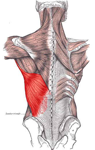
Latissimus dorsi
straighten/extends, pulls in (adducts), and rotates your arm inward behind you.
Erector spinae (spinalis, longissimus, lliocosstalis)
Straightens your spine and head, and bends your spine to the side.

External oblique and internal oblique
Squeezes your belly in, bends your trunk forward, and twists your waist.

Transversus abdominis
Deeply compresses/squeezes your abdomen inward like a tight belt.

Rectus abdominins
Bends your body forward (crunches) and compresses your belly.

External intercostals
Lifts your ribs up and outward when you inhale deeply.
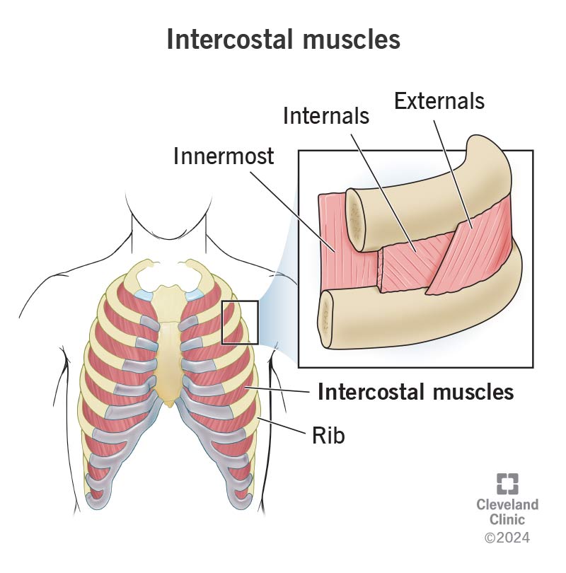
internal intercostals and innermost intercostals
Pulls your ribs down when you exhale hard (between cartilage, help lift ribs).

transversus Thoracis
Pulls ribs downward.

Levator ani and Coccygeus
Forms the pelvic floor muscle hammock that supports your lower pelvic organs and flexes your tailbone.

Skeletal System: Week 1
bone slide
a glass slide viewed under a microscope that shows bone tissue details.

Osteon
The main circular building block of compact bone that looks like a tree trunk cross-section.

Canaliculi
Tiny hair-like channels that connect bone cells so they can share food and messages.
Central canal
The tube in the middle of an osteon holding blood vessels and nerves.

Lamellae ( concentric and interstitial)
Bony rings inside an osteon (concentric) or leftover ring pieces between osteons (interstitial)

Osteocyte in Lacuna
A living bone cell (osteocyte) sitting inside its small protective space or “room” (lacuna)

Frontal bone
The forehead bone forming the upper eye sockets and front skull roof.
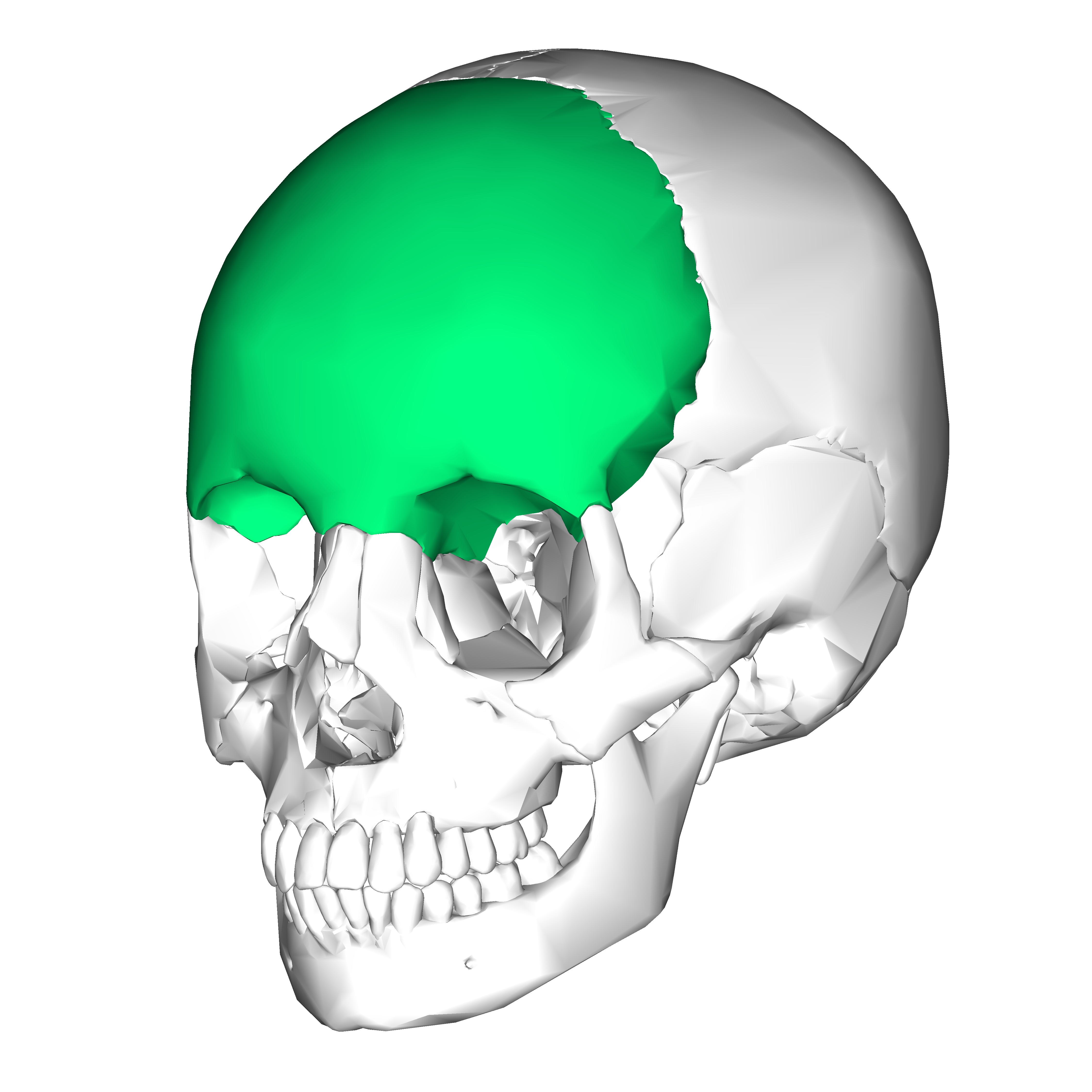
Glabella
The smooth, flat area of the forehead right between your eyebrows

Supraorbital foramen/notch
A small hole or notch above he eye socket for nerves and blood vessels to reach the forehead.

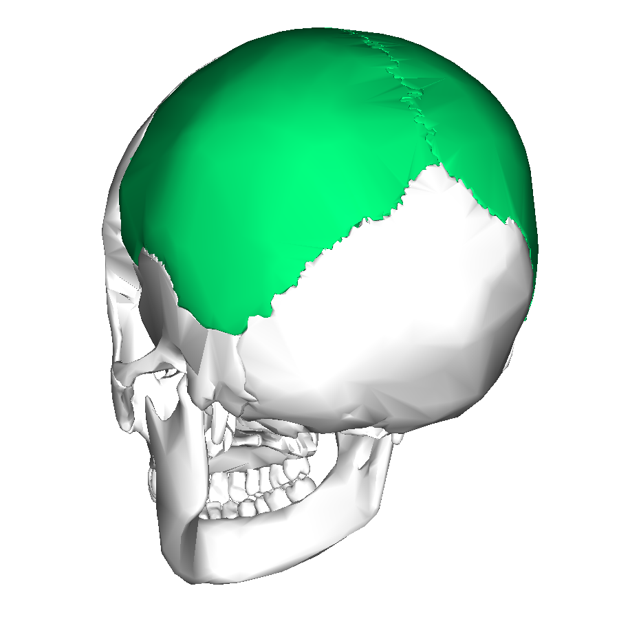
Parietal bone
Paired bones forming the top and upper sides of the skull roof.
Temporal bone
Paired bones forming the lower sides of the skull around the ear area.

Mastoid process
a large rounded bump behind your ear where neck muscles attach

Styloid process
A long, pointy bony spike beneath the ear anchoring tongue and throat muscles.

external auditory (acoustic) meatus
the outer ear canal tube leading inward toward the eardrum

Mandibular fossa
A shallow socket on the temporal bone where the lower haw bone snaps in.

Zygomatic process of the temporal bone
a bony extension reaching forward to help from the cheekbone arch.

internal acoustic meatus
An opening on the side of the skull for hearing and balance nerves.

Jugular foramen
A large hole at the skull base for the main jugular blood vessel and nerves.
