HN [5: Eyelid]
 Call Kai
 Learn
 Practice Test
 Spaced Repetition
 Match
 Flashcards
 Knowt Play
1/78

There's no tags or description
Looks like no tags are added yet.
79 Terms
Eyelid
Plays a big role in facial rejuvenation and visual function
Eyelid
Thinnest skin in the body to allow for uninterrupted and quick blinking
Anterior, Posterior Lamellae
The eyelid is divided into___
Skin, Orbicularis oculi
Anterior Lamellae is composed of___
Tarsus, Conjunctiva
Posterior Lamellae is composed of ___
Crow’s feet

Horizontal forehead furrows

Glabellar furrows

Orbital ocularis
A

Preseptal obicularis
B
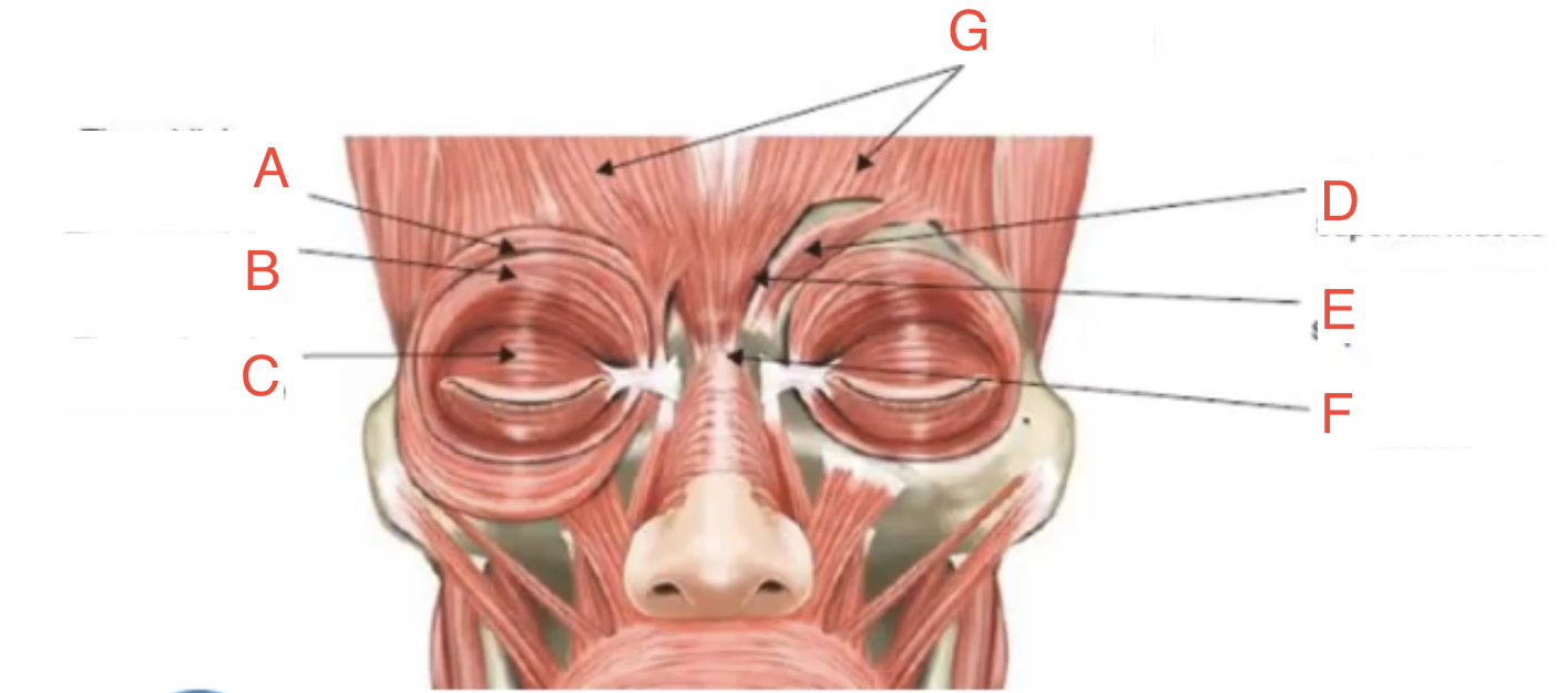
Pretarsal obicularis
C

Corrugator supercilli
D

Depressor supercilli
E
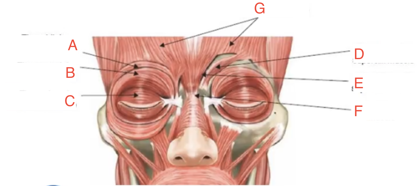
Procerus
F

Frontalis
G
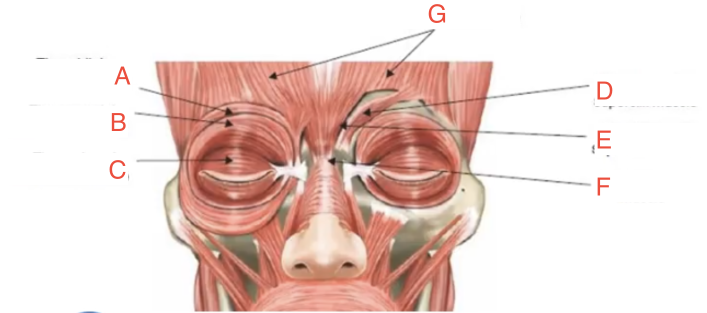
Orbicularis oculi, Corrugator supercilli, Procerus
Protractor of the eyelid muscle for closure
Pretarsal, Preseptal, Orbital
Orbicularis oculi muscle Divisions
Orbicularis oculi
Responsible for what can be seen as crow’s feet around one’s eyes
Corrugator supercillli
Attaches from the brow to the nasal bone
Procerus
Attaches from the frontal bone to the glabella, causing the glabellar furrows
Levator, Muller’s
Opening of the upper eyelid muscle
Levator
Primary elevator (retractor) of the upper lid
CN III/ Oculomotor
Nerve Supply of Levator
Levator
Extends beyond the rim as Whitnall’s ligament
Levator
Its fibrous aponeurosis gives out attachment extending to the skin, creating the lid crease
Eyelid Skin Crease
Separates the upper skin fold from the flat pre-tarsal component
Eyelid Skin Crease
Formed by attachments of the levator aponeurosis to the orbicularis and skin
Eyelid Skin Crease
Great surgical significance since it is a good incision site and hides and possible scar
Muller’s
Involuntary muscle of elevation
Muller’s
Subserved by the Sympathetic Nervous System
Muller’s
Accounts for about 1-2 mm of lid elevation
Muller’s
Located between conjunctiva and the levator, inserting to the superior border of the tarsus
Capsulopalpebral Fascia
Levator is analogous to ___ but for Lower Eyelid Retraction
Inferior Tarsal
Muller’s is analogous to ___ but for Lower Eyelid Retraction
Capsulopalpebral Fascia
Fibrous tissue extending from inferior rectur to inferior border of tarsus
Inferior Tarsal
Runs posterior to capsulopalpebral fascia
Inferior Tarsal
Extending from Lockwood’s ligament to inferior tarsal border
Tarsus or Tarsal Plates
Firm, dense plates of connective tissue that serve as structural support of the eyelids
Tarsus or Tarsal Plates
Have rigid attachments to the periosteum via the medial and lateral canthal tendons
Orbital Septum
Thin multi-layered sheet of fibrous tissue that arises from the periosteum over the superior and inferior orbital rims at the arcus marginalis
Orbital Septum
Attenuation or thinning of this structure tends to contribute to fat herniation in the aging eyeli
Preaponeurotic/Orbital Fat
Can be found anterior to the levator and posterior to the septum
Central, Medial
2 main fat pads of the Upper Lid
Nasal/ Medial, Central, and Temporal/ Lateral
3 fat pads of the Lower Lid
Suborbicularis Oculi Fat
Deep subcutaneous fat beneath the orbicularis running from the lower lid to the midface
Suborbicularis Oculi Fat
These fats are what we see as eye bags or dermatochalasis
Eyelid Coloboma
Eyelid Disorder: Embryologic cleft that is usually an isolated anomaly when occurs in medial upper eyelid
Eyelid Coloboma
Eyelid Disorder: When found in lower eyelid, it is frequently associated with other congenital conditions such as facial clefts of Goldenhar syndrome and lacrimal deformities
Eyelid Coloboma
Eyelid Disorder: A true one includes a defect in the eyelid margin
Blepharitis
Eyelid Disorder: Inflammation of the lid margin causing redness, itchiness and formation of dandruff-like scales called collarettes around base of lashes
Blepharitis
Eyelid Disorder: Due to bacteria or clogged oil glands
Blepharitis
Eyelid Disorder: Remedy is Lid hygiene with shampoo (mild) or antibiotic lid scrub
Eyelid Coloboma
Eyelid Disorder

Blepharitis
Eyelid Disorder

Hordeolum or Stye
Eyelid Disorder

Hordeolum or Stye
Eyelid Disorder: Acute, localized swelling of the eyelid due to bacterial infection
Hordeolum or Stye
Eyelid Disorder: May be internal (inflammation/infection in the Meibomian gland) or external (infection of Zeiss or Moll gland)
Hordeolum or Stye
Eyelid Disorder: External (infection of Zeiss or Moll gland) commonly involves eyelashes
Hordeolum or Stye
Eyelid Disorder: Remedy is Oral or topical antibiotics with warm compress
Chalazion
Eyelid Disorder
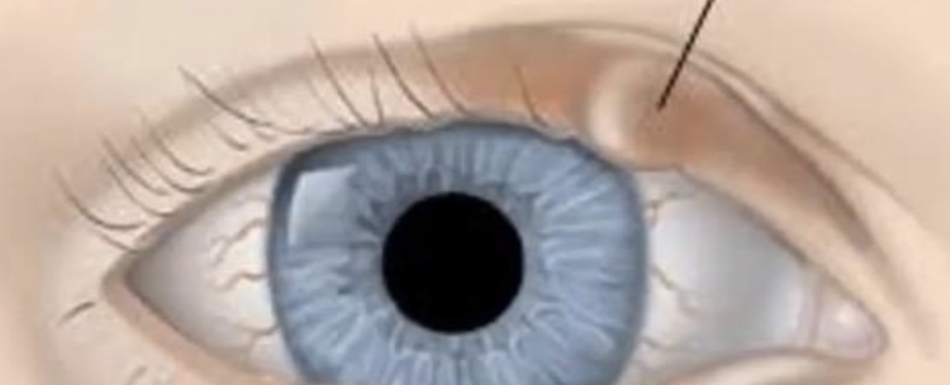
Chalazion
Eyelid Disorder: Is a non tender cyst noted along the eyelids
Chalazion
Eyelid Disorder: Usually develops when a Meibomian gland is clogged and develops inflammation
Chalazion
Eyelid Disorder: Sometimes it is an undertreated stye
Chalazion
Eyelid Disorder: Remedy is warm compress and proper drainage
Ectropion
Eyelid Disorder
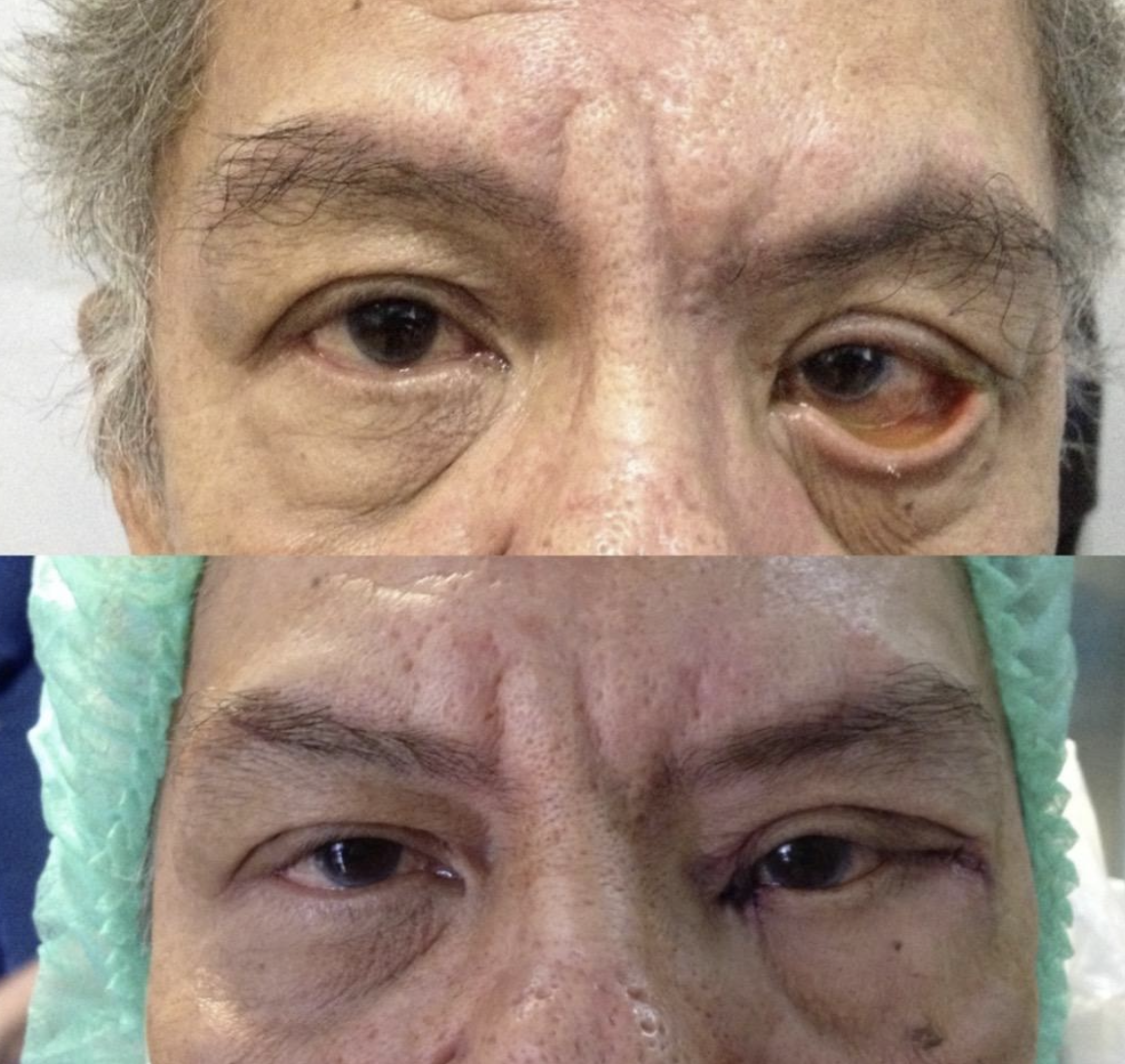
Ectropion
Eyelid Disorder: Eversion (outward turning) of lid margin
Ectropion, Entropion
Eyelid Disorder: Caused by natural aging process like lid laxity of medial & lateral canthal tendon
Ectropion
Eyelid Disorder: Also caused by mechanical* causes like a lid tumor and other conditions like facial paralysis
Entropion
Eyelid Disorder

Entropion
Eyelid Disorder: Inversion (inward turning) of lid margin that irritates the eye
Entropion
Eyelid Disorder: Also caused by overriding of the preseptal over pretarsal orbicularis and disinsertion of lower lid retractors
Trichiasis
Eyelid Disorder

Trichiasis
Eyelid Disorder: Inward growth of the lashes due to inflammation & trauma
Trichiasis
Eyelid Disorder: Only the lashes are turned in. Lid margins are in correct position
Trichiasis
Eyelid Disorder: To correct, epilation, electrocautery of follicles, or sometimes surgery is required
Ptosis
Eyelid Disorder

Ptosis
Eyelid Disorder: Drooping of the upper lid
Ptosis
Eyelid Disorder: Usually due to congenitally weak and fat infiltrated levator
Ptosis
Eyelid Disorder: In elderly, it is due to the lengthening of the levator aponeurosis