neuro unit two complete set
 Call Kai
 Learn
 Practice Test
 Spaced Repetition
 Match
 Flashcards
 Knowt Play
1/195

There's no tags or description
Looks like no tags are added yet.
196 Terms
biological rhythms
regular fluctuations in any living process- light is important to many of these rhythms
ultradian rhythms
repeat more than once a day (regular activity, feeding, hormone release)
infradian rhythms
repeat less than once a day (reproductive cycles)
circadian rhythms
have a span of about 24 hours, generated by an endogenous (internal/biological) clock that is highly accurate, only off by few minutes every day
what happened in Jean-Jacques deMairans experiment?
Observed the opening and closing of a heliotrope plant both when exposed to sun and when left in a dark cabinet- the leaves opened and closed regardless of the presence of light
how do ppl study internal clocks in animals?
FREE RUNNNING EXPERIMENTs in which the animal maintains its own schedule without human interference- the period of time when an animal will run on the wheel won’t be exactly 24 hours. but will be very close
phase shift
a shifting of a biological rhythm in response to a stimulus (like light)
entrainment
the process of shifting a biological rhythm to a stimulus
zeitgeber
the environmental cue that an animal syncs its internal clock to (sound, light, temp)
where is the biological clock locatesd
the suprachiasmatic nucleus (SCN) in the hypothalamus, above the optic chiasm (where the optic nerves cross the midline and become optic tracts) lesions of the SCN eliminate circadian rhythms of drinking, locomotion, and hormone secretion, in vitro SCN cells with show rhythm (daily periods of higher and lower activity) for days or weeks outside of the organism they were taken from
how do transplant studies prove that the SCN produces the circadian rhythm
Hamsters with 24 hour circadian rhythms had their SCN lesioned- in constant dim light setting showed no rhythm. Then got transplant from hamsters with 20 hour circ rhythms and began to display rhythm again, but instead of 24 hour previous, 20 hour transplant rhythm
pineal gland
in birds and amphibians is sensitive to light, thinner skull layer and photoreceptors in gland so is able to detect, releases melatonin in the absence of light- in mammals, rather than direct stim, must travel thru pathways for scn to get info
retinohypothalamic pathway
from eye to SCN, how circadian system interacts with light. Pathway contains retinal ganglion cells that project to the SCN- do not use rhodopsin or photopsin, use melanopsin, special light sensitive photopigment. SCN then activates/ deactivates pineal gland

molecular basis of SCN
SCN cells in mammals make two proteins: Clock anc Clye, which bind together to form a CLOCK AND CYCLE DIMER, dimer binds to DNA and promotes transcription of two genes: period (per) and cryptochrome (cry)*** important to regulating the circadian rhythm
How does the clock-cycle dimer and per cry protein cycle work
Clock and Cycle Dimers facilitate the transcription of per and cry genes, which then create per and cry proteins and also excite the SCN neurons, causing steady firing of AP’s. downstream these lead to wakefulness (melatonin suppression). When Per and Cry proteins accumulate, they bind to each other and inhibit the activity of the CC dimer, suppressing its excitatory influence and slowing its transcription of per and cry genes (thus stopping production of per and cry, then ceasing inhibition of clock and cycle, starting the cycle over again) CYCLE TAKES ABOUT 24 HOURS
how does the body determine the time of day
per and cry complex levels (fluctuate to impact wakefulness and sleepyness)
How does light impact the Clock-cycle per-cry shenanigans
Retinal ganglion cells detect light with melanopsin, flood cell w glutamate in presence of light, excitatory, leads to increased activation of SCN as well as increased transcription of the per gene via clock and cycle dimers, molecular clock becomes entrained to the photo (light) period and resets biological clock (why best way to combat jet lag is to go outside and stare at sun)
peripheral clocks
other organs outside of SCN can adapt to own internal/ external clocks , SCN is supposed to conduct them (like an orchestra)- cardiovascular clock, kidney clock w filtration and excretion ratesw
what can be induced by loss of sync between internal rhythms and external stimuli
diseases like cardiovasular damage
what happens when central and peripheral models fall out of sync
can contribute to the progression of disorders like cancer (shift work that changes is considered a high risk carcinogen bc body’s clock is being thrown off)
que quiere decir la orchestra model
the SCN is the conductor of peripheral clocks like melatonin, cortisol, body temp, and HR/ BP, without conductor, orchestra would play like shart

why is blue light before bedtime bad
light suppresses melatonin, melanopsin esp activated by short wavelengths like blue light (screens), can cause circadian delay or advancement by exciting the SCN artificially and REALLY effectively cir
circadian advancement
screen/blue light early in morning (activates SCN artificially early)
circadian delay
light in early evening artificially excites SCN again, and delays sleep part of circadian rhythm (melatonin production)
electroencephalogram (EEG)
electrodes all over scalp, track overall electrical current fluctuations in brain
electro-oculogram (EOG)
tracks eye movements in sleep
electromyogram
large muscle movements
what are the two distinct classes of sleep
REM- Small amplitude, fast EEG waves, complete muscle relaxation and rapid eye movements, NON-REM-Can be divided into 3 stages, has very different EEG waves
what does the brain activity of an awake person look like
BETA WAVES-mix of low amp waves with many fast frequencies
what is stage one sleep
starts with presleep alpha rhythm, which occurs during relaxation and drowsiness, time spent in alpha rhythm will decrease and waves of smaller amplitude and irregular frequency will start THETA WAVES, VERTEX SPIKES- sharp waves will appear, HR will drop, breathing will slow, muscles relax, eyes roll slowly, lasts several minutes

stage two sleep
waves of 12-14 hz will occur in bursts, called SLEEP SPINDLES- K COMPLEXES ALSO APPEAR- sharp negative EEG potentials

Stage 3 sleep (slow wave)
large amp, very slow waves called DELTA WAVES appear, occur at frequency of one wave per sec

REM sleep/ paradoxical sleep
small amplitude, high frequency activity, like an awake person except for presence of SAWTOOTH WAVES eyes move rapidly under closed lids, muscles relaxed, also called paradoxical sleep, mind looks awake but body is very asleep
Beta waves
person is awake
alpha waves
person is drowsy/ relaxed (nodding off in class)
theta waves
stage 1 sleep plus vertex spikes
K complexes and sleep spindles
stage two sleep
delta waves
stage ¾ sleep, deep sleep
fast, random wave +sawtooth waves
REM sleep
vertex spikes
stage one sleep plus theta waves
what happens in a typical night of young adult sleep (7-8) hours
around half is stage two sleep, 20% is REM sleep, cycles last from 90-110 mins, cycles in the first portion of night have more stage 3/ slow wave sleep (Growth hormone release), later cycles have less/ none SWS, more REM (memory consolidation)
impacts of sleep deprivation
irritability, difficulty concentrating, disorientation, prolonged hallucinations, compromises immune system which can lead to bacterial infections
fatal familial insomnia
inherited sleep disorder- person loses ability to sleep in midlife, leads to death within 7-24 months
what are the biological functions of sleep
restores body by restoring materials used up during day, like proteins (growth hormone released during slow wave sleep, essential for cell life repair and activation of the immune system) Sleep needed for immune system function, also helps with memory consolidation, sleep between learning and recall improves retention, consolidates declarative memory tasks, also “washes” you brain with CSF as blood vessels constrict, clears away dead cell material which is later peed out
4 neural systems underlie sleep
FOREBRAIN SYSTEM-arousal and slow wave sleep , BRAINSTEM SYSTEM-activates forebrain to wakefulness , PONTINE SYSTEM-REM sleep , HYPOTHALAMIC SYSTEM-coordinates other systems
basal forebrain
stims arousal and wakefulness in forebrain, going to sleep requires cholinergic neurons to be suppressed
reticular formation
wakes the forebrain up, targeting this will wake up sleeping animals, lesions of this area will produce persistent sleep
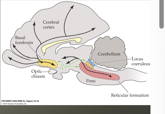
parts of brain active during REM sleep
Pontine Tegmentum (part of pons) and Locus Coeruleus

ACh neurons in the pontine tegmentum project to
TECTUM (rapid eye movements), LATERAL GENICULATE NUCLEUS (PGO Waves 2 v1, cause vibrant, intense dreams, MEDULLA NUCLEI: muscular paralysis, BASAL FOREBRAIN AREA: cortical activation
hypothalamus
contains several parts of brain known as sleep systems- Ventrolateral preoptic area (vlPOA) contains majority of GABAergic sleep neurons → receives input from the forebrain to induce slow wave sleep, inhibits reticular formation and orexin neurons in the hypothalamus → additionally contains OREXIN neurons, receives input from the SCN and promotes arousal in retic form , basal forebrain, and pons
how are the reticular formation and basal forebrain connected
reticular formation excites the basal forebrain which creates continuous wakefulness (like jumpstarting a car, retic form= jump starter, basal forebrain= engine that is now running)
narcolepsy
frequent, intense sleep attacks, go straight to REM sleep, may have CATAPLEXY, sudden loss of muscle tone without loss of consciousness, can be triggered by sudden intense emotional stimulus, DUE TO LOSS/ DAMAGE OF OREXIN NEURONS
Sudden Infant Death Syndrome
developmental defects of serotonin produce lesions in rostral reticular formation (waking) and medulla (breathing)- both have 5ht receptors involved in regulating breathing and waking, when disrupted, lead to insufficient arousal following life threatening co2 levels → baby suffocates because 5ht receptors required to detect suffocation and wake the baby do ont work
hormones
chemicals released by glands that travel through blood stream to act on target tissues
endocrine glands
release products INSIDE blood stream
exocrine glands
release products outside of bloodstream (sometimes outside of body) ie blood and sweat
peptide hormones
short string of amino acides (vasopressin (pair bonding)
amine hormones
modified version of a singular amino acid, aka monoamine hormones, ie epinephrine
steroid hormones
come from cholesterol, made from four rings of carbon atoms
where are hormones made?
PERIPHERAL SYSTEM: endocrine glands- adrenal and thyroid glands, gonads (testicles and ovaries) CENTRAL SYSTEM: neurohormones are produced by neuroendocrine cells (hypothalamus)
How do protein and amine hormones influence tissues?
protein and amine hormones bind onto specific receptors on the surface of a cell and activate chemical signals- second messengers- inside of the cell
how do steroid hormones impact the cells
steroid hormones cross the cell membrane and bind to receptors inside of the cell- steroid-receptor complex binds to specific regions of DNA and controls the expression of specific processes, slower than peptide/ amine hormones
pituitary gland
“master gland” releases important hormones and regulates other endocrine glands- two parts, anterior and posterior
pituitary stalk
connects the pituitary gland to the thalamuis
neuroendocrine cells
found in the pituitary, release hormones into the bloodstream
posterior pituitary gland
releases oxytocin and vasopressin into the bloodstream (social and parental behavior, mating/bonding) produce in neuroendocrine cells in the supraoptic and paraventricular nuclei of the hypothalamus, when AP in these cells reached terminal, neurohormones released right into thalamus
anterior pituitary
contains neuroendocrine cells that make and release TROPIC HORMONES: hormones that affect the release of other endocrine glands- hypotha;klamus regs the release of tropic hormones from the anterior pituitary using RELEASING HORMONES- axons of neuroendocrine cells that produce these hormones meet at MEDIAN EMINENCE-
tropic hormones
hormones that affect the release of other endocrine glands
Median eminence
contains blood vessels that form the HYPOTHALAMIC PITUITARY PORTAL SYSTEM- hypothalamic neurons make releasing hormones, which travel to the anterior pituitary, triggering the release of tropic hormones into circulation
how is cortisol released into the body
In response to stress or waking up rhythm, hypothalamus synthesizes and releases CRH (corticotropin releasing hormone) into the pituitary gland, which signals it to release the tropic hormone ACTH (adrenocorticotropic hormone) which travels through the bloodstream to the adrenal glands on top of the kidneys which release cortisol
how are sex hormones released in the body
the hypothalamus produces the releasing hormone GnRH (gonadotropin releasing hormone) which travels to the anterior pituitary triggering the release of the tropic hormones LH and FSH which travel to the gonads (testes+ovaries) and trigger the release of sex hormones testosterone and progesterone
differences between male and female brains
male brain is 8-10% heavier, sexually dimorphic nucleus of the preoptic area (SNDPOA)=2.5x larger in males, 2.2x more cells, interstitial nucleus of the hypothalamus (INAH) 2 and 3 cell groups have larger volume in men compared to women
role of hormones in sex diferentiation
genes effect gonad development- XY have SRY (sex determining region y for production of testosterone) X X do not, gonadal hormones initiate sexual diff of brain (masc or non masc brain) environmental factors can impact gene and hormone expression

Who are transgender male to female womens’ brains most similar to?
BIOLOGICAL WOMEN- environmental factors in pregnant women can alter the expression of SRY, leaving brain structure of MTF women most biologically similar to ppl AFAB
retina
layer of neurons at the back of the eye- sensory transduction occurs here
cornea
bends light entering eye (refraction)
lens
changes shape to focus light on the retina, ciliary muscles change the shape of the lens through the process of accommodation- causes nearer or further images to come into focus
photoreceptors
sensory neurons that detect light- two types, rods and cones
rods
photoreceptors that pick up low levels of light- night vision/ low illumination, rhodopsin is the photopigment receptor
cones
do not work in low light, but process color, pigment receptor is photopsin
bipolar cells
receive input from photoreceptors and then synapse onto ganglion cells whose axons form the optic nervegang
ganglion cells
synapse with bipolar cells, axons form the optic nerve, carry AP out of eye and into the brain
horizontal cells
contact photoreceptors and bipolar cells, can contribute to lateral inhibition and edge enhancement
amacrine cells
similar to horizontal cells, but lie between the bipolar and ganglion cells
What do all cell types with the exception of ganglion cells produce in the eye
graded potentials (ganglion cells produce APs)
scotopic system
rods, works in dim light and is insensitive to color, info from many receptors converge on individual ganglion cells
Photopic system
cones, requires more light, sensitive to different wavelengths and allows for color vision
visual acuity
measure of how much detail we can see- sharopest in center of visual field fo
fovea
central region of retina, contains high density of smaller, tightly packed cones with high acuity
where are rods located on the eye
periphery- none in the fovea
optic disk
where blood vessels and optic nerve leave the eye
blind spot
part of the eye that is sightless bc its where the optic disk is and there are no photoreceptors on the optic disk
what is the pathway from the eye to brain
light enters the eye, it is focused and refracted by the cornea and lens, it then hits the back of the retina where it is processed by rods and cones whose graded potentials communicate info to bipolar cells → ganglion cells → some/ all of optic nerve crosses midline at optic chiasm, then called the optic tract Lateral Geniculate nucleus in the thalamus- axons of LGN neurons form optic radiations, end in→ v1 (also called striate cortex)
what is cortical magnification
smaller portion of the retina is magnified and processed in a proportionately larger area of v1 → greater processing of details arriving at the fovea
what is blindsight
the inability to consciously perceive visual cues, but still discriminate based on them- when v1 pathway is damaged, there is sometimes primitive visual processing thru the thalamus sending visual info to be processed not externally (not thru v1)
what do photoreceptors do in response to light?
they HYPERPOLARIZE and release less glutamate (only system to do this)
on center bipolar cells
turning ON a light excites this cell because it receives LESS glutamate- bc glutamate is inhibitory, turning on a light hyperpolarizes the photoreceptor causing it to reduce or stop its release of glutamate, resulting in it releasing glutamate in response to the cessation of constant inhibition, then causing the synapsed ganglion cell to fire an AP
off center bipolar cell
turning OFF the light excites this cell because glutamate is excitatory and leads to depolarization (bc there is no light on, steady stream of glutamate flowing from photoreceptors, which would inhibit an on center bipolar cell, but excites an off center bipolar cell, BECAUSE IN THIS INSTANCE GLUTAMATE IS EXCITATORY
ganglion cells
synapse onto both oncenter and off center bipolar cells and are ALWAYS EXCITED BY GLUTAMATE
Summarize what happens to both on and off center bipolar neurons when it is DARK
Because no light is being shown, photoreceptors are depolarized and constantly releasing glutamate, on center bipolar cells are inhibited by the glutamate release and hyperpolarize, sending little to no glutamate to ganglion cells (NO ACTIVATION). Off center bipolar cells are excited by glutamate and depolarize, releasing glutamate onto ganglion cells= activation