PATHO WEEK 5 W/O NOTES
 Call Kai
 Learn
Learn Practice Test
 Spaced Repetition
 Match
Match Flashcards
 Knowt Play
1/120

There's no tags or description
Looks like no tags are added yet.
Name | Mastery | Learn | Test | Matching | Spaced | Call with Kai | Chat |
|---|
No analytics yet
Send a link to your students to track their progress
121 Terms
Surface area, membrane thickness, pressure gradient, gas solubility + molecular weight.
How does decreased alveolar surface area affect diffusion?
Less surface area causes less O₂ transfer and hypoxemia (emphysema).
How does a thick alveolar membrane affect diffusion?
O₂ crosses more slowly, causing hypoxemia (pneumonia, edema or fibrosis).
What do type I alveolar cells do?
Form the thin surface where gas exchange occurs.
What do type II alveolar cells do?
Produce surfactant, which reduces surface tension and keeps alveoli open.
Alveoli collapse (atelectasis), reducing ventilation and oxygenation.
Ventilation and perfusion do not match, causing impaired oxygenation.
What is dead space?
Air reaches the alveoli, but blood does not (ventilation without perfusion). Example: PE.
What is a shunt?
Blood reaches the alveoli, but air does not (perfusion without ventilation). Examples: pneumonia or atelectasis.
What is hypoxemia?
Can hypoxia occur without hypoxemia?
Why can uncontrolled atrial fibrillation cause dyspnea?
A rapid, irregular rhythm lowers cardiac output + tissue oxygen delivery.
What are early signs of hypoxemia?
Restlessness, anxiety, confusion, tachypnea + tachycardia.
What are late signs of hypoxemia?
Cyanosis, decreased LOC, bradycardia, hypotension + dysrhythmias.
How does the body compensate for chronic hypoxemia?
It increases ventilation, constricts pulmonary vessels + produces more RBCs (polycythemia).
Why does chronic hypoxemia cause polycythemia?
The kidneys release erythropoietin, which increases RBC production and O₂-carrying capacity.
What findings suggest chronic hypoxemia?
Clubbing, cyanosis + polycythemia.
Why does alveolar hypoxia cause pulmonary vasoconstriction?
Pulmonary vessels constrict to redirect blood away from poorly ventilated alveoli.
Why is chronic pulmonary vasoconstriction harmful?
Chronic vasoconstriction → pulmonary hypertension → right-sided heart failure.
What does increased PaCO₂ usually mean?
The patient is hypoventilating and retaining CO₂.
What findings show increased work of breathing?
Tachypnea, accessory-muscle use, retractions, nasal flaring + difficulty speaking. Think: working hard to move air.
What is acute respiratory failure?
What causes hypoxemic respiratory failure?
V/Q mismatch, shunting or impaired diffusion causes low PaO₂.
What causes hypercapnic respiratory failure?
Hypoventilation → CO₂ retention → high PaCO₂ + low pH.
What findings suggest worsening respiratory failure?
Confusion, exhaustion, shallow breathing, cyanosis + worsening ABGs.
Why is a decreasing respiratory rate dangerous in a struggling patient?
Respiratory muscles may be tiring → CO₂ rises → respiratory arrest may follow.

What is ARDS?
Severe inflammatory lung injury that causes leaky capillaries, fluid-filled alveoli + severe hypoxemia.
Sepsis, aspiration, major trauma, pancreatitis + severe infection.
What findings suggest ARDS?
Acute dyspnea, tachypnea, diffuse crackles, cyanosis + severe hypoxemia.
What is pneumonia?
Inflammation of the alveoli and bronchioles causes fluid or exudate to collect —> impair gas exchange.
What increases pneumonia risk?
Virulent organism + large exposure + weak host defenses
What happens in bacterial pneumonia?
Neutrophils + purulent exudate fill alveoli → consolidation + hypoxemia.
What findings suggest bacterial pneumonia?
Fever, productive cough, purulent sputum, crackles + focal consolidation.
What happens in viral pneumonia?
Viral injury causes interstitial inflammation and alveolar-cell damage → impaired diffusion.
What findings suggest viral pneumonia?
Dry cough, fever, fatigue, body aches + diffuse lung findings.
Why does pneumonia cause crackles?
Why does pneumonia cause hypoxemia?
Blood passes fluid-filled alveoli that receive little air (shunt/V-Q mismatch).
What is aspiration pneumonia?
Gastric/oral contents enter lungs → chemical inflammation + possible bacterial infection.
Who has a high aspiration risk?
A patient with decreased LOC, dysphagia, vomiting or a weak gag reflex.
What causes tuberculosis (TB)?
Mycobacterium tuberculosis, which spreads through airborne droplet nuclei.
Why is M. tuberculosis difficult to destroy?
Its waxy outer wall resists breakdown inside macrophages.
How does the immune system respond to TB?
Cell-mediated immunity forms granulomas to wall off bacteria, but inflammation damages lung tissue.
What findings suggest pulmonary TB?
Persistent cough, hemoptysis, fever, night sweats, fatigue + weight loss.
What increases TB transmission?
Prolonged contact in crowded, confined or poorly ventilated areas.
What does a positive TB skin test mean?
How do latent and active TB differ?
Latent = no symptoms/not contagious; active = symptoms + bacterial growth + contagious pulmonary disease.
What is asthma?
Chronic airway inflammation causing reversible bronchoconstriction + mucus production.
What happens during an asthma attack?
Why is expiration prolonged in asthma?
What findings suggest asthma?
Episodic wheezing, cough, chest tightness + prolonged expiration
Why can asthma worsen at night?
Why is a silent chest dangerous during an asthma attack?
Airflow is too limited to produce wheezing → severe obstruction.
What ABG change occurs early in an asthma attack?
Hyperventilation lowers PaCO₂, causing respiratory alkalosis.
What does rising PaCO₂ mean during severe asthma?

What is emphysema?
Alveolar walls are destroyed and lose elastic recoil, causing air trapping and reduced diffusion area.

Why does emphysema cause a barrel chest?
Loss of elastic recoil traps air → chronic lung hyperinflation.
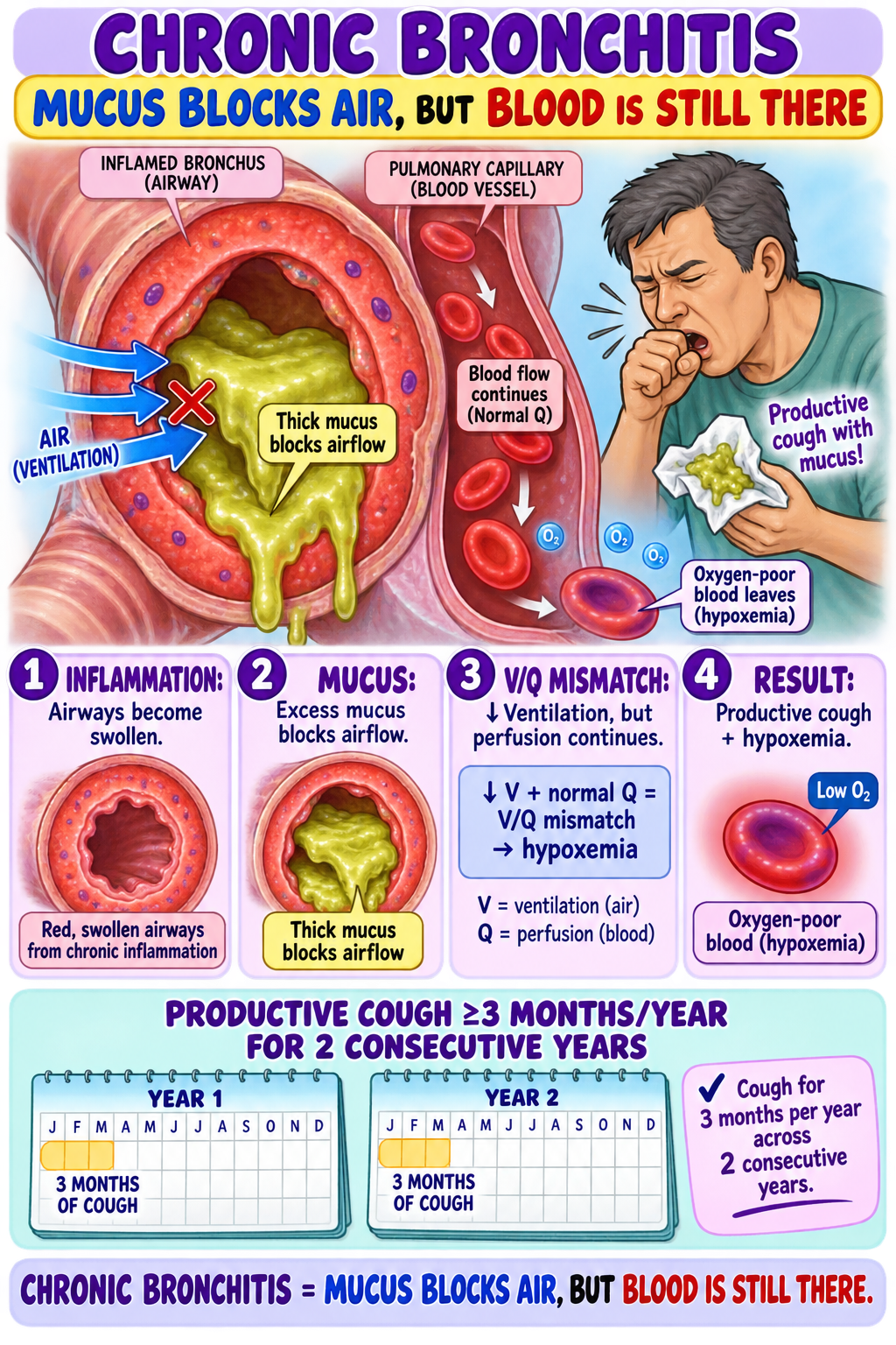
What is chronic bronchitis?
Airway inflammation and excess mucus cause a productive cough for at least 3 months in 2 consecutive years.
Mucus blocks airflow while blood flow continues, causing a V/Q mismatch.

What is bronchiectasis?
Repeated infection and inflammation destroy airway walls, causing permanent bronchial dilation.
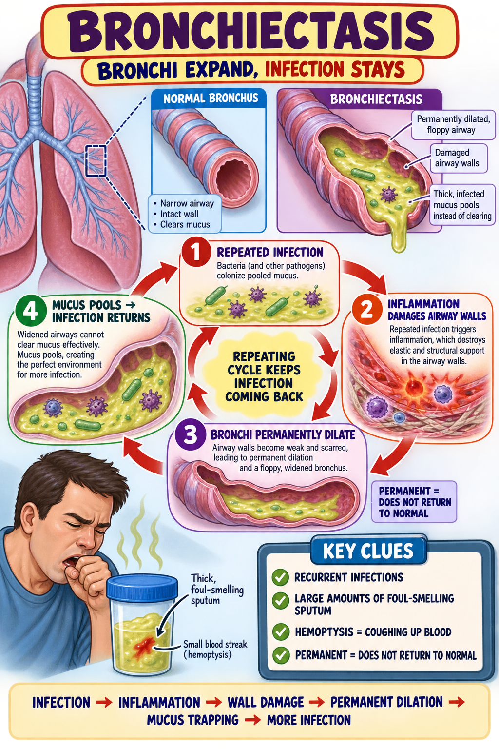
What findings suggest bronchiectasis?
Recurrent infections, large amounts of foul-smelling sputum + hemoptysis.s

What is restrictive interstitial lung disease?
Inflammation and fibrosis make the lungs stiff, reducing expansion and diffusion.
What findings suggest restrictive lung disease?
Progressive dyspnea, tachypnea, dry cough, clubbing + little or no wheezing.
How do obstructive and restrictive diseases differ?
Obstructive disease makes it difficult to get air out. Restrictive disease makes it difficult to expand the lungs.
What is a pulmonary embolism (PE)?
A venous clot travels through the right heart and blocks pulmonary blood flow.
The clot blocks perfusion to ventilated alveoli, creating dead space and a V/Q mismatch.
Sudden dyspnea, pleuritic chest pain, tachypnea, tachycardia + unexplained hypoxemia.
Hypoxic vasoconstriction, loss of pulmonary vessels, vascular obstruction or left-heart disease.
Exertional dyspnea, fatigue, chest discomfort, syncope + signs of right-heart strain.
JVD, peripheral edema, hepatomegaly + weight gain.
What are the main lung-cancer risk factors?
Smoking (strongest), asbestos exposure + family history.
How do small-cell and non-small-cell lung cancers differ?
What findings suggest lung cancer?
Persistent or changing cough, hemoptysis, recurrent pneumonia, chest pain + unexplained weight loss
What are paraneoplastic manifestations?
Tumor-released substances cause endocrine, neurologic or connective-tissue effects without direct metastasis.
What is a pleural effusion?
What findings suggest pleural effusion?
Dyspnea, decreased breath sounds, dull percussion + unequal chest expansion.
What is a pneumothorax?
Sudden chest pain, dyspnea, decreased breath sounds + hyperresonance on one side.
What is a tension pneumothorax?
Severe dyspnea, absent unilateral breath sounds, hypotension, JVD + tracheal deviation.