A&P ch 12: Central Nervous System

0.0(0)
Studied by 0 people
call kaiCall Kai
learnLearn
examPractice Test
spaced repetitionSpaced Repetition
heart puzzleMatch
flashcardsFlashcards
GameKnowt Play
Card Sorting

1/36

encourage image

There's no tags or description

Looks like no tags are added yet.

37 Terms

1
New cards

surface markings of cerebrum

Fissure: deep grooves

Longitudinal fissure

– Separates two hemispheres

▪ Transverse cerebral fissure

– Separates cerebrum and cerebellum

Sulcus: shallow grooves

 Gyrus: ridges

<p><span style="background-color: transparent;">Fissure: deep grooves</span></p><p><span style="background-color: transparent;">Longitudinal fissure</span></p><p><span style="background-color: transparent;">– Separates two hemispheres</span></p><p><span data-name="black_small_square" data-type="emoji">▪</span><span style="background-color: transparent;"> Transverse cerebral fissure</span></p><p><span style="background-color: transparent;">– Separates cerebrum and cerebellum</span></p><p><span style="background-color: transparent;">Sulcus: shallow grooves</span></p><p><span style="background-color: transparent;">&nbsp;Gyrus: ridges</span></p>
2
New cards

lobe vs cortex

Lobe: physical structure that can be SEEN and

TOUCHED

Cortex:  REGION within the brain that plays a role

in neural processing

3
New cards

gray matter and white matter

Gray matter: short, nonmyelinated neurons and cell

bodies

• White matter: myelinated and nonmyelinated axons

In CEREBRUM, grey matter is superficial to white matter

• EVERYWHERE ELSE IN CNS, grey matter is deep to

white matter

4
New cards

ventricles

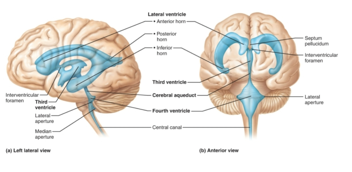
Fluid-filled chambers that are continuous to one another

and to central canal of spinal cord

– Filled with cerebrospinal fluid (CSF)

– Lined by ependymal cells (neuroglia cells)

• Paired lateral ventricles are large, C-shaped chambers

located deep in each hemisphere

• Each lateral ventricle is connected to the third ventricle via

interventricular foramen

• Third ventricle is connected to the fourth ventricle via

cerebral aqueduct
<p><span style="background-color: transparent;">Fluid-filled chambers that are continuous to one another</span></p><p><span style="background-color: transparent;">and to central canal of spinal cord</span></p><p><span style="background-color: transparent;">– Filled with cerebrospinal fluid (CSF)</span></p><p><span style="background-color: transparent;">– Lined by ependymal cells (neuroglia cells)</span></p><p><span style="background-color: transparent;">• Paired lateral ventricles are large, C-shaped chambers</span></p><p><span style="background-color: transparent;">located deep in each hemisphere</span></p><p><span style="background-color: transparent;">• Each lateral ventricle is connected to the third ventricle via</span></p><p><span style="background-color: transparent;">interventricular foramen</span></p><p><span style="background-color: transparent;">• Third ventricle is connected to the fourth ventricle via</span></p><p><span style="background-color: transparent;">cerebral aqueduct</span></p>
5
New cards

Frontal lobe

Handles executive functions such as planning, reasoning, problem-solving, emotional regulation, and personality

<p>Handles <strong>executive functions</strong> such as planning, reasoning, problem-solving, emotional regulation, and personality</p>
6
New cards

Parietal lobe

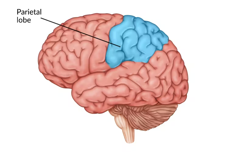
Integrates sensory information from across the body, including touch, temperature, pressure, and pain. It contains the somatosensory cortex and helps with spatial awareness and object recognition
<p>Integrates <strong>sensory information</strong> from across the body, including touch, temperature, pressure, and pain. It contains the <strong>somatosensory cortex</strong> and helps with <strong>spatial awareness</strong> and object recognition</p>
7
New cards

Temporal lobe

Processes auditory signals (hearing), language comprehension (such as Wernicke's area), memory storage via the hippocampus, and aspects of smell and emotion.

<p><span style="background-color: transparent;"> </span>Processes <strong>auditory signals</strong> (hearing), language comprehension (such as <strong>Wernicke's area</strong>), memory storage via the <strong>hippocampus</strong>, and aspects of smell and emotion.</p>
8
New cards

Occipital

Serves as the primary visual processing center, interpreting visual data from the eyes such as shape, color, depth, distance, and object identity.

<p> Serves as the primary <strong>visual processing center</strong>, interpreting visual data from the eyes such as shape, color, depth, distance, and object identity.</p>
9
New cards

Insula lobe

integrate internal bodily states with emotional, sensory, and cognitive experiences

10
New cards

Cerebral cortex

makes up abt 40% of brain

1. Contains three types of functional areas:

▪ Motor areas: control voluntary movement

▪ Sensory areas: conscious awareness of sensation

▪ Association areas: integrate diverse information

2. Each hemisphere is concerned with contralateral

(opposite) side of body

3. Lateralization (specialization) of cortical function can

occur in only one hemisphere

4. Conscious behavior involves entire cortex in one way

or another

<p><span><strong>makes up abt 40% of brain</strong></span></p><p><span>1. Contains three types of functional areas:</span></p><p><span data-name="black_small_square" data-type="emoji">▪</span><span> Motor areas: control voluntary movement</span></p><p><span data-name="black_small_square" data-type="emoji">▪</span><span> Sensory areas: conscious awareness of sensation</span></p><p><span data-name="black_small_square" data-type="emoji">▪</span><span> Association areas: integrate diverse information</span></p><p><span>2. Each hemisphere is concerned with contralateral</span></p><p><span>(opposite) side of body</span></p><p><span>3. Lateralization (specialization) of cortical function can</span></p><p><span>occur in only one hemisphere</span></p><p><span>4. Conscious behavior involves entire cortex in one way</span></p><p><span>or another</span></p>
11
New cards

 motor cortexes (in frontal lobe) and sensory cortexes (in parietal lobe)

MOTOR: Sends commands down the spinal cord and brainstem to move muscles

prefrontal cortex: decision making, the brain's "command center" for higher-level cognitive processes

Premotor cortex: sets up output, responsible for planning, selecting, and coordinating complex movement sequences

Primary motor cortex: releases signal, executes direct, voluntary muscle movements


SENSORY:

Primary somatosensory cortex: Acts as the main "landing spot" for raw tactile, thermal, nociceptive (pain), and proprioceptive signals.

Somatosensory association cortex: association area, Performs higher-level synthesis and integration. It combines elementary touch, temperature, and shape data with memory (via connections to the hippocampus and amygdala) and visual information to interpret the meaning of an object or experience.

<p>MOTOR: Sends commands down the spinal cord and brainstem to move muscles</p><p>prefrontal cortex: decision making,&nbsp;the brain's "command center" for higher-level cognitive processes</p><p>Premotor cortex: sets up output, responsible for planning, selecting, and coordinating complex movement sequences</p><p>Primary motor cortex: releases signal, executes direct, voluntary muscle movements</p><p></p><p>SENSORY:</p><p>Primary somatosensory cortex:&nbsp;Acts as the main "landing spot" for raw tactile, thermal, nociceptive (pain), and proprioceptive signals.</p><p>Somatosensory association cortex: association area, Performs higher-level synthesis and integration. It combines elementary touch, temperature, and shape data with memory (via connections to the hippocampus and amygdala) and visual information to interpret the <em>meaning</em> of an object or experience. </p>
12
New cards

Homunculus

knowt flashcard image
13
New cards

why the motor and somatosensory cortexes are so unequally broken up with regards (think homunculus)

the brain allocates cortical real estate based on functional density and evolutionary utility, rather than physical size

<p>the brain allocates cortical real estate based on functional density and evolutionary utility, rather than physical size</p>
14
New cards

Limbic system

Large part of emotional or affective brain

The limbic system is a group of structures deep inside the brain that controls emotions, memories, basic drives, and the fight-or-flight response. emotional responses turn into odors

Corpus Callosum: connects left and right hemisphere

<p><strong>Large part of emotional or affective brain</strong></p><p>The limbic system is a group of structures deep inside the brain that controls emotions, memories, basic drives, and the fight-or-flight response. emotional responses turn into odors</p><p>Corpus Callosum: connects left and right hemisphere</p>
15
New cards

Reticular formation

– Sends impulses to cerebral cortex to keep it

conscious and alert

– Filters out repetitive, familiar, or weak stimuli (~99%

of all stimuli is not relayed to consciousness)

– Inhibited by sleep centers, alcohol, drugs

– Severe injury can result in permanent

unconsciousness (coma)

<p>– Sends impulses to cerebral cortex to keep it</p><p>conscious and alert</p><p>– Filters out repetitive, familiar, or weak stimuli (~99%</p><p>of all stimuli is not relayed to consciousness)</p><p>– Inhibited by sleep centers, alcohol, drugs</p><p>– Severe injury can result in permanent</p><p>unconsciousness (coma)</p>
16
New cards

Hippocampus (shrimp shaped)

Location: deep into

temporal lobe

• Has a major role in learning and memory

– Helps to convert short term memory to long term

• Plastic and vulnerable structure that gets damaged by a variety of stimuli

• Affected in a variety of neurological and psychiatric disorders

• One of two places in the brain with active neurogenesis!

– Subgranular zone of dentate gyrus

<p><span>Location: deep into</span></p><p><span>temporal lobe</span></p><p><span>• Has a major role in learning and memory</span></p><p><span>– Helps to convert short term memory to long term</span></p><p><span>• Plastic and vulnerable structure that gets damaged by a variety of stimuli</span></p><p><span>• Affected in a variety of neurological and psychiatric disorders</span></p><p><span>• One of two places in the brain with active neurogenesis!</span></p><p><span>– Subgranular zone of dentate gyrus</span></p>
17
New cards

 Amygdala (you have 1 on each side)

recognizes angry or fearful facial

expressions, assesses danger, and elicits fear

response

<p>recognizes angry or fearful facial</p><p>expressions, assesses danger, and elicits fear</p><p>response</p>
18
New cards

Cingulate gyrus

plays role in expressing emotions via gestures and resolves mental conflict

19
New cards

Basal Nuclei (Ganglia)

– Influence muscle movements

– Play role in cognition and emotion

– Regulate intensity of slow or stereotyped movements

– Filter out incorrect/inappropriate responses

– Inhibit antagonistic/unnecessary movements

• Parkinson’s disease and Huntington’s disease are

disorders of the basal nuclei

<p>– Influence muscle movements</p><p>– Play role in cognition and emotion</p><p>– Regulate intensity of slow or stereotyped movements</p><p>– Filter out incorrect/inappropriate responses</p><p>– Inhibit antagonistic/unnecessary movements</p><p>• Parkinson’s disease and Huntington’s disease are</p><p>disorders of the basal nuclei</p>
20
New cards

The Diencephalon

– Thalamus

– Hypothalamus:

– Epithalamus:

21
New cards

Thalamus

Gateway to the cerebral cortex, act as relay station for information coming into cortex

<p>Gateway to the cerebral cortex, act as relay station for information coming into cortex</p>
22
New cards

Hypothalamus

Major control center of autonomic nervous system and endocrine system

– Plays essential role in homeostatic regulation of all body systems

<p>Major control center of autonomic nervous system and endocrine system</p><p>– Plays essential role in homeostatic regulation of all body systems</p>
23
New cards

Epithalamus

Most dorsal portion of diencephalon

• Forms roof of third ventricle

• Contains pineal gland (body)

– Extends from posterior border

– Secretes melatonin that helps regulate sleep-wake cycle

24
New cards

Brain Stem

Consists of three regions: midbrain, pons, and medulla oblongata

contains nuclei embedded in white matter

• Controls automatic behaviors necessary for survival

• Contains fiber tracts connecting higher and lower neural centers

• Nuclei are associated with 10 of the 12 pairs of cranial nerves

25
New cards

Pons

Ascending sensory tracts

• Descending motor tracts

• Pathways in and out of cerebellum

• Cranial nerves V, VI, VII, and VIII

– Sensory roles: hearing, equilibrium, taste, facial sensations

– Motor roles: eye movement, facial expressions, chewing,

swallowing, urination, and secretion of saliva and tears

<p>Ascending sensory tracts</p><p>• Descending motor tracts</p><p>• Pathways in and out of cerebellum</p><p>• Cranial nerves V, VI, VII, and VIII</p><p>– Sensory roles: hearing, equilibrium, taste, facial sensations</p><p>– Motor roles: eye movement, facial expressions, chewing,</p><p>swallowing, urination, and secretion of saliva and tears</p>
26
New cards

Medulla Oblongata

• All nerve fibers connecting the brain to the spinal cord pass through the

medulla

• Four pairs of cranial nerves begin or ending medulla—IX, X, XI, XII

•Cardiac center

•Vasomotor center

•Respiratory centers

•Reflex centers

•Reticular formation

<p>• All nerve fibers connecting the brain to the spinal cord pass through the</p><p>medulla</p><p>• Four pairs of cranial nerves begin or ending medulla—IX, X, XI, XII</p><p>•Cardiac center</p><p>•Vasomotor center</p><p>•Respiratory centers</p><p>•Reflex centers</p><p>•Reticular formation</p>
27
New cards

Cerebellum

• 11% of brain mass

• Located dorsal to pons and medulla

• Processes input from cortex, brain stem, and sensory

receptors to provide precise, coordinated movements

of skeletal muscles

• Also plays a major role in balance

<p>• 11% of brain mass</p><p>• Located dorsal to pons and medulla</p><p>• Processes input from cortex, brain stem, and sensory</p><p>receptors to provide precise, coordinated movements</p><p>of skeletal muscles</p><p>• Also plays a major role in balance</p>
28
New cards

meninges (from external to internal): dura

mater, arachnoid mater, and pia mater

– Cover and protect CNS

– Protect blood vessels and enclose venous sinuses

– Contain cerebrospinal fluid (C SF)

– Form partitions in skull

<p>– Cover and protect CNS</p><p>– Protect blood vessels and enclose venous sinuses</p><p>– Contain cerebrospinal fluid (C SF)</p><p>– Form partitions in skull</p>
29
New cards

Dura Mater

The tough, outermost layer. Its name means "hard mother" in Latin. It is made of thick, fibrous connective tissue and acts as a strong protective shield against physical trauma.

30
New cards

Arachnoid Mater

The middle, web-like layer. Its name comes from the spider-web-like appearance of the delicate fibers that stretch from it. Beneath it lies the subarachnoid space, which is filled with cerebrospinal fluid (CSF) to cushion the central nervous system.

31
New cards

Pia Mater

The delicate, innermost layer. Its name means "tender mother." It is a thin, microscopic membrane that closely hugs and contours directly to every fold, curve, and groove (sulci and gyri) of the brain and spinal cord, carrying blood vessels to nourish neural tissue.

32
New cards

general anatomy and structure of the spinal cord

• Spinal cord is enclosed in vertebral column

– Begins at the foramen magnum

– Ends at L1 or L2 vertebra

• Functions

– Provides two-way communication to and from brain and body

– Major reflex center: reflexes are initiated and completed at spinal cord

  • Cervical: 8 pairs (C1–C8)

  • Thoracic: 12 pairs (T1–T12)

  • Lumbar: 5 pairs (L1–L5)

  • Sacral: 5 pairs (S1–S5)

  • Coccygeal: 1 pair (Co1)


<p>• Spinal cord is enclosed in vertebral column</p><p>– Begins at the foramen magnum</p><p>– Ends at L1 or L2 vertebra</p><p>• Functions</p><p>– Provides two-way communication to and from brain and body</p><p>– Major reflex center: reflexes are initiated and completed at spinal cord</p><ul><li><p><span><strong>Cervical:</strong> 8 pairs (C1–C8)</span></p></li><li><p><span><strong>Thoracic:</strong> 12 pairs (T1–T12)</span></p></li><li><p><span><strong>Lumbar:</strong> 5 pairs (L1–L5)</span></p></li><li><p><span><strong>Sacral:</strong> 5 pairs (S1–S5)</span></p></li><li><p><span><strong>Coccygeal:</strong> 1 pair (Co1)</span></p></li></ul><p></p>
33
New cards

closer look at spinal cord

knowt flashcard image
34
New cards

motor (efferent) signals take out of the spinal cord vs the pathway that

sensory (afferent) signals take into the spinal cord

Sensory (afferent) signals enter the spinal cord through the dorsal (posterior) root

motor (efferent) signals exit the spinal cord through the ventral (anterior) root

<p>Sensory (<strong>afferent</strong>) signals enter the spinal cord through the <strong>dorsal (posterior) root</strong></p><p> motor (<strong>efferent</strong>) signals exit the spinal cord <mark>through the </mark><strong><mark>ventral (anterior) root</mark></strong></p>
35
New cards

Ascending Spinal Cord Tracts (sensory)

• Conduct sensory pathways upward through a chain of three neurons:

– First-order neuron

▪ Conducts impulses from cutaneous receptors and proprioceptors

– Second-order neuron

▪ Interneuron (spinal cord to brain)

– Third-order neuron

▪ Also an interneuron

▪ Axon extends to somatosensory cortex

<p>• Conduct sensory pathways upward through a chain of three neurons:</p><p>– First-order neuron</p><p><span data-name="black_small_square" data-type="emoji">▪</span> Conducts impulses from cutaneous receptors and proprioceptors</p><p>– Second-order neuron</p><p><span data-name="black_small_square" data-type="emoji">▪</span> Interneuron (spinal cord to brain)</p><p>– Third-order neuron</p><p><span data-name="black_small_square" data-type="emoji">▪</span> Also an interneuron</p><p><span data-name="black_small_square" data-type="emoji">▪</span> Axon extends to somatosensory cortex</p>
36
New cards

Descending Pathways (motor)

Deliver efferent impulses from brain to spinal cord

• Motor pathways involve two neurons:

– Upper motor neurons

▪ Pyramidal cells in primary motor cortex

– Lower motor neurons

▪ Ventral horn motor neurons

▪ Innervate skeletal muscles

<p> Deliver efferent impulses from brain to spinal cord</p><p>• Motor pathways involve two neurons:</p><p>– Upper motor neurons</p><p><span data-name="black_small_square" data-type="emoji">▪</span> Pyramidal cells in primary motor cortex</p><p>– Lower motor neurons</p><p><span data-name="black_small_square" data-type="emoji">▪</span> Ventral horn motor neurons</p><p><span data-name="black_small_square" data-type="emoji">▪</span> Innervate skeletal muscles</p>
37
New cards

 ventral/dorsal roots and horns

Dorsal Horn:

Posterior (back) region of the spinal gray matter

Receives and processes incoming sensory information

Sensory interneurons and relay neurons


Dorsal Root

Afferent root extending from the dorsolateral spinal cord

Carries sensory signals into the spinal cord from the body

Afferent (sensory) nerve fibers

Dorsal Root Ganglion (DRG)

A swelling on the dorsal root just outside the cord

Houses the cell bodies of unipolar sensory neurons

Sensory neuron cell bodies

Ventral Horn

Anterior (front) region of the spinal gray matter

Sends motor commands out to control skeletal muscles

Somatic motor neurons (alpha and gamma)

Ventral Root

Efferent root emerging from the ventrolateral spinal cord

Carries motor commands away from the spinal cord

Efferent (motor and visceral) nerve fibers


<p>Dorsal Horn: </p><table style="width: 0px;"><colgroup></colgroup><tbody><tr><td colspan="NaN" rowspan="1" style=""><p>Posterior (back) region of the spinal gray matter</p></td><td colspan="NaN" rowspan="1" style=""><p>Receives and processes incoming sensory information</p></td><td colspan="NaN" rowspan="1" style=""><p>Sensory interneurons and relay neurons</p></td></tr></tbody></table><p></p><table style="width: 0px;"><colgroup></colgroup><tbody><tr><td colspan="NaN" rowspan="1" style=""><p><strong>Dorsal Root</strong></p></td><td colspan="NaN" rowspan="1" style=""><p>Afferent root extending from the dorsolateral spinal cord</p></td><td colspan="NaN" rowspan="1" style=""><p>Carries sensory signals <em>into</em> the spinal cord from the body</p></td><td colspan="NaN" rowspan="1" style=""><p>Afferent (sensory) nerve fibers</p></td></tr></tbody></table><table style="width: 0px;"><colgroup></colgroup><tbody><tr><td colspan="NaN" rowspan="1" style=""><p><strong>Dorsal Root Ganglion (DRG)</strong></p></td><td colspan="NaN" rowspan="1" style=""><p>A swelling on the dorsal root just outside the cord</p></td><td colspan="NaN" rowspan="1" style=""><p>Houses the cell bodies of unipolar sensory neurons</p></td><td colspan="NaN" rowspan="1" style=""><p>Sensory neuron cell bodies</p></td></tr><tr><td colspan="NaN" rowspan="1" style=""><p><strong>Ventral Horn</strong></p></td><td colspan="NaN" rowspan="1" style=""><p>Anterior (front) region of the spinal gray matter</p></td><td colspan="NaN" rowspan="1" style=""><p>Sends motor commands out to control skeletal muscles</p></td><td colspan="NaN" rowspan="1" style=""><p>Somatic motor neurons (alpha and gamma)</p></td></tr><tr><td colspan="NaN" rowspan="1" style=""><p><strong>Ventral Root</strong></p></td><td colspan="NaN" rowspan="1" style=""><p>Efferent root emerging from the ventrolateral spinal cord</p></td><td colspan="NaN" rowspan="1" style=""><p>Carries motor commands <em>away from</em> the spinal cord</p></td><td colspan="NaN" rowspan="1" style=""><p>Efferent (motor and visceral) nerve fibers</p></td></tr></tbody></table><p></p>



