HEENT Eye Disorders Pt 2
 Call Kai
 Learn
Learn Practice Test
 Spaced Repetition
 Match
Match Flashcards
 Knowt Play
1/114

There's no tags or description
Looks like no tags are added yet.
Name | Mastery | Learn | Test | Matching | Spaced | Call with Kai | Chat |
|---|
No analytics yet
Send a link to your students to track their progress
115 Terms
Retinal pigment epithelium
single layer of pigmented cells sitting between photoreceptors and the choroid. It is a highly active
supportive system that keeps the photoreceptors alive and functioning. When it breaks down, it is central to diseases like macula degeneration.
Macula
small specialized central region of the retina responsible for sharp, detailed central and color vision. Where light is most focused
Fovea
the center of the macula, highest visual acuity of the entire eye
Bruchs Membrane
thin, acellular, multilayered structure that sits between the retinal pigment epithelium and the choroid. It is a barrier and its breakdown is central to age related macular degeneration
Optic Disk
the circular or oval spot at the back of the eye where nerve fibers leave the retina to form the optic nerve
Optic cup
the small, pale, central depression or pit located inside the optic disc at the back of the eye where nerve fibers and blood vessels exit
Retina
- Loosely lines inner globe, attaching anteriorly to the ciliary body
- Convert light energy into electrical signal
Retinal Disorders
- Macular Degeneration
- Retinal Detachment
- Retinopathy
- Retinal vascular occlusion
Age-Related Macular Degeneration
1. Nonexudative form (dry) - this is more common
2. Exudative form (wet)
Nonexudative/Dry Macular Degeneration
Accumulation of extracellular deposits called drusen underneath the retinal pigment epithelium

How does drusen cause vision loss?
Retinal pigment epithelium will detach and become atrophic,
causing visual loss by interfering with photoreceptor function
Dry Macular Degeneration: Clinical Features
PAINLESS Vision Loss
Straight lines appear wavy
Scotoma: a central blind spot
Peripheral vision REMAINS INTACT
**NO HEMORRHAGE, NO EXUDATE, NO NEOVASCULARIZATION**
How to monitor Dry Macular Degeneration at home
amsler grid testing; if new lines appear wavy they should have urgent eval and ensure it has not converted to wet
Dry Macular Degeneration Treatment
- No definitive treatment; progression can be slowed
- AREDS2 Vitamin: Vit C, E, Leutein, Zinc, Copper, Zeaxanthin
- Smoking cessation
Abnormal Amsler Grid

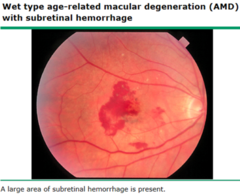
Exudative Macular Degeneration (Wet)
- Abnormal new blood vessel (choroidal neovascularization) growth occurs beneath the retina
- Leaking/bleeding from these vessels cause blurring or sudden vision loss
Wet Macular Degeneration: Signs and Symptoms
- Sudden progressive central vision loss
- Metamorphopsia: straight lines appear wavy or distorted
- Central Scotoma: blind or blurry spot in center of vision
Wet Macular Degeneration Treatment
Anti-VEGR intravitreal injections
• Avastin
• Lucentis
• Eylea
• Vabysmo
Retinal Detachment
Separation of the retina from the underlying retinal pigment
epithelium and choroid

How do most cases of retinal detachment form?
one or more retinal holes or tears
Retinal Detachment symptoms
- Sudden onset of flashers or floaters
- normal central vision until the macula is affected
- "dark curtain" over part of their visual field
How does the retina detach?
- Once a break has developed in the retina, liquified vitreous is free to enter the subretinal space, separating the retina from the pigment epithelium.
Retinal Detachment Risk Factors
hx of myopia, hx of cataract extraction or trauma
How is Retinal Detachment diagnosed?
made by fundoscopy of the dilated eye
Retinal Detachment Treatment
- Urgent referral to ophthalmology
- Surgical repair: permanent adhesion with laser photocoagulation to the retinal pigment epithelium
Retinopathy
- Any disease or damage to the retina
- Can be asymptomatic or with gradual or sudden vision loss
Most common types of Retinopathy
diabetic retinopathy and hypertensive retinopathy
Non-proliferative Diabetic Retinopathy
- "background" retinopathy
- Retinal capillaries leak proteins, lipids or red cells into the retina
- In macula, macular edema occurs

Non-proliferative Diabetic Retinopathy Treatment
- Optimize BP, glucose
- VEGF inhibitor therapy
Proliferative Diabetic Retinopathy
- Less common, more severe
Capillary occlusion causes retinal ischemia and release of VEGF; this is turn stimulates new vessel growth with vision loss from preretinal hemorrhages, fibrosis and retinal traction
Proliferative Diabetic Retinopathy treatment
Invitreal injection of VEGF inhibitor
Diabetic Retinopathy Screening
fundus photography including
dilated examinations of retina
Hypertensive Retinopathy
Disease of the retina due to high blood pressure.
Hypertensive Retinopathy findings
- AV Nicking
- Microaneurysms and flame hemorrhages
- Cotton wool spots (retinal ischemia)
Hypertensive Retinopathy diagnosis
fundoscopic exam
Central Retinal Artery Occlusion
- "Eye stroke"
- An embolus or thrombus suddenly blocks the central retinal artery cutting off blood supply to the inner retina causing sudden, monocular, painless, vision loss

How long can retina tolerate an eye stroke before irreversible damage?
90 minutes
Central Retinal Artery Occlusion: Risk Factors
- Carotid artery atherosclerosis with embolization (most common)
- Cardiac emboli (atrial fibrillation, valvular disease, endocarditis)
- Giant cell arteritis (GCA)
- Hypercoagulable states
- Vasculitis
- HTN
- T2DM
Central Retinal Artery Occlusion symptoms
- Profound, monocular vision loss
- Only vision in temporal field
- PALE SWELLING OF REINA WITH CHERRY RED SPOT AT FOVEA
- BOX CARRING OF RETINAL VESSELS
How to diagnose Central Retinal Artery Occlusion
- ESR and CRP
- MRI of brain (rule out cerebral infarction (stroke))
- Duplex (ultrasound) of carotids
- EKG
- Echo
- CT/MR for carotid dissection
- Screen for DM, hyperlipidemia
Central Retinal Artery Occlusion
Treatment
- Emergency ER referal
- Oxygen, IV acetazolamide (IOP lowering agent), Anterior chamber paracentesis, early thrombolysis
- Check carotids for blockage
- If afib, need anticoagulation
- If valvular disease or PFO, need surgery
Branch Retinal Artery Occlusion
- Occlusion at branch rather than main trunk artery
- Partial vision loss

Branch Retinal Artery Occlusion treatment
Urgent opthal eval, stroke level work up
Central Retinal Vein Occlusion
Blockage of central retinal vein by thrombus, causing blood and
fluid to back up in the retina

Central Retinal Vein Occlusion Risk factors
- Glaucoma (major)
- Atherosclerosis (major)
- age
- DM
- HTN
- Elevated HCT
- Increased blood viscosity
Central Retinal Vein Occlusion symptoms
- Painless vision loss; usually unilateral
- causes hemorrhage and edema rather than ischemia
Central Retinal Vein Occlusion diagnosis
Fundoscopy
Central Retinal Vein Occlusion findings
- "Blood and thunder fundus"
- Retinal cotton wool spots
- Retinal veins will appear distended and torturous
- Fundus is congested and edematous
Central Retinal Vein Occlusion Treatment
No direct treatment
treat underlying complications
Branch Retinal Vein Occlusion
- Partial vision loss; can be at the time of occlusion of fovea is involved or can be later
- Hemorrhages, micro-aneurysms, venous dilation and tortuosity and cotton wool spots are confined to the area drained by the obstructed vein

What is the most common type of retinal occlusion?
Branch Retinal Vein Occlusion
Branch Retinal Vein Occlusion
Treatment
1st Line: Anti-VEGF intravitreal injections
2nd Line: Intravitreal corticosteroids
3rd Line: Focal/grid laser photocoagulation
Last Line: Panretinal or sector photocoagulation if neovascularization develops in
the ischemic territory
Neuro-opthalmologic Disorders
- Nystagmus
- Optic Neuritis
- Papilledema
Primary symptom associated with nystagmus
Vertigo
Primary direction of movement associated with nystagmus
Horizational (BPPV, neuritis,
labyrinthitis)
Optic Neuritis
Inflammation of the optic nerve causing subacute, painful, monocular vision loss

What disease is inflammatory optic neuropathy commonly associated with?
demyelinating disease, particularly multiple sclerosis
Other diseases associated with optic neuritis
Sarcoid, post viral infection, autoimmune- SLE and sjorgens, spread of inflammation by meninges, orbital tissues and paranasal sinuses
Optic Neuritis: Clinical Features
- Unilateral vision loss
- Pain behind eye
- Central field vision loss, color vision loss, pupil defect
- Improves in 2-3 weeks
Optic Neuritis Imaging
MRI if no MS for periventricular white matter demylination
Optic Neuritis Treatment
- In acute demyelinating optic neuritis: IV methylprednisolone 1g daily x 3 days followed by tapering course of oral prednisolone.
- monoclonal antibodies, cell-based therapies
When does optic neuritis have a poorer prognosis?
Optic neuritis due to sarcoid, SLE, herpes zoster
Papilledema
Bilateral optic disc swelling from raised intracranial pressure

Papilledema symptoms
- Brief episodes of graying out
- Dimming or completely black out vision, lasting only seconds.
- Also occurs with rapid positional change
- Headache, nausea, tinnitus, diplopia
Papilledema causes
-Brain mass
- Cerebral edema from head trauma, hypoxic brain injury
- Obstruction of CSF flow in ventricles
- Obstruction of cerebral venous flow
- CSF overproduction
- Idiopathic intracranial hypertension aka pseudotumor cerebri
Papilledema imaging
- MRI (exclude intracranial lesion)
- Lumbar Puncture (elevated pressure and normal CSF = pseudotumor cerebri, which is idiopathic intracranial hypertension
Papilledema Treatment
- Reduce elevated inter-cranial pressure
- If mass, surgical management
Orbital and Periorbital Cellulitis
severe infection involving the fat and muscle within the orbit
Orbital Cellulitis symptoms
- Fever
- Proptosis
- Restriction of extraocular movements
- Painful purple-red eyelid swelling
- Usually caused by infection of paranasal sinuses or dental infection -> S pneumoniae, H influenzae & S aureus

Orbital Cellulitis imaging
CT
CBC
Cultures
Opthal referral
Orbital Cellulitis Treatment
- Immediate IV antibiotics
- Anaerobic infections -> nafcillin, with metronidazole or clindamycin
- MRSA -> Vanco or Clindamycin
- PCN allergy -> Vanco, levofloxacin and metronidazole
Periorbital Cellulitis
- More common, mild infection in front of the eye socket membrane.
- This occurs anteriorly to the orbital septum

Periorbital Cellulitis symptoms
- Edema
- Painless
- Circumferential erythema of the eyelids and periorbital skin
- Normal eye movement
Periorbital Cellulitis causes
- bug bite, skin scratch, local face/eyelid wound
- S aureus and Streptococcus
Periorbital Cellulitis treatment
Oral antibiotics
Vision Abnormalities
- Amaurosis fugax
- Amblyopia
- Glaucoma
- Strabismus
- Presbyopia
Amaurosis Fugax "Fleeting blindness"
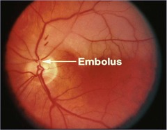
transient ischemic attack of the retina
How does Amaurosis Fugax happen?
Interruption of blood flow (due to embolus becoming stuck in retinal arteriole) to the retina for more than a few seconds will result in transient monocular blindness
How does a Amaurosis Fugax look like to the patient?
Curtain descending affecting only a portion of visual field
Amaurosis Fugax Diagnosis
Urgent neuroimaging to assess for cerebral infarction and ID source of emboli
What are patients with Amaurosis Fugax at an increased risk of?
stroke, MI and other vascular events
Amaurosis Fugax Treatment
- Oral aspirin/Plavix
- Statins to reduce LDL cholesterol
- Carotid Endarterectomy/stenting
- Lifestyle changes
Amblyopia
Eye fails to acquire its potential visual acuity because images are not properly projected onto the fovea during a formative period of cerebral development.
Amblyopia causes
strabismus, ptosis, cataract, refractive errors
Strabismus "crossed eyes"
Any ocular misalignment in which only one eye fixates with the fovea on object
6 ocular muscles
- superior rectus
- medial rectus
- inferior rectus
- lateral rectus
- superior oblique
- inferior oblique
Esotropia
inward turning, convergent axes

Exotropia
outward turning, divergent axes

Hypertropia
upward turning

Hypotropia
downward turning

Strabismus: Causes and Risk Factors
Family history
Cerebral palsy
Down syndrome
Hydrocephalus
Brain tumors
Head injuries
Graves Disease
Strabismus Diagnosis
Light reflex
Cover test
Light Reflex
The corneal light from a penlight held along a toy that the child focuses on should appear symmetric
Cover test
In an abnormal test, when the dominant eye is covered, the weaker eye will move to focus on an object
Strabismus Treatment
Esotropia - surgery
Exotropia - patching and exercise
Strabismus Complications
- amblyopia (decreased visual acuity in less dominant eye)
- diplopia
- contracture of the ocular eye muscles
Presbyopia
Normal age related condition, the eyes natural lens gradually loses its flexibility, making it hard to focus on close up objects
Presbyopia age onset
40 yo
Presbyopia symptoms
blurred near vision, holding phones, books, menus farther away to read them
Presbyopia Treatment
- Reading glasses
- Bifocals are split lenses used to visualize out of distinct sections
of the glasses