Exam 4 (Ch. 31-35)
 Call Kai
 Learn
 Practice Test
 Spaced Repetition
 Match
 Flashcards
 Knowt Play
1/78

Earn XP
Description and Tags
Comprehensive vocabulary flashcards covering dental radiography interpretation, including descriptive terminology, identification of dental materials and restorations, classification of dental caries, assessment of periodontal bone loss, and diagnosis of traumatic and periapical lesions.
79 Terms
Radiolucent
The portion of a processed dental image that appears dark or black because the imaged structure is less dense and permits the passage of the x-ray beam (e.g., dental caries).
Radiopaque
The portion of a processed dental image that appears light or white because the imaged structure is dense and absorbs radiation (e.g., metallic restorations).
Unilocular Radiolucent Lesion
A radiolucent lesion exhibiting only one compartment; these lesions tend to be small and nonexpansile.
Unilocular Lesion, Corticated Border
A thin, well-demarcated radiopaque rim of bone outlining a radiolucent lesion. Usually indicative of a benign, slow-growing process.
Unilocular Lesion, Noncorticated Border
A radiolucent lesion periphery that appears fuzzy or poorly defined without a radiopaque rim of bone. May represent either a benign or a malignant process.
Multilocular Radiolucent Lesion
A radiolucent lesion that exhibits multiple compartments, is frequently expansile, and typically represents a benign lesion with aggressive growth potential.
Radiolucent Lesions
May appear in a periapical, inter-radicular, edentulous, or pericoronal location
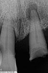
Periapical Location
Lesion located around the apex of a tooth root
Example: periapical cyst secondary to pulpal necrosis


Inter-radicular Location
Lesion located between the roots of adjacent teeth.
Example: lateral periodontal cyst


Edentulous Zone
Lesion located in an area without teeth. A variety of radiolucent or radiopaque lesions may occur in this area.
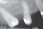

Pericoronal Location
Radiolucent lesion located around the crown of an impacted tooth.
Example: dentigerous cyst


Alveolar Bone Loss
Loss of supporting bone in the maxilla or mandible that surrounds and supports the teeth, appearing radiolucent on dental images.

Radiopaque Lesions
Can be described as a focal opacity, target lesion, multifocal confluent, irregular, ground glass, or mixed lucent-opaque. Occur in bone and in soft tissue. May appear in the same places as radiolucent lesions (periapical, inter-radicular, edentulous, or pericoronal locations)

Focal Opacity
A well-defined, localized radiopaque lesion viewed on a dental image
Example: condensing osteitis


Target Lesion
A well-defined, localized radiopaque area surrounded by a uniform radiolucent halo
Example: benign cementoblastoma


Multifocal Confluent Pattern
Multiple radiopacities that appear to overlap or flow together. Multifocal confluent radiopacities involving multiple quadrants of the jaws typically represent benign fibro-osseous disorders.
Example: osteitis deformans, florid osseous dysplasia

Irregular/Ill-Defined Opacity
A radiopacity exhibiting an irregular, poorly defined pattern that may indicate a malignant condition
Examples: osteosarcoma or chondrosarcoma

Ground Glass Opacity
A granular or pebbled radiopacity resembling pulverized glass or the texture of an orange peel
Examples: fibrous dysplasia, osteitis deformans, osteopetrosis


Mixed Lucent-Opaque Lesion
A lesion that exhibits both radiopaque and radiolucent components, commonly representing calcifying tumors
Example: compound odontoma
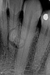

Soft Tissue Opacity
A well-defined radiopaque area located within soft tissue
Examples: sialolith, calcified lymph node

Radiopaque
Metallic restorations appear completely _______ on a dental image.
Radiolucent, radiopaque
Nonmetallic restorations may vary in appearance from ______ to slightly ______, depending on the density of the material.

Amalgam Restoration
Appears as a distinct, dense radiopacity with irregular outlines.


Amalgam Overhang
An extension of amalgam seen beyond the crown portion of a tooth in the interproximal region that disrupts cleansing contours, traps food and plaque, and contributes to bone loss.
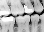

Amalgam Fragments
Fragments of amalgam inadvertently embedded in adjacent soft tissue during tooth restoration, appearing on images as dense radiopacities with irregular borders.


Gold Restoration
A cast metallic restoration that appears completely radiopaque on dental images and exhibits a smooth, regular marginal outline (unlike amalgam resorations).


Stainless Steel Crown
A radiopaque restoration with smooth and regular margins that is not as densely radiopaque as amalgam or gold, appearing see-through in some areas.


Post and Core Restoration
A radiopaque restoration placed in the pulp canal and coronal space of an endodontically treated tooth to retain and support a cast crown.


Porcelain Restoration
A nonmetallic restoration that appears slightly radiopaque on dental images, closely resembling the radiopacity of natural dentin. All porcelain crown and bridges. Porcelain-fused-to-metal crown and bridges may have greater radiopacity due to the metal.


Composite Restoration
A nonmetallic tooth-colored restoration whose radiographic appearance varies from radiolucent to slightly radiopaque depending on the composition of the composite material.
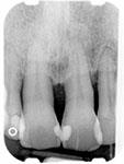
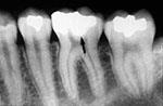
Base Materials
Cavity liners placed on the floor of a cavity preparation to protect the pulp, appearing radiopaque on images but less radiodense than amalgam.
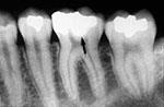

Metallic Pins
Metallic retention devices placed into dentin to enhance retention of amalgam or composite restorations, appearing as distinct radiopaque cylinders.
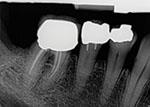

Gutta Percha
A claylike endodontic material used to fill and seal pulp canals, appearing radiopaque on dental images.

Silver Points
Rigid endodontic points used historically to fill root canals that appear highly and uniformly radiopaque, distinctly more radiodense than gutta percha.
Dental Caries
The localized destruction of tooth structure by microorganisms, appearing radiolucent on dental images because the demineralized area has decreased density.

Interproximal Caries
Caries located between two adjacent surfaces, typically seen on dental images at or just below the contact point.

Incipient Interproximal Caries
An early interproximal carious lesion extending less than halfway through the thickness of enamel.

Moderate Interproximal Caries
An interproximal carious lesion extending more than halfway through enamel but not involving the dentinoenamel junction (DEJ).

Advanced Interproximal Caries
An interproximal carious lesion extending to or through the DEJ and into dentin, but not extending more than half the distance toward the pulp.

Severe Interproximal Caries
An interproximal carious lesion extending through enamel and dentin more than half the distance toward the pulp.
Incipient Occlusal Caries
Early caries on the chewing surface of posterior teeth that cannot be visualized on a dental image and must be detected clinically using an explorer.
Moderate Occlusal Caries
Occlusal caries extending into dentin that appears on a dental image as a thin, radiolucent line under the occlusal enamel.

Severe Occlusal Caries
Extensive occlusal caries extending into dentin that appears as a large radiolucency spreading beneath the enamel of the occlusal surface.

Buccal and Lingual Caries
Carious lesions involving the buccal or lingual surfaces of teeth that, when visible on dental images, appear as small, circular radiolucent areas.

Root Surface Caries
Caries involving only the roots of teeth, appearing on dental images as a cupped-out or crater-shaped radiolucency below the cementoenamel junction (CEJ).

Recurrent Caries
A carious lesion occurring adjacent to an existing dental restoration.

Rampant Caries
Advanced and severe carious lesions affecting a significant number of teeth in the dentition.

Cervical Burnout
A collar-shaped or wedge-shaped radiolucent artifact seen between the CEJ and alveolar bone, caused by anatomical differences in tissue thickness and concavity.

Attrition
The mechanical wearing down of tooth structure on the incisal or occlusal surfaces, distinguishable clinically from carious lesions.

Abrasion
The mechanical wearing away of tooth structure resulting from the friction of a foreign object (most frequently improper toothbrushing), appearing clinically as hard, highly polished dentinal defects.

Periodontal Ligament Space
A thin radiolucent line appearing between the root of the tooth and the lamina dura that is continuous and of uniform thickness in healthy teeth.
Lamina Dura
A dense radiopaque line that surrounds the tooth root and lines the tooth socket in healthy teeth.

Alveolar Crest
The coronal border of alveolar bone, normally located about 1.5 to 2mm apical to the CEJ of adjacent healthy teeth (pointed and sharp in anterior teeth, flat and smooth in posterior teeth).
Periodontal Disease
A group of diseases that affect the tissues surrounding teeth, ranging from superficial gingival inflammation to the destruction of supporting bone and the periodontal ligament.

Horizontal Bone Loss
A pattern of bone loss in which the resorption occurs in a plane parallel to an imaginary line connecting the cementoenamel junctions of adjacent teeth.

Vertical Bone Loss
An angular pattern of bone loss in which the remaining alveolar bone is not parallel to the line connecting adjacent cementoenamel junctions.
Severity of Bone Loss
Classification of periodontal bone loss measured in millimeters: slight bone loss is 1 to 2mm, moderate bone loss is 3 to 4mm, and severe bone loss is 5mm or greater.
Stage I Periodontal Disease
Periodontal disease characterized by radiographic bone loss limited to the coronal third of the root (<15%).
Stage II Periodontal Disease
Periodontal disease characterized by radiographic bone loss in the coronal third of the root ranging from 15% to 33%.
Stage III Periodontal Disease
Periodontal disease characterized by radiographic bone loss extending into the mid-third of the root, with vertical bone loss and furcation involvement.
Stage IV Periodontal Disease
Advanced periodontal disease characterized by radiographic bone loss extending to the mid-third of the root and beyond, accompanied by vertical bone loss, furcation involvement, and tooth mobility.

Dental Calculus
Mineralized bacterial plaque appearing on dental images as pointed or irregular radiopaque projections extending from proximal root surfaces.

Crown Fracture
A traumatic fracture most often involving anterior teeth, which may affect the enamel, dentin, and/or pulp of the crown.

Root Fracture
A traumatic break most frequently occurring in the maxillary central incisor region; it appears as a radiolucent line when the x-ray beam is parallel to the fracture plane.

Jaw Fracture
A fracture most commonly observed in the mandible that appears on dental images as a radiolucent line, best visualized with a panoramic image.
Luxation
The abnormal displacement of teeth resulting from trauma, divided into intrusion (displacement into bone) and extrusion (displacement out of bone).

Dental Avulsion
The complete traumatic displacement of a tooth from the alveolar bone, visualized radiographically as an empty tooth socket.

Physiologic Resorption
The normal biological shedding of primary teeth accompanied by progressive root resorption.

External Resorption
Resorption along the periphery of the root surface (most often the apex), causing root blunting and shortening; associated with trauma, reimplantation, chronic inflammation, or abnormal forces.

Internal Resorption
Resorption occurring within the crown or root of a tooth precipitated by trauma, pulp capping, or pulp polyps, appearing as a round-to-ovoid radiolucency in the midcrown or midroot pulp space.
Pulpal Sclerosis
Diffuse calcification of the pulp chamber and canals associated with aging, having minimal clinical significance unless endodontic intervention is indicated.

Pulpal Obliteration
The extensive deposition of secondary dentin within the pulp cavity obliterating the pulp chamber and canals, rendering the tooth nonvital without requiring treatment.

Pulp Stones
Dystrophic calcifications appearing as round, ovoid, or cylindrical radiopacities within the pulp chamber or canals, requiring no clinical treatment.

Periapical Granuloma
A localized mass of chronically inflamed granulation tissue at the apex of a nonvital tooth, representing the most common sequela of pulpitis and presenting as a widened periodontal ligament space at the root apex.

Periapical Cyst
An asymptomatic, slow-growing lesion resulting from cystic degeneration of a periapical granuloma at the apex of a nonvital tooth; it is the most common tooth-related cyst.

Periapical Abscess
A localized collection of pus around the apex of a nonvital tooth; acute abscesses are painful and may not show radiographic changes, while chronic abscesses appear as round or ovoid apical radiolucencies.

Periodontal Abscess
A localized collection of pus resulting from infection within periodontal tissues, presenting as a radiolucency along the lateral aspect of the root with deep, throbbing pain.

Condensing Osteitis
A well-defined radiopacity seen below the apex of a tooth with a history of long-standing pulpitis (most commonly the mandibular third molar), not attached to the tooth root.

Hypercementosis
Excess deposition of cementum along the root surfaces resulting from supereruption, inflammation, or trauma, causing the apex to appear enlarged and bulbous while the tooth remains vital.