Comparative Vertebrate Anatomy
 Call Kai
 Learn
Learn Practice Test
 Spaced Repetition
 Match
Match Flashcards
 Knowt Play
1/334

There's no tags or description
Looks like no tags are added yet.
Name | Mastery | Learn | Test | Matching | Spaced | Call with Kai | Chat |
|---|
No analytics yet
Send a link to your students to track their progress
335 Terms

Identify the animal in the following photo:
Amphioxus
Cephalachordata
Lancelet

What is structure A in the following diagram?
Dorsal fin
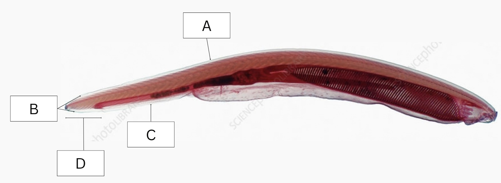
What is structure B in the following diagram?
Caudal fin

What is structure C in the following diagram?
Ventral fin

What is structure D in the following diagram?
Post-anal tail

What is A in the following diagram?
Velar tentacles
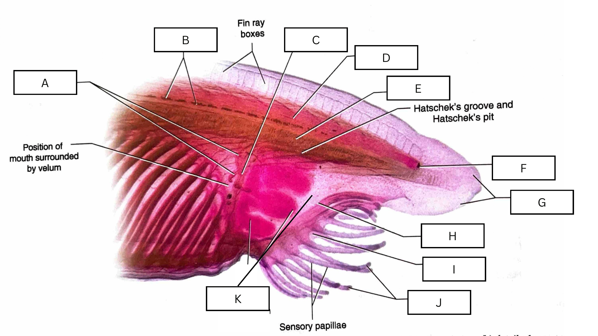
What is B in the following diagram?
Hesse ocelli

What is C in the following diagram?
Velum

What is D in the following diagram?
Dorsal hollow nerve cord
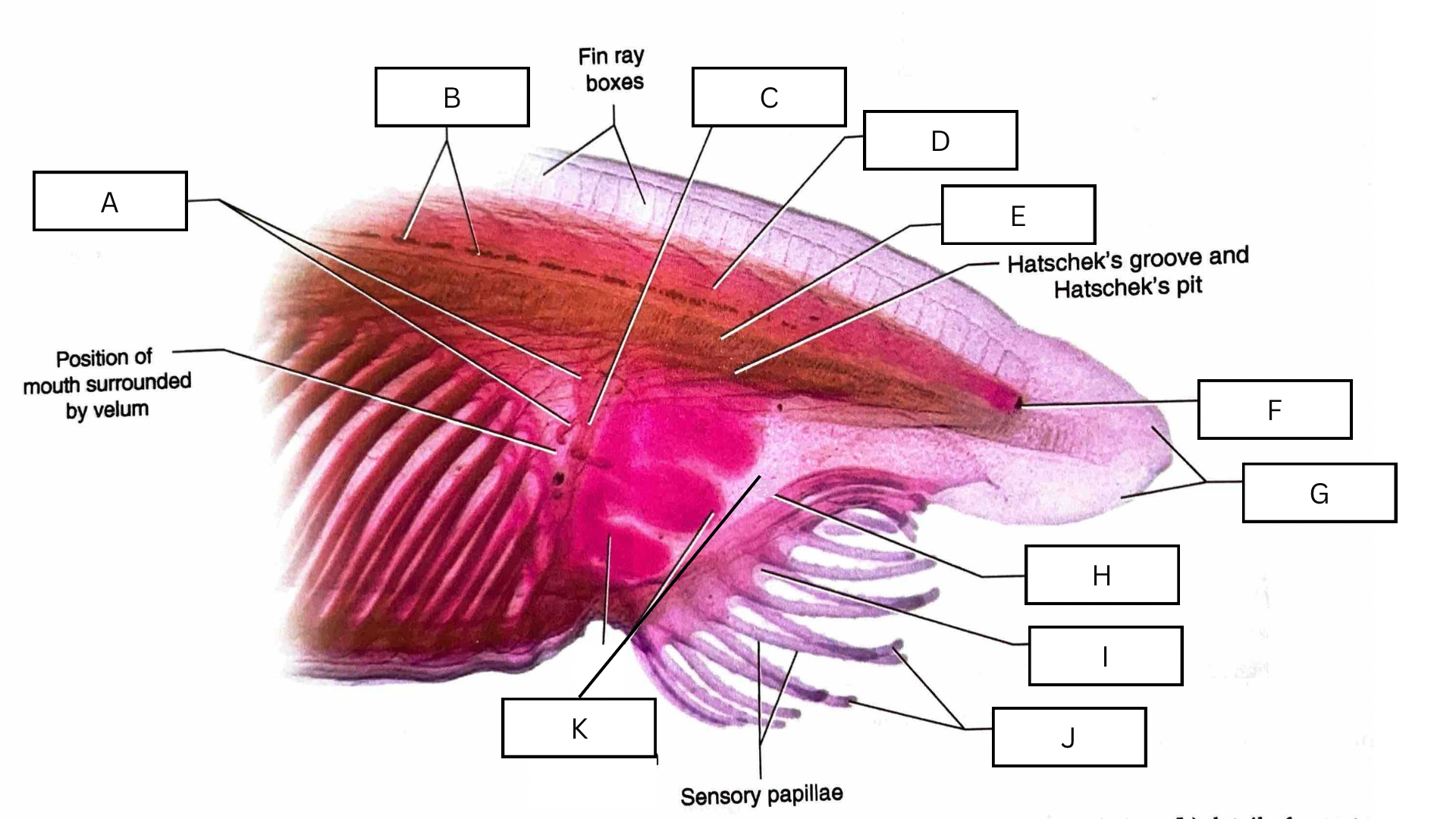
What is E in the following diagram?
Notochord

What is F in the following diagram?
Frontal eye

What is G in the following diagram?
Rostrum / Rostral fin

What is H in the following diagram?
Oral hood

What is I in the following diagram?
Inhalant aperture

What is J in the following diagram?
Oral cirri

What is K in the following diagram?
Oral cavity (internal)

What is A in the following diagram?
Hindgut

What is B in the following diagram?
Iliocolon

What is C in the following diagram?
Midgut

What is D in the following diagram?
Esophagus

What is E in the following diagram?
Hesse ocelli

What is F in the following diagram?
Anus

What is G in the following diagram?
Atriopore

What is H in the following diagram?
Hepatic cecum

What is I in the following diagram?
Pharynx

What is shown in the following diagram?
Cross section of amphioxus

What is A in the following diagram?
Dorsal hollow nerve cord

What is B in the following diagram?
Notochord

What is C in the following diagram?
Iliocolon

What is D in the following diagram?
Metapleural fold
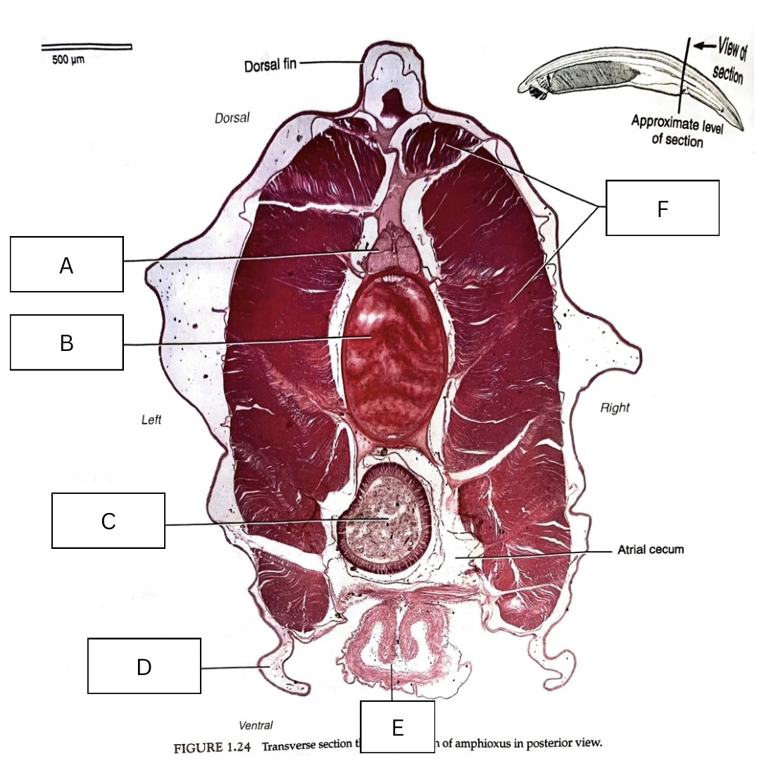
What is E in the following diagram?
Atriopore
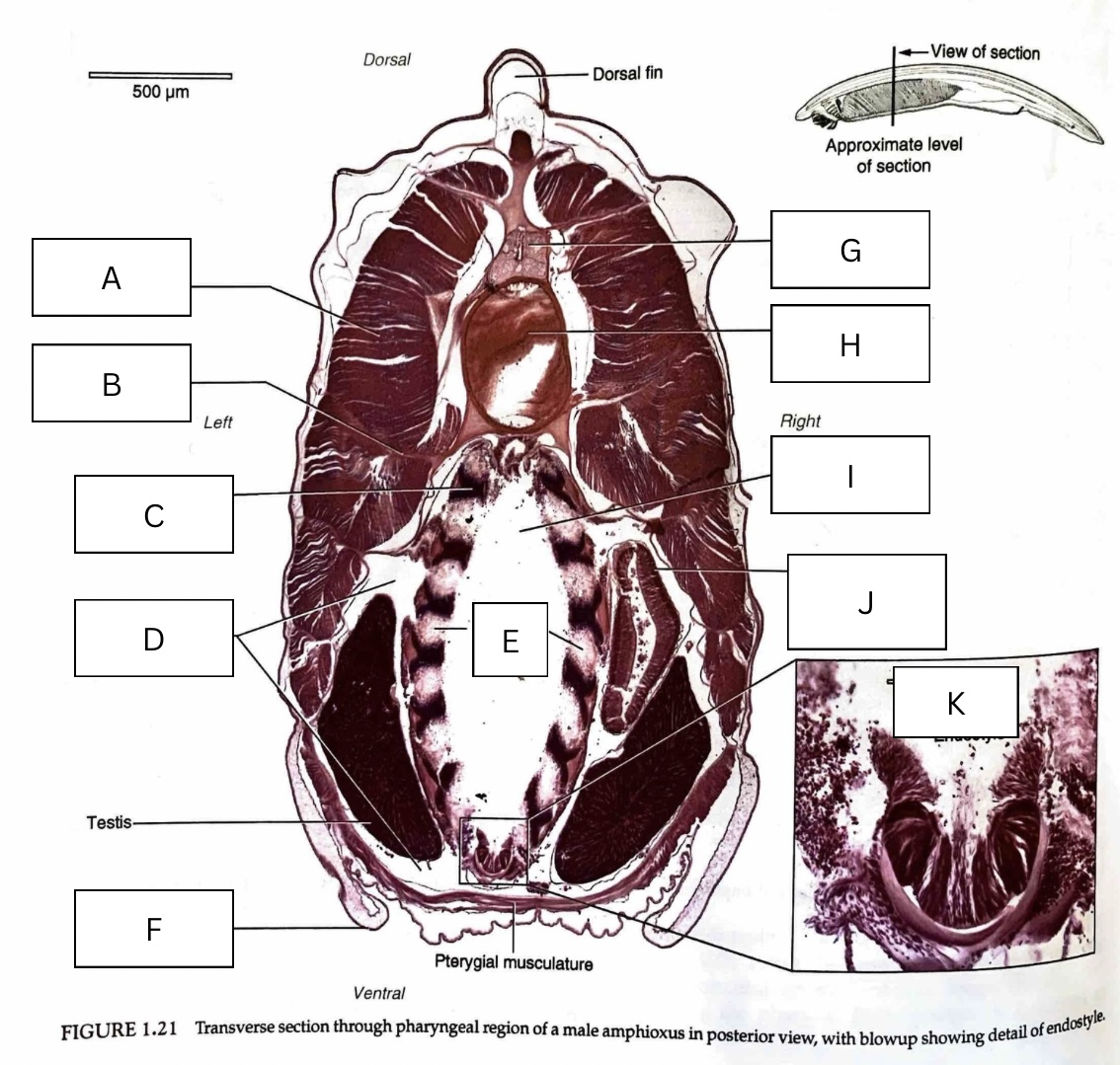
What is A in the following diagram?
Myomere

What is B in the following diagram?
Myoseptum
Myosepta (pleural form)

What is C in the following diagram?
Gill bar

What is D in the following diagram?
Atrium

What is E in the following diagram?
Gill slits

What is F in the following diagram?
Metapleural fold

What is G in the following diagram?
Dorsal hollow nerve cord

What is H in the following diagram?
Notochord

What is I in the following diagram?
Pharynx
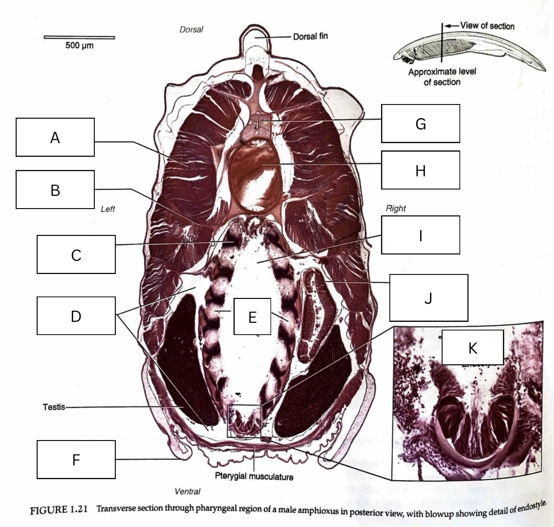
What is J in the following diagram?
Hepatic cecum

What is K in the following diagram?
Endostyle

What is A in the following diagram?
Gonads

What is A in the following diagram?
Epibranchial groove

Identify the animal in the following photo:
Urochordata / Tunicate
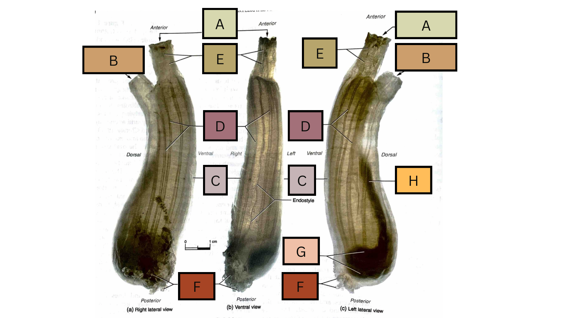
What is structure A in the following diagram?
Oral siphon

What is structure B in the following diagram?
Atrial siphon

What is structure C in the following diagram?
Tunic
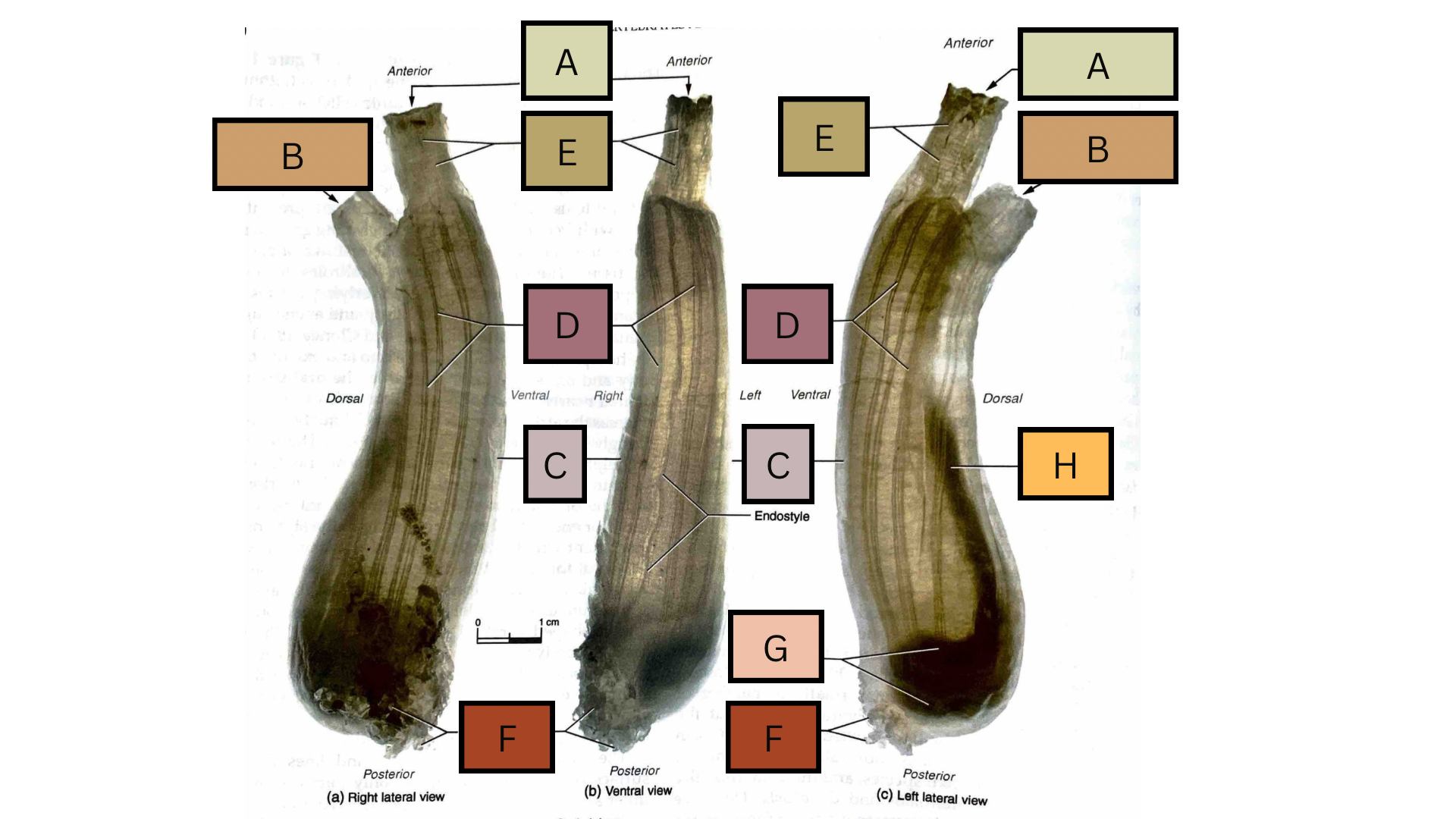
What is structure D in the following diagram?
Longitudinal muscles

What is structure E in the following diagram?
Circular muscles
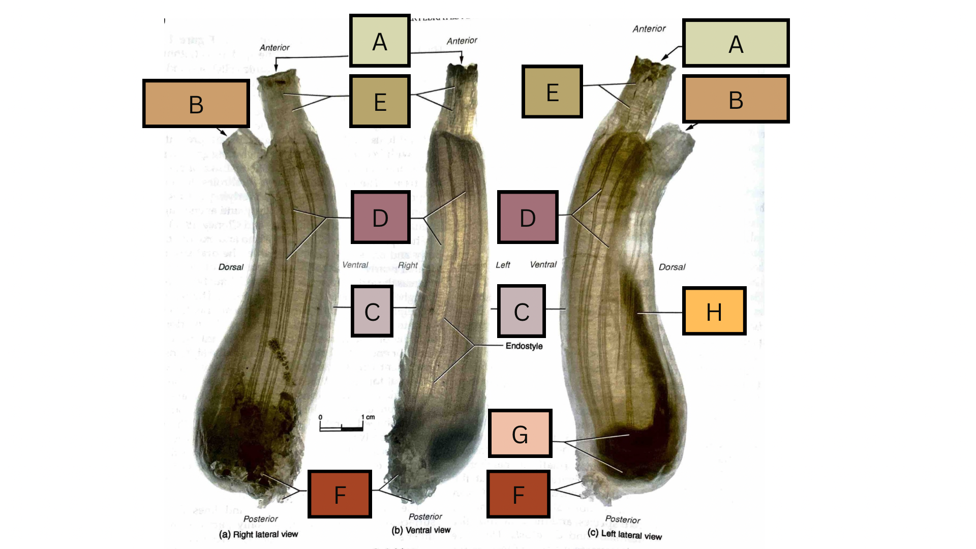
What is structure F in the following diagram?
Attachment villi

What is structure G in the following diagram?
Internal organs

What is structure H in the following diagram?
Rectum

What animal skeleton is shown in the following diagram?
Petromyzontidae / Lamprey

What is A in the following diagram?
Chondocranium

What is B in the following diagram?
Annular cartilage
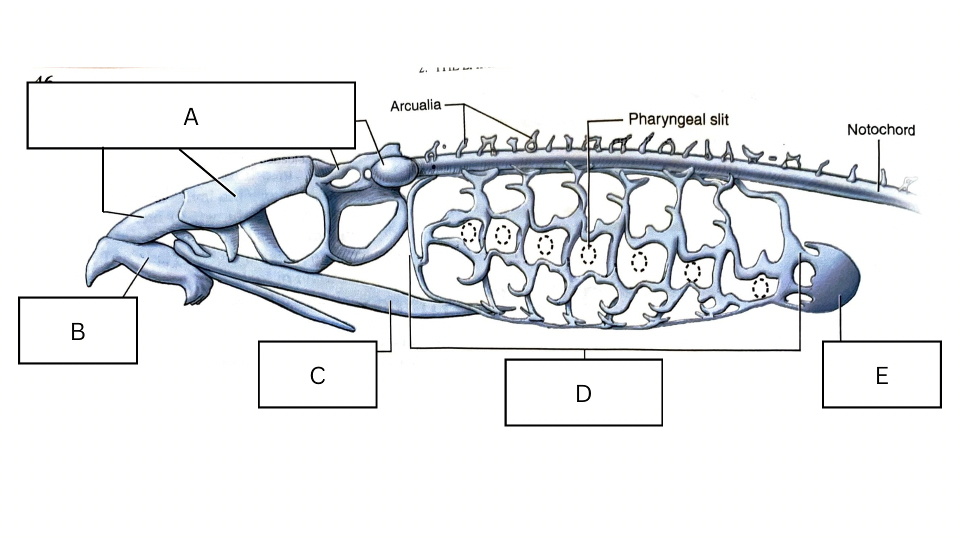
What is C in the following diagram?
Lingual cartilage
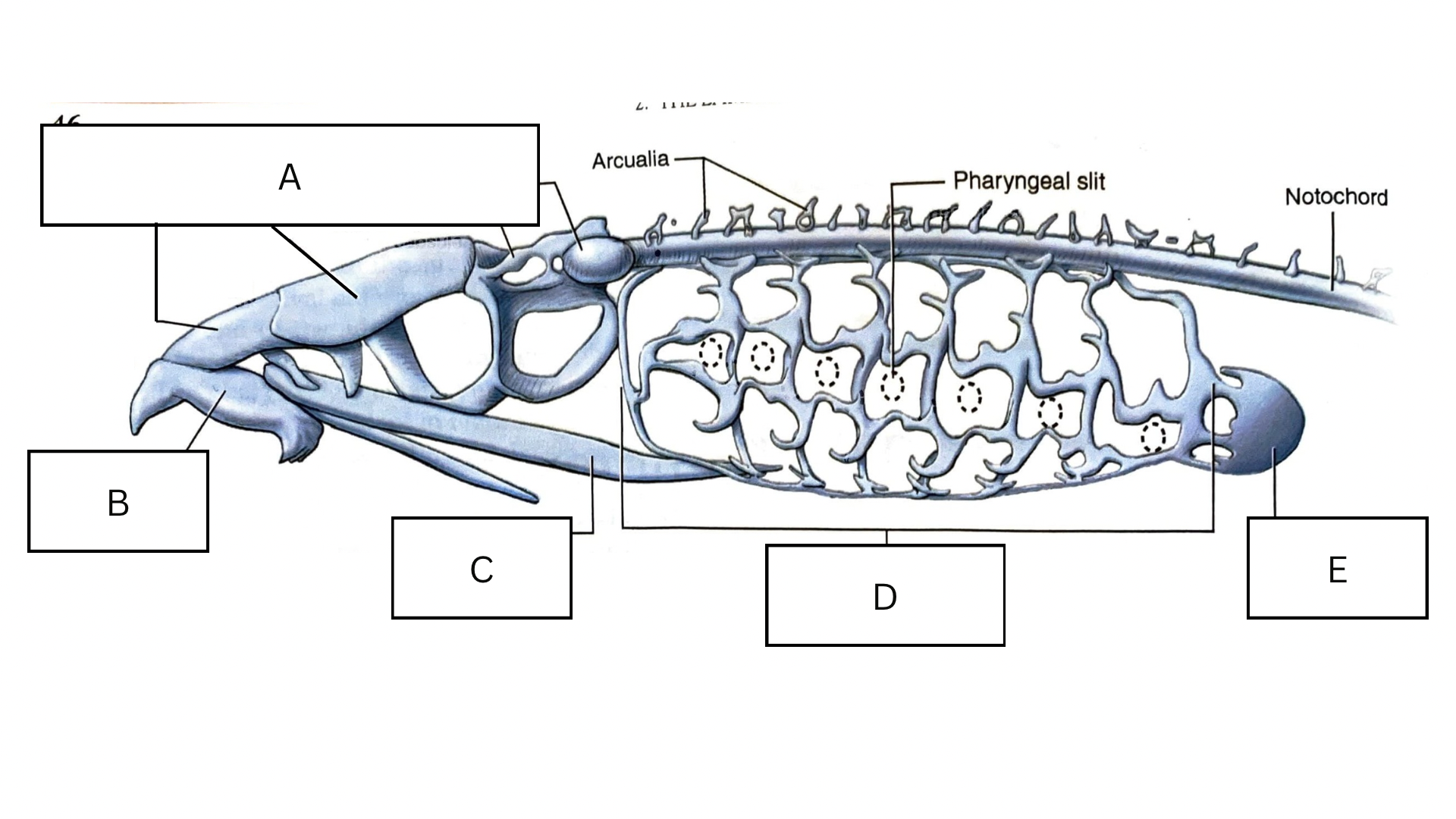
What is D in the following diagram?
Branchial basket
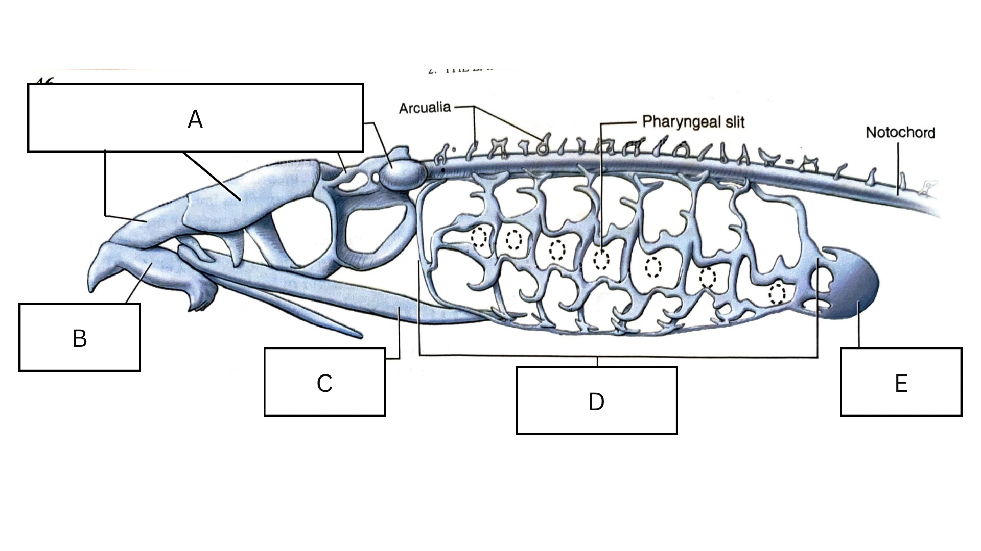
What is E in the following diagram?
Pericardial cartilage

What animal does this mouth belong to?
Petromyzontidae / Lamprey

What is A in the following diagram?
Teeth

What is B in the following diagram?
Mouth

What is C in the following diagram?
Oral disk

What is D in the following diagram?
Tongue

What is E in the following diagram?
Buccal papillae
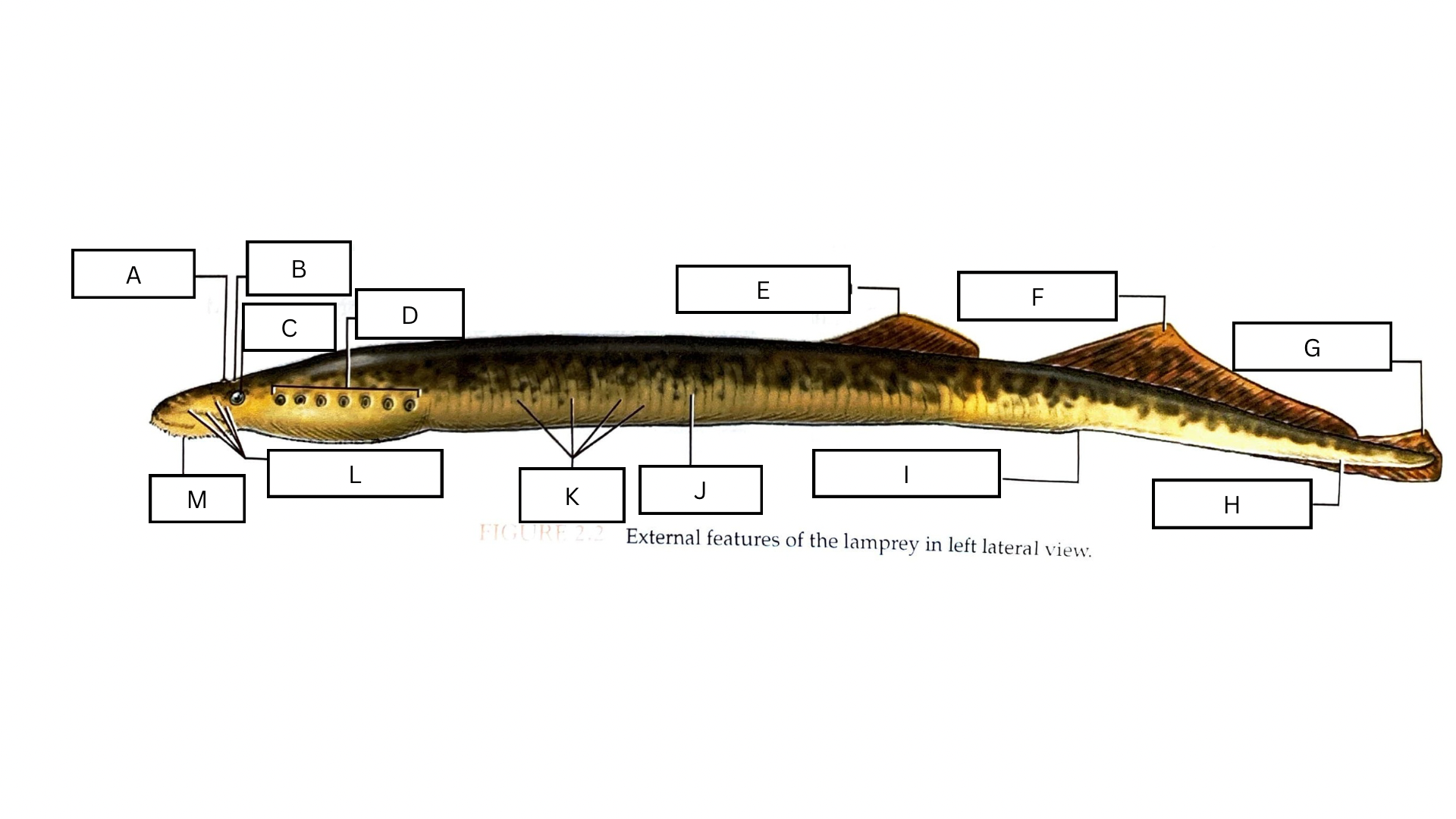
What is A in the following diagram?
Naris

What is B in the following diagram?
Pineal eye complex

What is C in the following diagram?
Eye
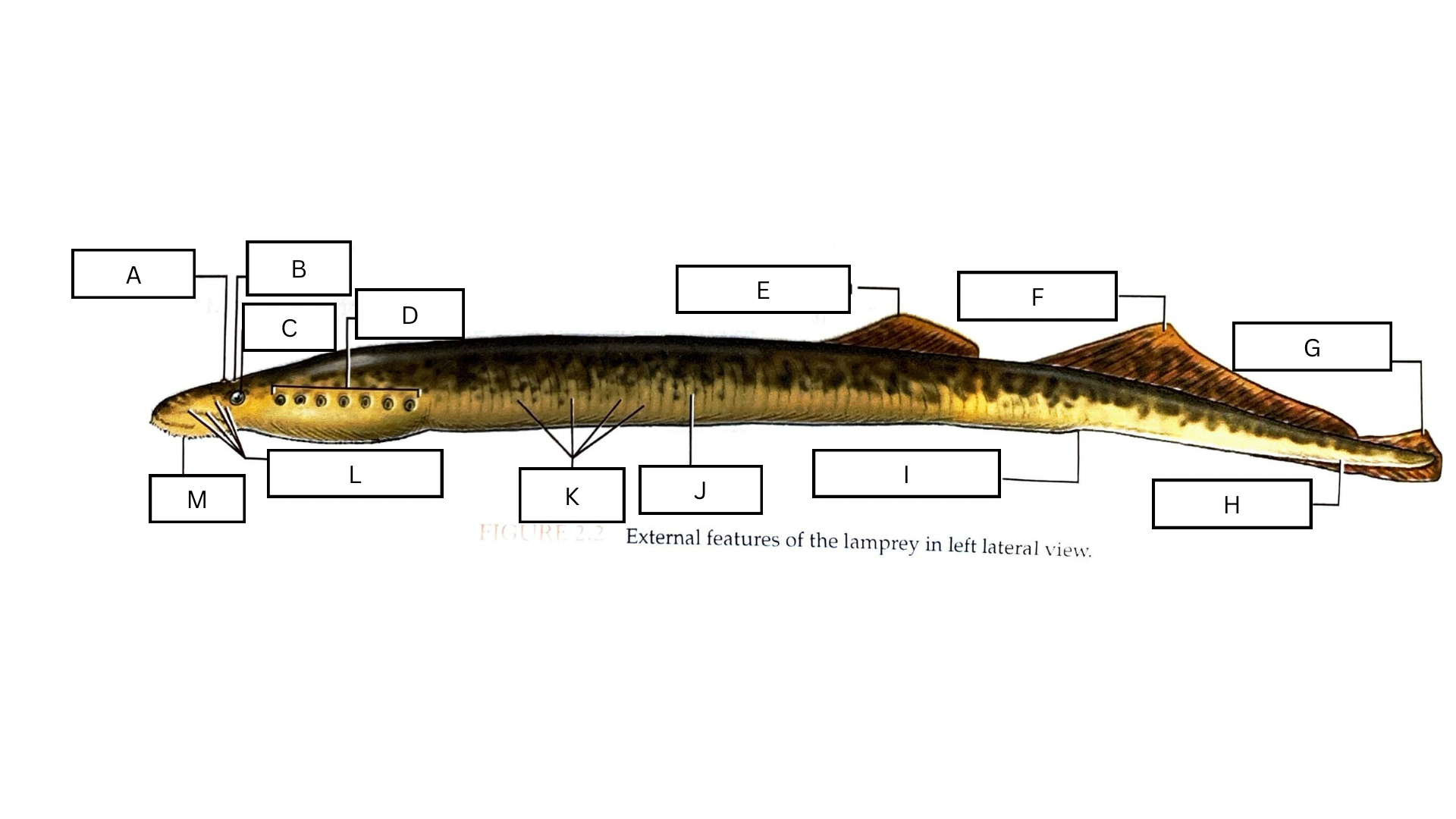
What is D in the following diagram?
External pharyngeal slits

What is E in the following diagram?
Anterior dorsal fin

What is F in the following diagram?
Posterior dorsal fin

What is G in the following diagram?
Caudal fin
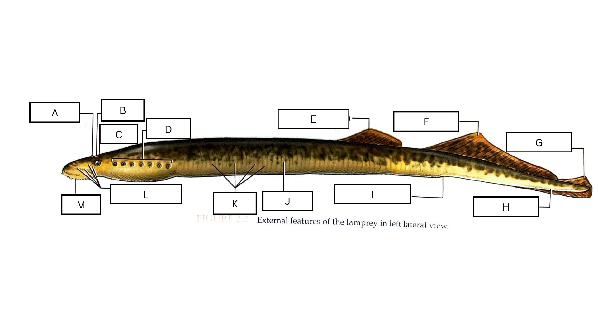
What is H in the following diagram?
Tail

What is I in the following diagram?
Cloacal aperture
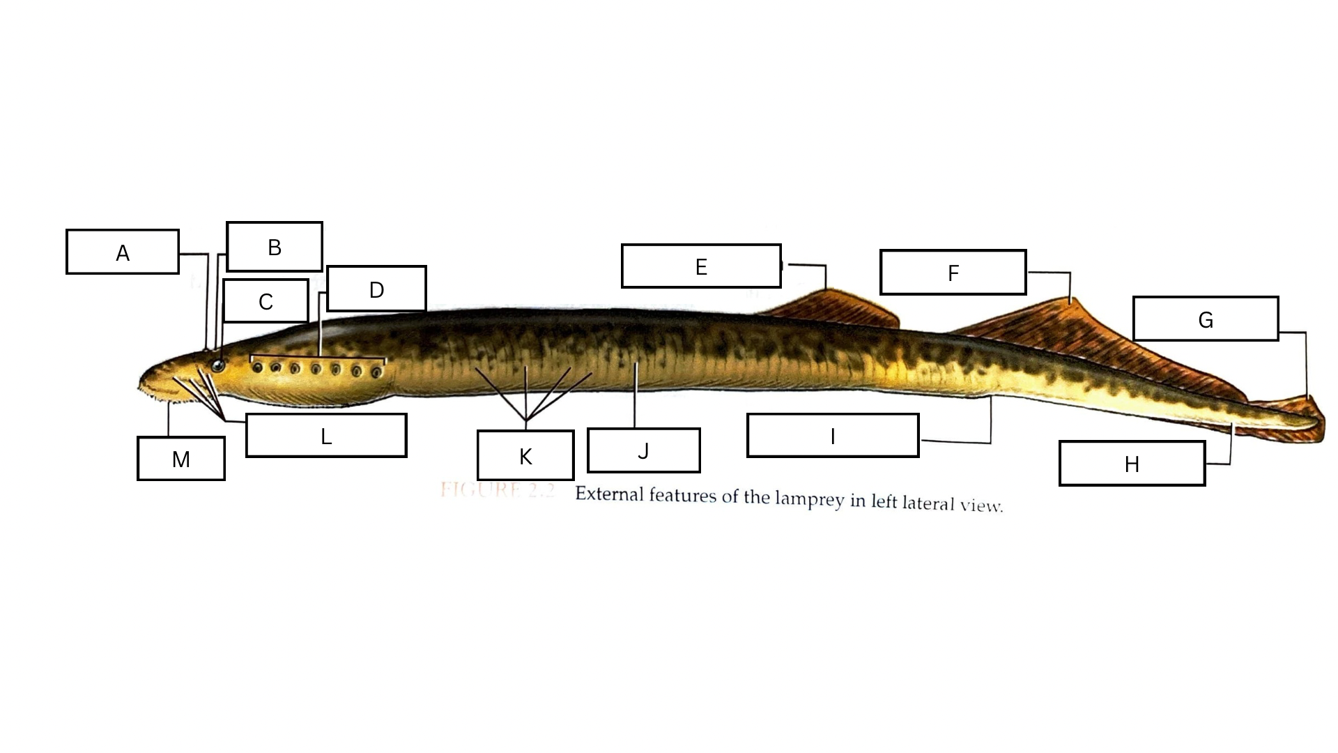
What is J in the following diagram?
Trunk

What is K in the following diagram?
Myomeres

What is L in the following diagram?
Lateral line system + pores
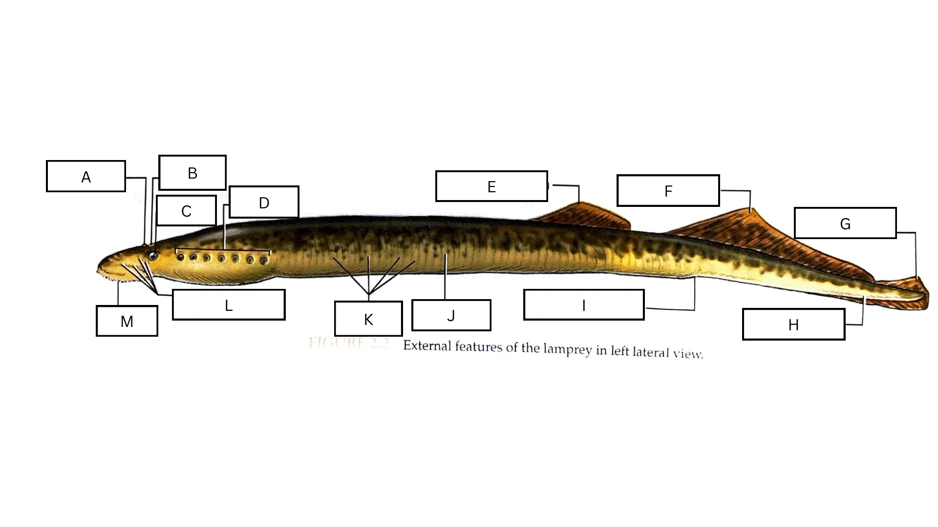
What is M in the following diagram?
Oral funnel

What is A in the following diagram?
Spinal cord

What is B in the following diagram?
Notochord

What is C in the following diagram?
Common cardinal vein

What is D in the following diagram?
Ovary

What is E in the following diagram?
Dorsal aorta
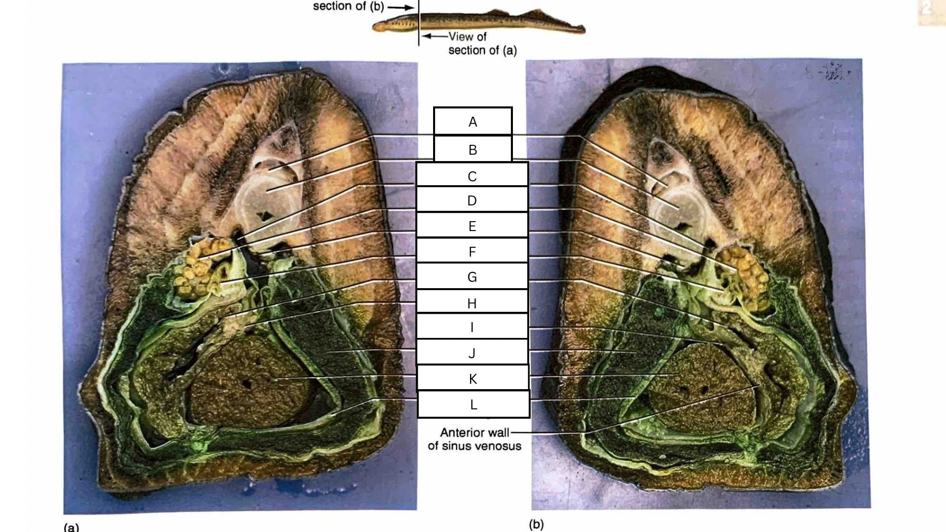
What is F in the following diagram?
Intestine

What is G in the following diagram?
Atrium

What is H in the following diagram?
Sinoatrial aperture

What is I in the following diagram?
Sinus venosus
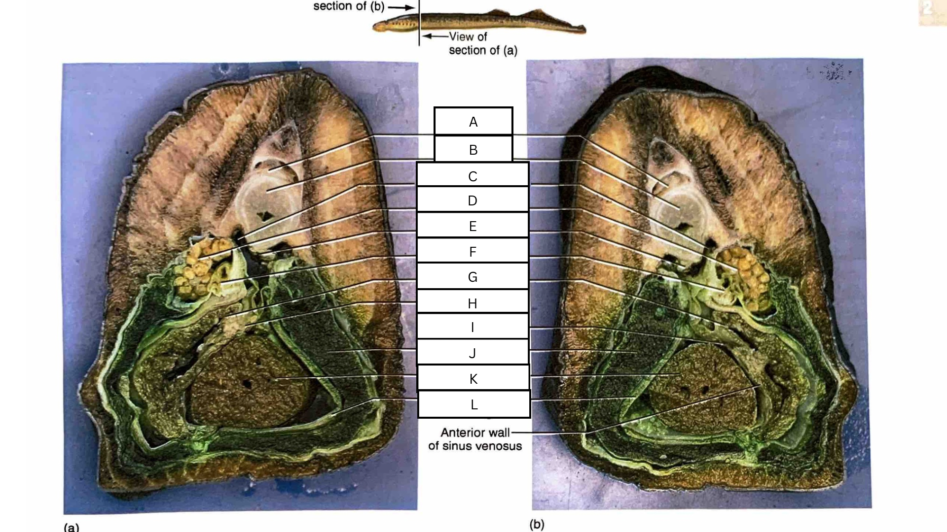
What is J in the following diagram?
Liver

What is K in the following diagram?
Ventricle

What is L in the following diagram?
Pericardial cartilage

What is A in the following diagram?
Dorsal aorta

What is B in the following diagram?
Kidney

What is C in the following diagram?
Archinephric duct
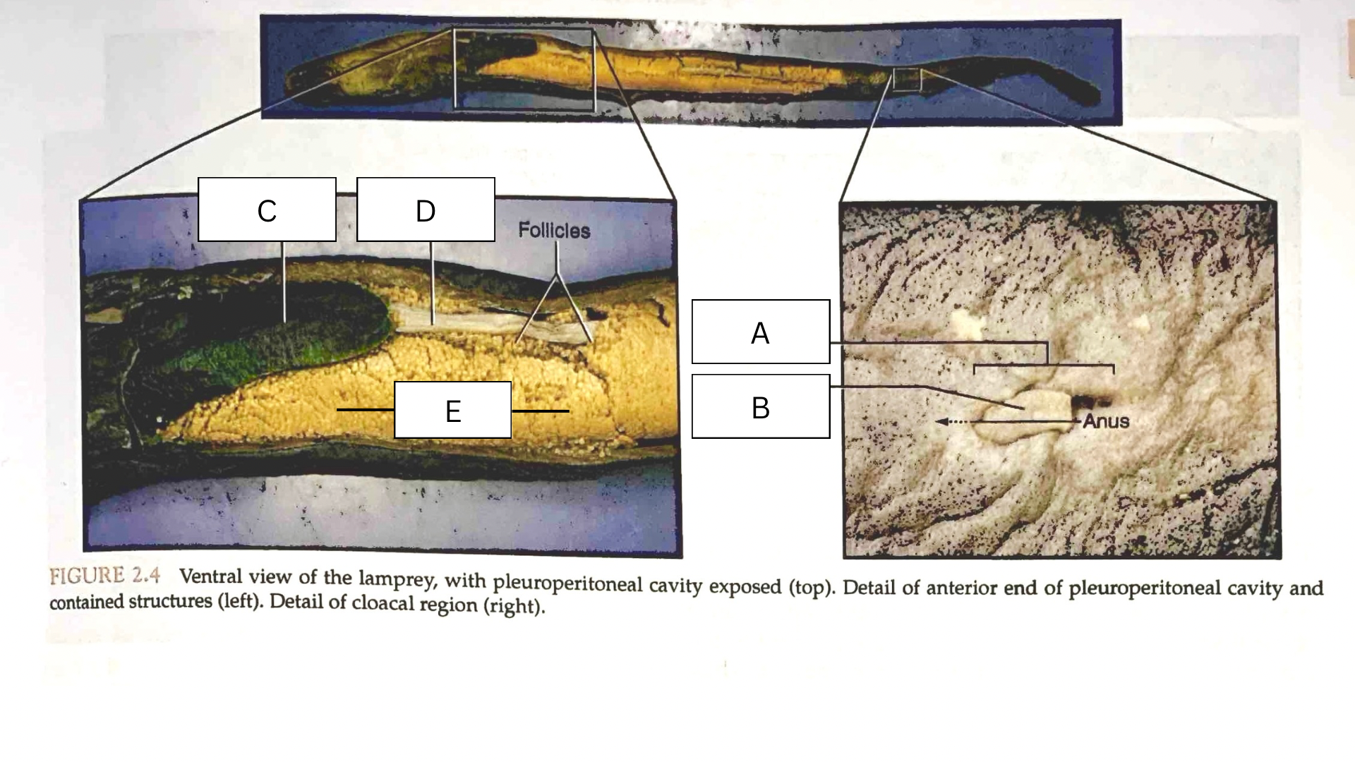
What is A in the following diagram?
Cloaca
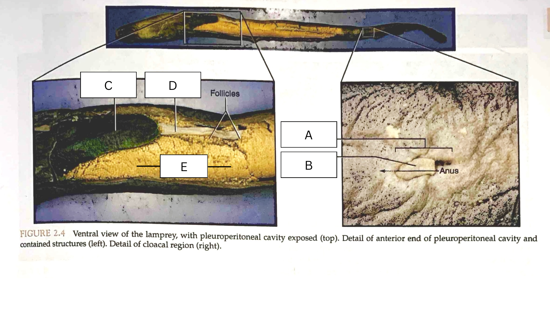
What is B in the following diagram?
Urogenital papilla

What is C in the following diagram?
Liver

What is D in the following diagram?
Intestine

What is E in the following diagram?
Ovary
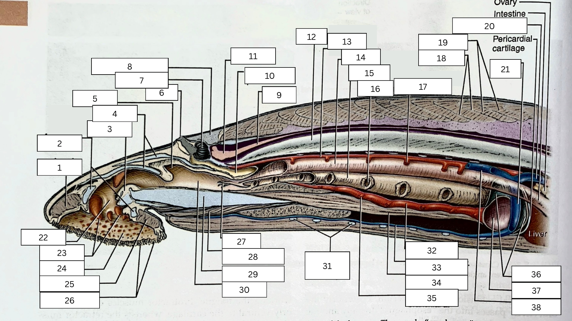
What is structure #1 in the sagittal section of the lamprey?
Annular muscle

What is structure #2 in the sagittal section of the lamprey?
Annular cartilage