Blood Bank: ABO Blood Group Review
 Call Kai
 Learn
 Practice Test
 Spaced Repetition
 Match
 Flashcards
 Knowt Play
1/57

There's no tags or description
Looks like no tags are added yet.
58 Terms
Landsteiner’s Law
Individuals will form immune Abs (naturally occurring Abs) to ABO blood group Ags that they do not possess
Oligosaccharide
The precursor substance (proteins and lipids) is formed on an (——) chain (the basic structure
Fucose
The H gene codes for the enzyme that adds the sugar (——) to the terminal sugar of a precursor substance
N-acetyl-D-galactosamine
N-acetylgalactosaminyl transferase
Terminal sugar and enzyme for the A gene
D-galactose (Gal)
D-galactosyl transferase
Terminal sugar and enzyme for the B gene
L-fucose
L-fucosyl transferase
Terminal sugar and enzyme for the H gene
Lipid
Glucose
N-acetylglucosamine (GlcNac)
Galactose
Fucose (H)
N-acetylgalactosamine/D-galactose (A/B)
What is the structure of the ABH antigen on an RBC
Protein
N-acetylgalactosamine
N-acetylglucosamine
Galactose
Fucose (H)
N-acetylgalactosamine/D-galactose (A/B)
What is the structure of soluble ABH antigen
O > A2 > B > A2B > A1 > A1B
List the order of blood groups from most amount of H antigen to least
FUT 2 (Se gene)
The presence of absence of the ABH Ag in secretions is indirectly controlled by the secretory gene (——); controls 2-L-fucosyltransferase
Autosomal dominant (80% of population)
The Se gene is inherited as (——) and is responsible for the expression of the H Ag on glycoprotein structures found in body secretions
Amorphic
The se allele is (——), where nothing is expressed; individuals do not convert Ag precursors to H Ag and has neither soluble H Ag nor A or B Ag in body fluids
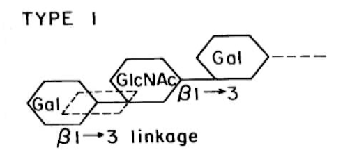
Type I β1→3 linkage
What type of linkage does soluble ABH Ags have?
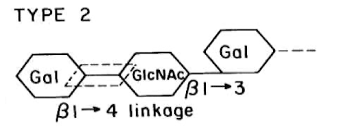
Type II β1→4 linkage
What type of linkage does ABH Ags on RBCs have?
RBCs - Glycolipids
Soluble - Glycoproteins
Basic backbone for RBC Ags vs soluble Ags
FUT 1 (by H gene)
Gene that controls the 2-L-fucosyltransferase in RBC antigens
3-6 months (reverse typing only by 6 month)
10
ABO antibodies are naturally occurring, first appearing by (——), and adult levels are reached by age (——)
groups A and B: IgM
group O: primarily IgM, anti-A,B = IgG
How do the Ab types differ based on blood group?
Intravascular hemolysis
ABO Abs can bind C’, causing (——)
Unbranched straight: H1 and H2
Complex branched: H3 and H4
Describe the different forms of H ag
H1 → Aa Ag
H2 → Ab Ag
A1 enzyme > A2 enzyme
The straight chain H’s correlate with which type A subgroup? By which enzymes?
H3 → Ac Ag
H4 → Ad Ag
A1 enzyme is ok, A2 very poorly
The complex H’s correlate with which type A subgroup? By which enzymes?
Forward grouping
Lectins (degree of Ag expression)
Transferase activity
Reverse grouping
Adsorption-elution tests
Saliva studies
Family/pedigree studies
How are ABO subgroups differentiated? (7)
Bombay Oh (Hnull)
Absence of H, A, and B Ags on RBCs due to the mutation of Fut-1 (H) gene producing a silenced gene incapable of coding for the H transferase. No agglutination with reagents anti-A, anti-B, or H lectin
⍺-2-L-fucosyltransferase
N-acetylgalactosaminyl transferase and D-galactosyl transferase
Bombay individuals lack (——) enzymes in their serum, but they can have (—2—) in their serum depending on their ABO genotype
H-deficient, nonsecretor (Bombay Oh)
(H deficient phenotypes) genetically homozygous for nonfunctional H (hh) and Se (sese) genes (no Fut1, no Fut2)
H-deficient, secretor (para-bombay Oho, OhA, OhB, OhAB) or H+w
(H deficient phenotypes) Genetically homozygous for nonfunctional H (hh), at least one functional Se gene. No Fut1, normal Fut2
H-partial or mostly deficient, nonsecretor (Para-Bombay Oh, Ah, Bh, ABh) or H+w
(H deficient phenotypes) The RBCs of these individuals have a slight amount of ABH Ags that are primarily detected by adsorption and elution studies. Mainly of European descent; Genetically weak variant H (hh) (weak Fut1) and sese
Septicemia
Gastrointestinal disorders
Carcinoma of the colon or rectum
Wound infections
Acquired B is usually found in type A patients who have… (4)
E. coli
Proteus vulgaris
Deacetylation of GalNac to D-Galactosamine (similar to D-galactose)
Acquired B can be caused by enzymes produced by certain strains of (—2—), which causes (—-)
Leukemia (e.g., CLL)
Hemolytic disease that induces stress hematopoiesis (e.g., thalassemia)
Hodgkin’s disease, malignant non-Hodgkin’s disease
Stomach carcinoma
Pancreas carcinoma
Immunodeficient disease (e.g., congenital agammaglobulinemia)
Diseases that cause weak ABO Ag expression (6)
Anti-A1 lectin
Anti-A,B
Anti-H
A1 and A2 reagent RBCs
With suspected A subgroups, test the blood with… (4)
Adsorption-Elution procedure
For suspected B subgroups, test the blood with…
Acquired B (B(A) phenotype due to disease)
Rouleaux
Polyagglutination
Wharton’s jelly
What causes extra Ags in forward grouping? (4)
Lower GI tract disease
Cancer of colon/rectum
Intestinal obstruction
Things that cause in-vivo acquired B (3)
Bacterial contamination (E.coli/P.vulgaris membranes have determinants similar to B Ag)
What causes in-vitro acquired B?
In-vivo = type A only
In-vitro = both type O and A
In which blood groups can acquired B happen?
Check ABO record
Check diagnosis for bacterial infection
If polyclonal used, retest ABO w/ monoclonal anti-A and anti-B
If monoclonal used, use manufacturer that doesn’t react w/ acquired B
Ensure autologous control is negative
Test RBCs with ant-B reagent at pH=6.0
(Usually not necessary) If secretor, check secretor studies
Resolutions for acquired B phenomenon (7)
Type A (or O) washed red cells (remove traces of donor anti-B)
What should group A acquired B patients receive for their transfusion?
Polyagglutination
Agglutination of RBCs with human antisera no matter what blood type due to either bacterial infection or expression of hidden T antigens react with antisera
Rouleaux
Stacking caused by extra serum proteins; occurs in diseases such as multiple myeloma, macroglobulinemia, or can be caused by infusion of macromolecular substances such as dextran or polyvinyl pyrrolidone (PVP), which are used as blood volume expanders
Wharton’s Jelly
Gelatinous substance derived from connective tissue that is found in cord blood and may cause false agglutination (remember: only forward typing is performed on cord blood)
Group O transfusion
Transplant (bone marrow/stem cell recipient)
A3 phenotype
Mixed field agglutination reactions are caused by… (3)
Newborns
Elderly
Hypogammaglobulinemia
What causes weak or missing Abs in reverse typing (3)
Determine pt age, medical diagnosis
Incubate serum for 15 min (RT)
If negative, place serum testing at 4°C for 5 min with autologous control (mini-cold panel)
How to resolve weak or missing Abs in reverse typing (3)
Cold Abs (I, H, M, N, P, Le)
Rouleaux
Anti-A1 in an A2 or A2B individual
Transfusion of incompatible plasma
IV Ig infusion
What causes extra Abs in reverse typing (5)
Cold auto adsorption of specimen (break IgM bonds with 2-ME or DTT)
Resolution for cold Abs (extra Ab in reverse typing)
Type patient RBCs with anti-A1 lectin
Repeat reverse group with A2 cells
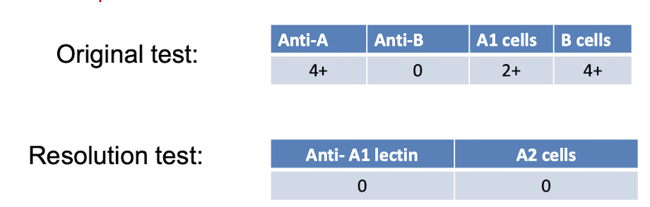
How to resolve Anti-A1 discrepancy (2)
Test with anti-H lectin
Resolution for typing Bombay phenotype
Multiple myeloma
Waldenstrom’s macroglobulinemia (WM)
Hydroxyethyl starch (HES), dextran, etc.
Rouleaux are associated with which conditions? (3)
Forward: wash cells
Reverse: saline replacement technique
How to resolve a typing discrepancy caused by rouleaux (2)
Chimerism (transfusion, transplant, feto-maternal hemorrhage)
Newborn infant
Elderly
Hypogammaglobulinemia
Which conditions cause weak reacting or missing antibodies? (4*)
Subgroups of A or B
Leukemia (excess of B)
Acquired B phenomenon (septicemia, intestinal obstruction, cancer of colon/rectum
Which conditions cause weak reacting or missing antigens (3*)
Elevated globulin level (MM, WM, plasma dyscrasias, Hodgkin lymphoma)
Plasma expanders (dextran, polyvinyl pyrrolidone)
Wharton’s jelly
Which conditions cause protein/plasma abnormalities leading to rouleaux formation (3*)
Exposure of hidden erythrocyte T antigen (polyagglutination)
Cold and warm autoantibody (AIHA)
Transfused foreign antigen
Unexpected ABO iso-agglutinin and alloantibody
Antibody other than anti-A and anti-B (e.g., acriflavin Ab)
cis-AB individuals
Miscellaneous causes for discrepancies in typing (6)
Incorrect ID of specimen/tubes/slides
Mix-up in samples
Cells suspension too heavy/light
Clerical/recording error of results
Failure to add reagent
Failure to add sample
Missed observation of hemolysis
Uncalibrated centrifuge
Over/under centrifugation
Contaminated reagents
Warming during centrifugation
Common technical errors resulting in ABO discrepancies (11)
Lyophilized powedered thrombin
Pink top tube
Serum that does not clot may be due to low platelet count, anticoagulant therapy, or factor deficiencies; serum that does not clot completely before testing is prone to developing fibrin clots that may mimic agglutination. How is this fixed? (2)
C’ binding (anti- A, B, H, Lea, Jka, etc.)
Bacterial contamination
Physical causes (water, mechanical strain, extreme temperature)
Causes for hemolysis detected in serum/plasma after centrifugation (3)