Cell Bio Exam III Guide
 Call Kai
 Learn
 Practice Test
 Spaced Repetition
 Match
 Flashcards
 Knowt Play
1/86

There's no tags or description
Looks like no tags are added yet.
87 Terms
Cell Signaling
Cell releases a signaling molecule called a ligand which binds to a receptor on another cell, the receptor becomes activated, proteins inside the cell relay the message, and the cell produces a response - this facilitates communication of cells between one another
Ligand
Signaling molecule that binds to a receptor → activates response pathways within the cell
Receptor
Protein that receives the ligand signal
Effector
Protein that becomes activated by the receptor/signaling activity
Second Messenger
Small intracellular molecules that relay the activated message/signal to downstream proteins
Target Protein
Protein that carries out the cellular signal response
Endocrine Signaling
A cell releases hormones into the bloodstream, they travel a distance to target cells → ie. insulin traveling through the blood stream

Paracrine Signaling
A cell releases signaling molecules that bind to nearby cell sites → growth factors acting on neighboring cells

Autocrine Signaling
A cell releases a signal that then binds to the receptors on itself → Immune cells releasing signals that stimulate themselves

Synaptic Signaling
A neuron releases cell signals across a synapse (gap between nerves) to communicate with a target cell → ie. neuron to neuron communication
Ligand Binding and Disassociation Constant
Kd = [R][L]/[R*L] → tells you how strongly ligand binds to its target receptor
R = unbound receptors
L = unbound ligand
R*L = receptor-ligand complex
High Kd
Weak binding → ligand and receptor associate less strongly
Low Kd
Strong binding → ligand and receptor associate strongly
[L] = Kd
At this stage, when the concentration of unbound ligand equals the disassociation content, it signals 50% of ligand are occupied by receptors
cAMP
Second messenger protein made by adenyl cyclase and activates PKA
cGMP
Second messenger protein made by guanylyl cyclase and activates downstream targets, including PKG → can regulate ion channels
IP3
Second messenger protein made by phospholipase C and releases Ca2+ from the ER
DAG
Second messenger protein made by phospholipase C and activates PKC
cAMP Signaling Pathway
Ligand binds to a G coupled protein receptor (GCPR), the G protein activates adenylyl cyclase, adenylyl cyclase converts ATP to cAMP, cAMP activates protein kinase K (PKA), PKA phosphorylates downstream target proteins, and these proteins generate a cellular response

IP3 Pathway
An enzyme called phospholipase C (PLC) breaks down membrane phospholipids and releases two second messengers. This one travels through the cytoplasm, binds to receptors on the ER, opens calcium channels to be released into the cytoplasm, and cytosolic calcium levels increase → this can bind to and activate calmodulin and activate PKC

DAG Pathway
An enzyme called phospholipase C (PLC) breaks down membrane phospholipids and releases two second messengers. This one remains associated with the membrane, and activates protein kinase C (PKC) → PKC then phosphorylates other proteins

G Protein Coupled Receptors (GPCR)
Membrane proteins that communicate with trimeric G proteins → trimeric G proteins obtain an alpha, beta, and gamma subunit
GPCR Activation
A signaling molecule binds to the GPCR, the receptor activates the G protein, GDP on the alpha subunit is converted to GTP, the alpha subunit separates from the beta-gamma complex, the activated subunits interact with effector proteins, secondary messengers are produced, and the cell generates a response → G proteins are activated when GDP is converted to GTP, and they inactivate when GTP is hydrolyzed to GDP
Gas
G protein whose effector is adenylyl cyclase and produces cAMP
Gaq
G protein who’s effector is PLC, and produces IP3 and DAG
Gai
G protein that can regulate cGMP phosphodiesterase and produces cGMP regulation
Gas Inactivation
HYdolyzes GTP into GDP and the signaling pathways shut down
Phosphodiesterase (PDE)
Enzyme that breaks down and inactivates the cAMP pathway → converts cAMP into AMP, less cAMP means less PKA activation, and signaling responses decrease (cAMP DOWN)
B-arrestin
Binds to the activated receptor in GCPR’s and contributes to receptor desensitization → prevents signaling (receptor inhibition)
Prolonged Exposure to Enephrin
Continuous exposure causes desensitization of B2-adrenoreceptors and Gas-coupled receptors → the cell becomes less responsive to the ongoing signal
Receptor Tyrosine Kinase
A membrane receptor that becomes activated when a growth factor binds to it → signal growth, division, survival, and differentiation
How RTKs Work
A growth factor binds to an RTK, two receptor molecules come together - this is called dimerization, the kinase domains become activated, the receptors autophosphorylate tyrosine residues on each other, these residues act as docking sites for signaling proteins, proteins with SH2 domains bind to these sites (protein domain that recognizes and binds to phosphorylated tyrosines), downstream signaling pathways become activated
HER 1,3,4 Receptors
All RTK’s that bind to ligands. After ligand binding and dimer forming, it creates an active kinase domain dimer → after activation, phosphotyrosine residues act as binding surfaces for many proteins with their SH2 domains

HER2
An RTK that cannot bind to any ligand but rather forms heterodimers with HER1 HER3 and HER4
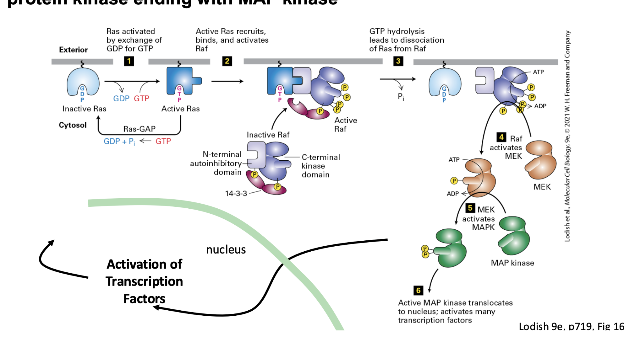
Ras/MAP Kinase Signaling Pathway
Ras (binding protein activated by exchange of GDP for GTP) → Raf (protein kinase activated downstream by Ras) → Mek (kinase activated by Raf) → MAP Kinase (Activated by Mek and translocates to the nucleus to activate many transcription factors) → Activation of transcription factors → gene transcription in the nucleus
Ras/MAP Kinase Purpose
Carries a signal from a growth factor bound to a receptor on the cell surface all the way to the nucleus, where it can change gene expression
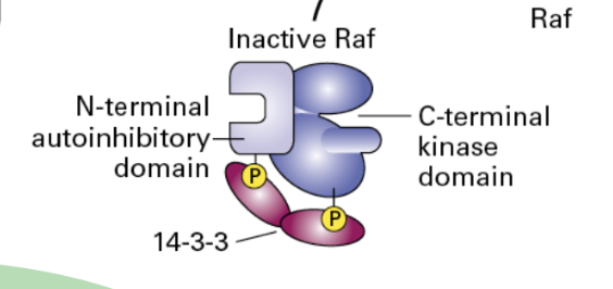
Inactive Raf
N-terminal is auto inhibitory domain, and C-terminal is the kinase domain
Erythropoietin
A cytokine-like signaling protein that regulates red blood cell production
Short Term Regulation
In order to stop signal transduction via the erythropoietin receptor JAK2 kinase gets deactivated by detachment of SHP1

Long Term Regulation
Newly produced SOCS proteins shut down signaling by binding to JAK kinases and inhibiting their activity, binding to cytokine receptors and preventing STAT recruitment, and recruiting ubiquitin ligases that target signaling proteins for degradation

JAK/STAT
A cytokine binds to its receptor, receptor associated JAK proteins become activated, JAK phosphorylates the receptor and STAT proteins, STAT proteins bind to the phosphorylated receptor, STAT proteins become phosphorylated and form dimers, STAT proteins then enter the nucleus and influence gene transcription
TGF-Beta/Smad Pathway
A process involved in regulating cell growth and differentiation. First TGF-Beta binds to its receptor, the receptor complex activates the type I receptor (RI), activated RI phosphorylates Smad2 and Smad3, Smad2/3 (R Smads) associate with Smad4 (co-Smad)and generate the Smad complex, the Shad complex enters the nucleus and regulate gene transcription

Inhibitory Smad(s)
Smad7 - binds to TGF-Beta receptor site → Ski and Snow also inhibit TGF-Beta/Smad signaling - binds to Smad complex in the nucleus
Notch Signaling
A contact dependent pathway where the ligand on one cell binds to a receptor on the neighboring cell. The notch receptor is cleaved. Its intracellular portion (NICD) enters the nucleus, and regulates gene expression

Wnt Signaling
Wnt binds to frizzled, this process inhibits the B-catenin destruction pathway and B-catenin accumulates. From here, B-catenin enters the nucleus, and gene transcription is activated

Without Wnt
B-catenin is targeted for degradation - a destruction complex forms to mark B-catenin for breakdown. Therefore, levels of B-catenin remain low → gene transcription for Wnt target genes do not get activated

Hedgehog Signaling
…
Inactive NF-kB
It is composed of p65 and p50 subunits, it is bound to its inhibitor IkBa, and this prevents NF-kB from entering the nucleus and activating genes.
NF-kB Signaling Pathway
Pathway for immunoresponse (B cells) and inflammation. An inflammatory factor such as TNF-a or IL-1 activate this pathway. IkBa becomes phosphorylated and an E3 adds a ubiquitin to the IkBa - now IkBa is targeted for protein degradation. Here, NF-kB (p65 and p50) is released, enters the nucleus, and permits gene transcription

Proteosomal Degradation
Signaling pathways that have proteins who obtain the ability to be targeted for degradation and influence how the rest of the cycle proceeds.
Wnt → B-catenin targeted → inhibits Wnt signaling
Hedgehog → signaling components targeted → regulates pathway activity
NF-kB → IkBa targeted → releases NF-kB and activated gene transcription in the nucleus
Cell cycle → cyclins and regulators targeted → controls cycle progression
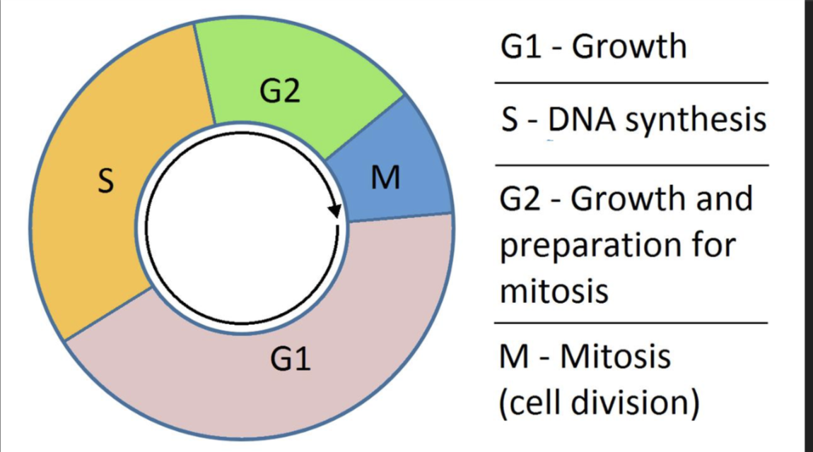
The Cell Cycle
G1: Growth, S: DNA is replicated, G2: Cells prepares for division and checks for proper DNA replication, M: Mitosis and cell division
CDKs
Cyclin dependent kinases that regulate progression through the cell cycle by phosphorylating target proteins → need cyclins to become active
Cyclins
Determine CDK activity → 4 types: G1 cyclins, G1/S cyclins, S cyclins, and Mitosis cyclins. Their activity rises before they are needed, and fall after their job is completed
CDK1 and Cyclin B
Needed during M phase/mitosis → controls progression into mitosis
CDK2 and Cyclin E/Cyclin A
Needed for G1 to S phase transition and S phase → Helps initiate DNA replication and regulate S phase progression

CDK4/6 and Cyclin D
Needed for G1 phase → helps the ell progress through G1, towards the G1/S transition
Whi5 Activity
A transcription repressor in yeast that prevents transcription of genes needed for progression into S phase (inhibits) → the cell DOES NOT proceed into S phase

Inactive Whi5
When this is phosphorylated, it is inactive and genes required to push the cell into S phase are transcribed → the cell DOES move forward into S phase

Rb Activity
A regulatory protein that acts as a tumor repressor in metazoans (multicellular animals). When active, it binds to the transcription factor E2F. This binding prevents E2F from activating genes required for S phase progression. Therefore, the cell cycle is restricted there
Rb Inactivity
When phosphorylated, this becomes inactive. From here, E2F is released and activates the genes needed for S phase progression → cell cycle proceeds towards DNA replication
Rb Mutation
When it does not function properly, it cannot properly restrain E2F and the cell will proceed uncontrollably through S phase into mitosis. Uncontrollable division can lead to tumor formation → ie. retinoblastoma, a tumor that develops in the eye
APC/C
Anaphase promoting complex/cyclosome. This is an E3 ubiquitin ligase whose job is to attach ubiquitin to certain cell cycle regulatory proteins and target them for degradation → specializes in sister chromatid segregation, exit from mitosis, and degradation of cyclins and other regulatory proteins
Protein Targeted by APC/C
APC/C adds ubiquitin to it, the protein is recognized by the proteasome, the protein is degraded, and its regulatory activity decreases
Cdc14 and Cdh1
An APC/C pathway example in yeast. Cdc14 has the power to activate Cdh1 by dephosphorylating it. When active, Cdh1 helps the APC/C target S-phase and mitotic cyclins for degradation

Sic1 and the Start of S-Phase
A cyclin dependent kinase inhibitor (CKI). Before S phase it binds to and inhibits S-phase CDK - DNA replication is prevented. To promote S-phase, G1/S CDK phosphorylate this so it can be targeted for degradation by SCF E3 ubiquitin ligase. S-phase CDK is released from inhibition and DNA replication proceeds (Sic1 is a brake on S-phase CDK. Destroying Sic1 releases the brake)

G2 Arrest
Incomplete DNA replication holds the cell from proceeding through mitosis
Cdc25-phosphatase
Removes inhibitory phosphate groups from CDK1 and activates it → promoting cell entry into M phase (X)
Wee1 Kinase
Adds inhibitory phosphate groups to CDK1 → preventing premature cell entry into M phase (Y)

Cohesins
Proteins that hold sister chromatids together
Separase
Enzyme that cleaves cohesin proteins so that sister chromatids can separate during anaphase → and removes remaining cohesins from the centromere
Shugosin (Sgo1) and PP2A
During G2 Sgo1 recruits PP2A to the centromere on sister chromatids. This helps protect centromeric cohesins. All together they prevent premature separation prior to anaphase
Plk1 and Aurora B
Proteins that phosphorylate cohesins and remove them from sister chromatid arms to enable separation
ATM and ATR
Protein kinases involved in responding to DNA damage or replication problems → signal responses that STOP cell cycle progression
p21
A cyclin dependent kinase inhibitor (CKI) whose job is to inhibit CDK activity and arrest the cell cycle
Cdc25
Activates CDKs through dephosphorylation → when checkpoint signaling inhibits this, CDKs remain inactive and prevent cell cycle progression
p53
A tumor repressor protein that can stop the cell cycle, allow time for DNA repair, or trigger apoptosis within a cell
Stem Cells
Cells that have the ability to self renew - divide and produce more, and pluripotentency - the ability to differentiate into many different cell types
Embryonic Stem Cells
Found in the inner mass of the blastocyte. The bastocyte is an early developmental stage that occurs around 4-7 days after fertilization - they are pluripotent
Induced Pluripotent Stem Cells
Adult somatic cells that have been reprogrammed into a pluripotent state - they are pluripotent
Adult Stem Cells
Found in tissues throughout the body
Pluripotent
Term meaning a stem cell can develop into an array of three primary germ layers - ectoderm, mesoderm, or endoderm (can also generate cells from all three layers)
Ectoderm
Encompasses nervous system and epidermis
Mesoderm
Encompasses muscle, bones, and blood
Endoderm
Encompasses digestive and respiratory tract linings
Stem Cell Niches
The environment that keeps certain stem cells functioning instead of differentiating Maintain self-renewal → prevent unwanted differentiation, regulate stem-cell activity, and help control when stem cells divide and differentiate
Multipotent
Can produce several related cell types - via stem cells
Unipotent
can produce only one particular cell type - via stem cells