RAD 265 Sectional Anatomy Unit 1 Exam
 Call Kai
 Learn
 Practice Test
 Spaced Repetition
 Match
 Flashcards
 Knowt Play
1/301

Earn XP
Description and Tags
intro to sec. anat., cranium, facial bones, and brain.
302 Terms
the fleshy part of the hand at the base of the thumb
thenar
pertaining to the palm of the hand or flexor surface of wrist or sole of foot
volar
toward the nose
rostral
posterior surface of elbow area of the arm
cubital
what is the location of this internal landmark: aortic arch
2.5 cm below jugular notch
what is the location of this internal landmark: aortic bifurcation
L4-L5
what is the location of this internal landmark: carina
T4-T5, sternal angle
what is the location of this internal landmark: carotid bifurcation
upper border of thyroid cartilage
what is the location of this internal landmark: celiac trunk
4 cm above transpyloric plane
what is the location of this internal landmark: circle of willis
suprasellar cistern
what is the location of this internal landmark: common iliac vein bifurcation
upper margin of sacroiliac joint
what is the location of this internal landmark: conus medullaris
T12-L1, L2
what is the location of this internal landmark: heart; apex
5th intercostal space
what is the location of this internal landmark: heart; base
level of 2nd and 3rd costa cartilages behind sternum
what is the location of this internal landmark: inferior mesenteric artery
4 cm above bifurcation of abdominal aorta
what is the location of this internal landmark: inferior vena cava
L5
what is the location of this internal landmark: portal vein
posterior to pancreatic neck
what is the location of this internal landmark: renal arteries
anterior to L1, inferior to superior mesenteric artery
what is the location of this internal landmark: superior mesenteric artery
2 cm above transpyloric plane
what is the location of this internal landmark: thyroid gland
thyroid cartilage
what is the location of this internal landmark: vocal cords
midway between superior and inferior border of thyroid cartilage
what is the location of this external landmark: thyroid cartilage
C5
what is the location of this external landmark: jugular notch
T2-T3
what is the location of this external landmark: sternal angle
T4-T5
what is the location of this external landmark: xiphoid process
T10
what is the location of this external landmark: costal margin
L3
what is the location of this external landmark: level of umbilicus
L3-L4
what is the location of this external landmark: iliac crest
L4
what is the location of this external landmark: ASIS
S1
Coronal images are displayed as if
The viewer and patient are facing the same direction.
The patient is facing right or left.
The viewer and patient are face to face.
The patient is behind the viewer.
The viewer and patient are face to face.
The inguinal region refers to the:
cheek of the face
buttocks
groin area
posterior elbow
groin area
The midpoint of the shades of gray displayed in a digital image is called the:
window width
window level
image matrix
pixeling
window level
The number of shades of gray displayed in a digital image is called the:
window width
window level
image matrix
pixeling
window width
The regional term for the forearm is:
brachial
antebrachial
axillary
cubital
antebrachial
The term that is used interchangeably for anterior is:
dorsal
posterior
ventral
antecubital
ventral

What is letter A
(anterior view of skull)
temporal bone

what is letter F
(anterior view of skull)
sphenoid bone (greater wing)

what is letter J
(lateral view of skull)
occipital bone

what is letter L
(lateral view of skull)
external auditory meatus

what is letter E
(superior view of cranial fossae)
sphenoid bone

what letter is G
(superior view of cranial fossae)
ethmoid bone

what is letter M
(superior view of cranial fossae)
foramen ovale

what is letter D
(lateral view of inner skull)
ethmoid bone
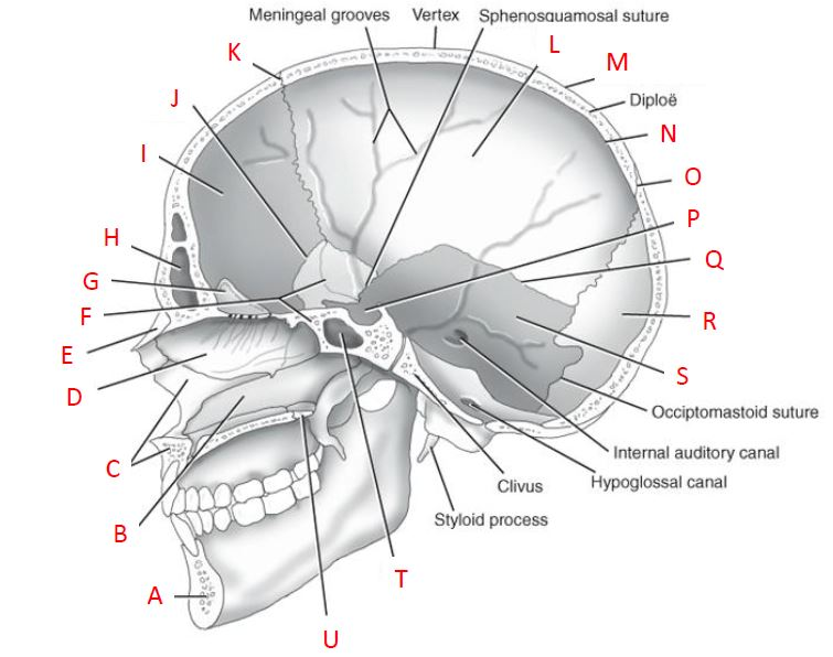
what is letter G
(lateral view of inner skull)
crista galli

what is letter K
(lateral view of inner skull)
coronal suture

what is letter L
(lateral view of inner skull)
parietal bone

what is letter N
(lateral view of inner skull)
internal table

what is letter P
(lateral view of inner skull)
sella turcica

what is letter B
(sagittal view of facial bones)
ethmoid bone

what is letter C
(sagittal view of facial bones)
sphenoid bone

what is letter D
(sagittal view of facial bones)
frontal bone

what is letter E
(sagittal view of facial bones)
parietal bone

what is letter F
(sagittal view of facial bones)
occipital bone

what is letter H
(sagittal view of facial bones)
mandibular condyle

what is letter I
(sagittal view of facial bones)
angle (gonion)

what is letter J
(sagittal view of facial bones)
mandibular notch

what is letter K
(sagittal view of facial bones)
coronoid process

what is letter A
(coronal CT of frontal bone)
perpendicular plate of ethmoid

what is letter B
(coronal CT of frontal bone)
nasal bone

what is letter C
(coronal CT of frontal bone)
frontal sinus

what is letter D
(coronal CT of frontal bone)
maxilla

what is letter A
(coronal CT of osteomeatal unit)
lateral wall of orbit
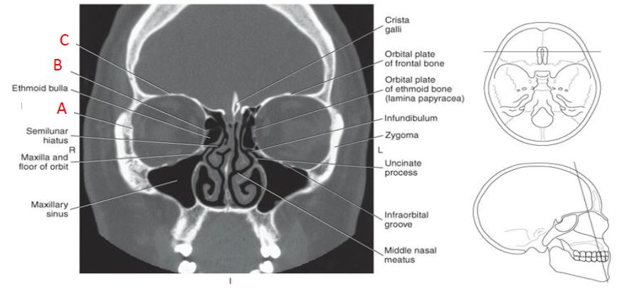
what is letter B
(coronal CT of osteomeatal unit)
medial wall of orbit
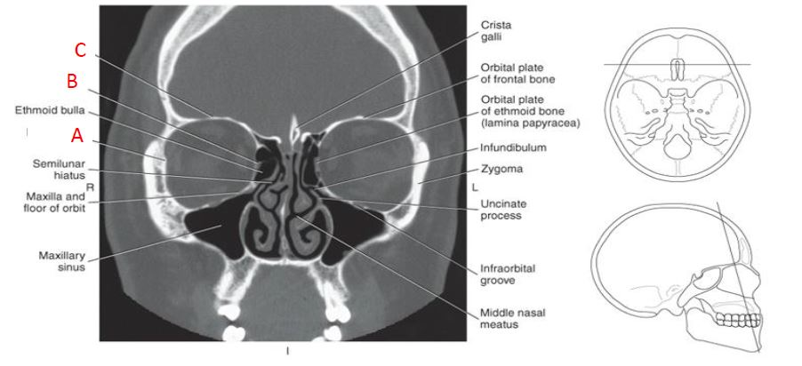
what is letter C
(coronal CT of osteomeatal unit)
roof of orbit
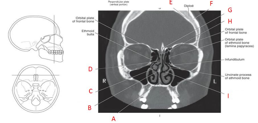
what is letter G
(coronal CT of ethmoid bone with crista galli)
crista galli

what is letter H
(coronal CT of ethmoid bone with crista galli)
cribform plate

what is letter I
(coronal CT of ethmoid bone with crista galli)
middle nasal meatus

what is letter A
(coronal CT of sphenoid bone)
pterygoid hamulus on medial pterygoid płate

what is letter C
(coronal CT of sphenoid bone)
greater wing of sphenoid bone

what is letter D
(coronal CT of sphenoid bone)
anterior clinoid process of sphenoid bone

what is letter E
(coronal CT of sphenoid bone)
sphenoid sinus

what is letter G
(coronal CT of sphenoid bone)
ptergoid process (lateral pterygoid plate)

what is letter A
(coronal CT of mandibular rami)
pterygoid process of sphenoid bone (lateral plate)

what is letter B
(coronal CT of mandibular rami)
greater wing of sphenoid bone

what is letter C
(coronal CT of mandibular rami)
anterior clinoid process
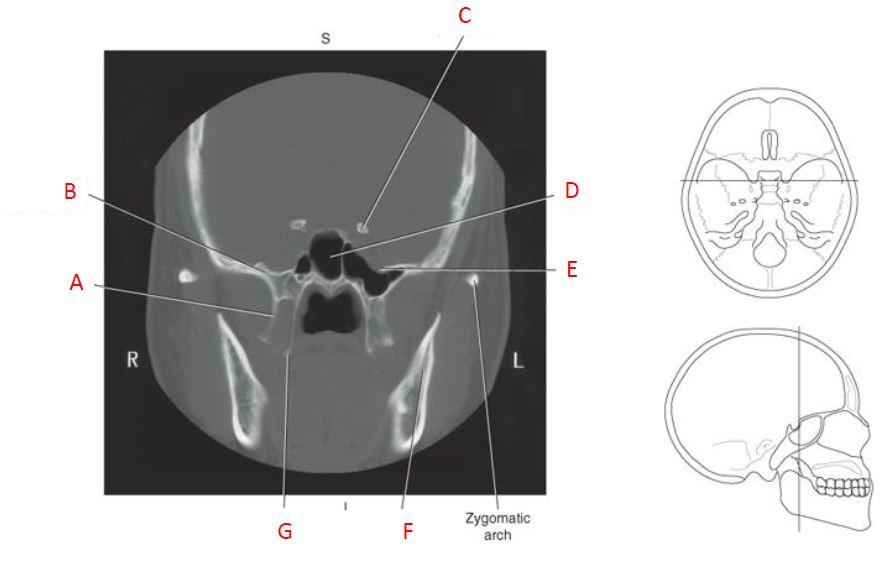
what is letter D
(coronal CT of mandibular rami)
sphenoid sinus

what is letter E
(coronal CT of mandibular rami)
foramen rotundum

what is letter G
(coronal CT of mandibular rami)
pterygoid hamulus of sphenoid bone

what is letter A
(coronal CT of dorsum sella)
parietal bone

what is letter B
(coronal CT of mandibular rami)
posterior clinoid process

what is letter C
(coronal CT of mandibular rami)
temporal bone
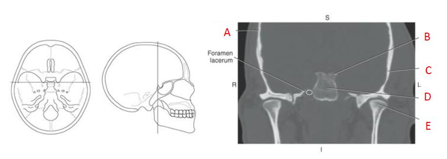
what is letter D
(coronal CT of mandibular rami)
dorsum sella

what is letter E
(coronal CT of mandibular rami)
condyle process of mandible

what is letter B
(sagittal CT reformat of temporal bone)
mastoid air cells

what is letter C
(sagittal CT reformat of temporal bone)
external auditory meatus

what is letter D
(sagittal CT reformat of temporal bone)
madibular condyle

what is letter A
(sagittal CT reformat of frontal sinus)
inferior nasal conchae

what is letter B
(sagittal CT reformat of frontal sinus)
ethmoid air cells
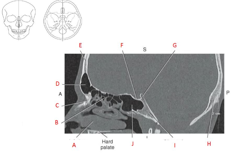
what is letter C
(sagittal CT reformat of frontal sinus)
nasal bone

what is letter D
(sagittal CT reformat of frontal sinus)
frontal sinus

what is letter E
(sagittal CT reformat of frontal sinus)
frontal bone
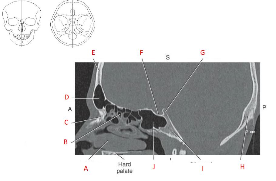
what is letter F
(sagittal CT reformat of frontal sinus)
sella turcica

what is letter G
(sagittal CT reformat of frontal sinus)
dorsum sella

what is letter J
(sagittal CT reformat of frontal sinus)
sphenoid sinus

what is letter A
(axial CT of coronal suture)
squamous portion of temporal bone

what is letter B
(axial CT of coronal suture)
frontal bone

what is letter C
(axial CT of coronal suture)
coronal suture
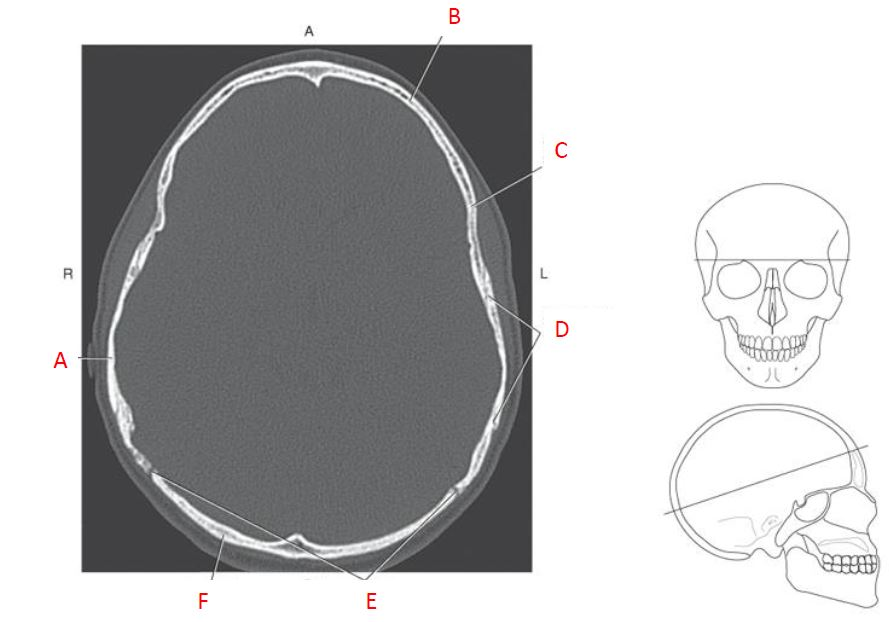
what is letter D
(axial CT of coronal suture)
squamous suture

what is letter E
(axial CT of coronal suture)
lambdoidal suture

what is letter F
(axial CT of coronal suture)
occipital bone