Nervous System BIOL 3310 Anatomy and Physiology 1 Lecture Exam 3
 Call Kai
 Learn
 Practice Test
 Spaced Repetition
 Match
 Flashcards
 Knowt Play
1/146

Earn XP
Description and Tags
147 Terms
Endocrine system
communicates by means of chemical messengers, hormones, secreted into the blood
Nervous system
employs chemical and electrical means to send messages from cell to cell
What are the steps the nervous system carries out?
receive information, process information, issue commands
Central nervous system (CNS)
brain and spinal cord enclosed in bony covering
Peripheral nervous system (PNS)
all nervous system except for brain and spinal cord, composed of nerves and ganglia
Nerves
bundle of nerve fibers (axons) wrapped in fibrous connective tissue
Ganglion
knot-like swelling in a nerve where neuron cell bodies are concentrated
Sensory (afferent) division of PNS
carries sensory signals from various receptors to the CNS
informs the CNS of stimuli within or around the body
Somatic sensory division
carries signals from receptors in the skin, muscles, bones, and joints
Visceral sensory division
carries signals from the viscera of the thoracic and abdominal cavities
Motor (efferent) of PNS
carries signals from CNS to motor gland and muscle cells that carry out the body’s response
Somatic motor division
carries signals to skeletal muscles
output produces muscular contractions
Somatic reflexes
involuntary muscle contractions
Visceral motor division (autonomic nervous system)
carries signals to glands, cardiac muscle, and smooth muscles
involuntary, and responses of the system and its receptors are visceral reflexes
Sympathetic division
tends to arouse the body for action
accelerating heart beat and respiration, while inhibiting digestive and urinary systems
Parasympathetic division
tends to have calming effect
slows heart rate and breathing
stimulates digestive and urinary systems
Excitability (irritability)
response to stimuli
Conductivity
neurons response to stimuli by producing electrical signals that are quickly conducted to other cells at distant locations
Secretion
electrical signals reach the end of nerve fibers, chemical neurotransmitter is secreted that crosses the gap and stimulates the next cell
Sensory (afferent) neurons
specialized to detect stimuli
transmit information about them to the CNS
begin in almost every organ in the body, end in CNS
Afferent
conducting signals towards the CNS
Interneurons (association) neurons
lie entirely within the CNS
receive signals from many neurons and carry out the integrative function
90% of all neurons
lie between and interconnect the incoming sensory pathways, and the outgoing motor pathways of the CNS
Integrative function
process, store and retrieve information and make decisions that determine how the body will respond to stimuli
Motor (efferent) neuron
send signals out to muscle and gland cells
Effectors
muscle and gland cells
Motor
lead to muscle
Efferent
conduct signals away from CNS
Soma (neurosoma, cell body, perikaryon)
single, centrally located nucleus with large nucleolus
What is included in the cytoplasm of a neuron?
contains mitochondria, lysosomes, Golgi complex, numerous inclusions, extensive rough endoplasmic reticulum and cytoskeleton
What is the cytoskeleton of a neuron comprised of?
dense mesh of microtubules and neurofibrils
Neurofibrils
bundles of actin filaments
Nissl bodies
compartments of rough ER, dark staining
What does the soma of a neuron lack?
centrioles, cannot divide cellularly
Inclusions
glycogen granules, lipid droplets, melanin, and lipofuscin
Lipofuscin
golden brown pigment produced when lysosomes digest worn-out organelles
What happens to lipofuscin with age?
accumulates, act as wear-and-tear granules, most abundant in old neurons
Dendrites
vast number of branches coming from a few thick branches from soma
What is the primary site for receiving signals from other neurons?
dendrites
Axon (nerve fiber)
originates from axon hillock
Axon hillock
mound on one wide of the soma
What is the shape of an axon?
cylindrical, relatively unbranched for the majority of its length
Axon collaterals
branches of axon
Axoplasm
cytoplasm of axon
Axolemma
plasma membrane of axon
How many axons can one neuron have?
one
What encloses an axon?
schwann cells and myelin sheath
Terminal arborization
distal end of axon, extensive complex of fine branches
Synaptic knob (terminal button)
little swelling that forms a junction with the next cell
Synaptic vesicles
inside synaptic knob, contain neurotransmitter

Multipolar neuron
one axon, multiple dendrites
most common
most neurons in brain and spinal cord (CNS)
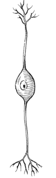
Bipolar neuron
one axon, one dendrite
composes olfactory cells, retina, inner ear

Unipolar neuron
single process leading away from soma
sensory from skin and organs to spinal cord

Anaxonic neuron
many dendrites but no axon
aid in visual processes
Axonal transport
two-way passage of proteins, organelles, and other material along an axon
Anterograde transport
movement down an axon away from soma
Retrograde transport
movement up an axon towards soma
Microtubules
guide materials along axon
Fast axonal transport
20-400mm/day
Fast anterograde transport
up to 400mm/day
transports organelles, enzymes, synaptic vesicles, small molecules
Fast retrograde transport
recycled materials and pathogens, rabies, herpes simplex, tetanus, polio
delay between infection and symptoms is time needed for transport up the axon
Slow axonal transport (axoplasmic flow)
0.5-10mm/day
always anterograde
moves enzymes, cytoskeletal components, new axoplasm down the axon during repair and regeneration of damaged axons
damaged nerve fibers regen at pace of slow axonal transport
Neuroglia (glial cells)
support and protect the neurons
bind neurons together and form framework for nervous tissue
covers neurons not in contact with other neurons to prevent incorrect signaling
Which neuroglia are found in the CNS?
oligodendrocytes, ependymal cells, microglia, astrocytes
Which neuroglia are found in the PNS?
schwann cells, satellite cells
Oligodendrocytes
form myelin sheaths in CNS
each arm-like process wraps around a nerve fiber forming an insulating layer that speeds up signal conduction
Ependymal cells
lines internal cavities of the brain
cuboidal epithelium with cilia on apical surface
secretes and circulates cerebrospinal fluid
Microglia
small, wandering macrophages formed with white blood cells called monocytes
thought to perform a complete checkup on the brain tissue several times a day
wander in search of cellular debris to phagocytize
Astrocytes
most abundant glial cell in CNS
cover entire brain surface and most non synaptic regions of the neurons in the gray matter of the CNS
What are the functions of astrocytes?
form supportive framework for nervous tissue
have extensions that contact blood capillaries that stimulate them for form a tight seal
convert blood glucose to lactate and supply nourishment
secrete nerve growth factors
communicate electrically with neurons and may influence synaptic signaling
regular chemical composition of tissue fluid by absorbing excess neurotransmitters and ions
astrocytosis/sclerosis
Perivascular feet
extensions of astrocytes that form tight seal on blood capillaries
Blood-brain barrier
seal astrocytes form between sarcoplasm and blood capillaries from rest of the body
Lactate
nourishes neurons
Nerve growth factors
secreted by astrocytes, promote neuron growth and synapse formation
Astrocytosis (sclerosis)
hardened scar tissue that fills space formally occupied by now damaged neuron
Schwann cells
envelope nerve fibers in PNS, wind repeatedly around a nerve fiber to form myelin sheath
assist in regeneration of damaged fibers
Satellite cells
surround the neurosomas in ganglia of PNS
provide electrical insulation around the soma
regulate chemical environment of the neurons
Brain tumors arise from
meninges
metastasis of non-neuronal tumors in other organs of the body
most come from glial cells that are mitotically active through life
Gliomas
rapid growing, highly malignant
cannot be treated with chemo, require direct radiation or surgery
Meninges
protective membrane of CNS
Myelin sheath
insulating layer around a nerve fiber
formed by oligodendrocytes in CNS and Schwann cells in PNS
consists of plasma membrane of glial cells, 20% protein, 80% lipid
Myelination
production of the myelin sheath
begins in 14th week of fetal development, proceeds rapidly during infancy and completed in late adolescence
What is the process of myelination in the PNS?
Schwann cells spiral around single nerve fiber, lay down ~100 layers of membrane
no cytoplasm between the layers
neurilemma thickest layer, contains nucleus and most of the cytoplasm and endoneurium
What is the process of myelination of the CNS?
oligodendrocytes myelinate several fibers nearby
cannot migrate
myelination spirals inward
no neurilemma or endoneurium
Nodes of Ranvier
gap between segments
Internodes
myelin covered segments from one gap to another
Initial segment
short section of nerve fiber between the axon hillock and the first glial cell
Trigger zone
the axon hillock and the initial segment
Multiple sclerosis
oligodendrocytes and myelin sheaths in the CNS deteriorate
myelin replaced by hardened scar tissue
nerve conduction disrupted
onset between 20-40, fatal 25-30 years post diagnosis
may be autoimmune triggered by virus
Tay-Sachs disease
hereditary disorder of infants, typically of eastern European Jewish ancestry
abnormal accumulation of glycolipid called GM2 in the myelin sheath
normally decomposed by lysosomal enzyme
enzyme missing in individuals homozygous for Tay-Sachs allele
accumulation of ganglioside (GM2) disrupts conduction of nerve signals
blindness, loss of coordination, and dementia
fatal before age 4
What factors influence the speed at which a nerve signal travels?
diameter of the fiber or presence/absence of myelin
How does the diameter of a fiber influence speed of conduction?
larger fibers have more surface area and conduct signals more rapidly
How does myelin influence speed of conduction?
speeds up signal conduction
Where are slow conduction speed signals found?
stomach and dilation of the pupils
Where are fast conduction speed signals found?
skeletal muscles and transport sensory signals for vision and balance
Speed of small, unmyelinated fibers
0.5-2.0m/sec
Speed of small, myelinated fibers
3.0-15.0m/sec
Speed of large, myelinated fibers
up to 120m/sec
What part of the nervous system is capable of regeneration?
PNS
What are the steps to nerve regeneration?
Axon distal to injury degrades, macrophages clean up
cell body swells, ER breaks up, nucleus moves off center
axon stump sprouts multiple growth processes
Schwann cell neurolemma, endoneurium, and basal lamina form a regeneration tube towards original destination
once contact is reestablished with original target, the neurosoma shrinks and returns to original appearance
How long does nerve regeneration take?
very slow, may take up to 2 years
some may connect with the wrong muscle fibers
damaged CNS axons usually unable to regenerate