CPPS 302 Cardiovascular System Cardiac arrhythmias
 Call Kai
 Learn
 Practice Test
 Spaced Repetition
 Match
 Flashcards
 Knowt Play
1/44

There's no tags or description
Looks like no tags are added yet.
45 Terms
What is overdrive suppression
The phenomenen when pacemaker cells with higher intrinsic firing rate suppress the automaticity of slower latent (ectopic) pacemaers, preventing them from firing independently
What happens to a latent (backup) pacemaker when it is forced to fire faster than its natural (intrinsic) rate
Its ability to generate its own electrical impulses becomes reduced (suppressed), especially after being forced to fire rapidly for a prolonged period
The backup (latent) pacemaker becomes temporarily less able to start its own heartbeat after following a faster pacemaker
What is the role of the Na/K ATPase pump in override suppression
Increased Na influx during frequent excitation increases Na/K ATPase activity. The pump moves 3 Na out and 2 K in, creating a net outward movement of positive charge and causing hyperpolarization
How does hyperpolarization suppress the automacity of latent pacemaker cells
Hyperpolarization makes the maximum diastolic potential more negative, needing more time for the If (funny current) to bring the memrane potential to threshold
What is the sequence of events involved in overdrive suppression
Higher frequency of excitation → increased Na influx → inreased Na/K ATPase activity → hyperpolarization → more negative maximum diastolic potential → longer time to reach threshold → decreased pacemaker automaticity
What happens to a laten pacemaker’s firing rate when its maxiumum diastolic potential becomes more negative (the most negative membrane potential a pacemaker cell reaches during its relaxation phase)
Its firing rate decreased because it takes longer for the membrane potential to reach threshold and initate another action potential
What is a cardiac re-entry loop, and what can it cause
A phenomenon where an electrical impulse re-excited cardiac myocytes it has already activated within the same cardiac cycle. It can cause tachycardia or fibrillation
What heart conditions are commonly associated with re-entry loops
Coronary arterial stenotic disease
Myocardial ischemia
Myocardial infarction
Accessory electrical pathways
What are the three essential conditions required for re-entry to occur
Closed conduction loop
unidirectional block
sufficiently slow conduction
What is a unidirectional block in a re-entry loop
A region where an electrical impulse can travel in one direction but not the opposite direction
Why is slow conduction necessary for re-entry to occur?
It gives previously activated cardiac cells enough time to recover from their refractory period, allowing the returning impulse to re-excite them
What happens during normal conduction versure abnormal conduction in a re-entry loop
Normal: impulses travel through both pathways, meet, and stop because the tissue is refractory
Abrnomal (re-entry): The impulse travles through the unblocked pathway, returns through the recovered blocked pathway, and re-excited the cells causing an extra beat

What is the relationship between the effective refractory period and propagation time for re-entry to occur
The effective refractory period must be shorter than the propagation time around the loop, allowing cells to recover before the impulse returns
What is a normal sinus rhythm, and what is its heart rate range
A cardiac rhythm controlled by the SA node, with a normal resting heart rate of 60-100 bpm
What is the difference between sinus bradycardia vs sinus tachycardia
Sinus bradychardia: Sinus rhythm with HR below 60 bpm
Sinud tachycardia: Sinus rhythm with HR above 100 bpm
What is an arrhythmia, and what is another name for it
An abnormal heart rhythm, also known as a dysrhythmia
What are the possible symptoms or consequences of cardiac arrhythmias
They range form benign palpitations (rapid or irregular heartbeats) to a drastic decrease in cardiac output
What is an escape beat, and how does it develop into an escape rhythm?
An escape beat occurs when a latent (backup) pacemaker initiates a heartbeat because the SA node fails or becomes too slow. Repeated escape beats produce an escape rhtyhm.
SA node fails to control heartbeat, so another pacemaker can take over to keep the heart beating
What is a junctional escape rhythm, and what can cause it
A rhythm where the AV junction takes over as the primary pacemaker because SA node automaticity is reduced.
Causes include:
SA node ischemia
Excessive parasympathetic activity. HR is typically 40-60 bpm
What is an ectopic beat
A heartbeat initiated by an electrical impulse originating somehwere other than the SA node, such as the AV junction or ventricles
In juncitonal escape rhythm, which two directions can the electrical impulse travel, and what are their efffects
Towards ventricles → ventricualr depolarization and contraction
Backward toward the atria → atrial depolarization, potentially producing an inverted P waves in lead II, III, aVF
What is a ventricular escape rhythm, and what typically causes it
A rhtyhm where a lower ventricular pacemaker takes over when impulses from the SA/AV nodal region fail to reach the ventricles, such as during complete AV block. It usually produces bradycardia
What ECG findings are associated with ventricular escape rhythm
Slow HR
P waves and QRS complex may be dissoaciated (AV dissociation)
Ofen a wide QRS complex when impulses spread slowly through ventricular myocardium rather than the normal conduction system
Why does a ventricular escape beat often produce a wide QRS complex
Becayse impulses originating in the ventiruclar myocardium spread through slower cell-to-cell conduction rather than the rapid His-Purkinje system, prolonging ventricular depolarization
Ventricular Tachycardia
Occurs when abnormal electrical impulses originating in the ventricles cause the heart to beat rapidly
What is ventricular tachycardia and what are its characteristic ECG findings
A rapid abnormal rhythm originating in the ventircles, typically producing a heart rate over 120 BPM and wide QRS complexes
What conditions can cause ventricualr tachycardia, and what mechanism may be involved
Ischemic heart disease or myocardial infarction can disrupt ventricular conduciton, allowing a re-rentry mechanism to develop and repeatedly stimulate the ventricles
What are common symptoms of ventricular tachycardia
Palpittions
Chest pain
Syncope (fainting)
What is ventricular flutter, and how can it develop
A highly unstable, extremly rapid ventricular arrhythmia (often over 200 bpm) that can develop form ventricular tachycardia and may deteriorate into a ventricular fibrillation
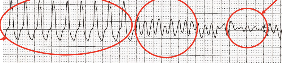
IDENTIFY THE following heart rhythms

What is a heart block, and which structures can be affected?
Impaired electrical conduction through the AV conduction system, including the AV node, bundle of his, and right and left branches. It can cause cardiac arrhythmias
What is a 1st degree AV block, and what is its characteristic ECG finding?
Delayed conduction between atrial and ventricular depolarization, producing prolonged PR interval greater than 0.20s
Every P wave is still followed by a QRS complex

What are the clinical characteristics of 1st degree AV block
Benign and asymptomatic, but it may progress to a higher degree AV bllock
What is second degree AV block
Intermittent failure of AV electrical conduction from the atria to the ventricles, resulting in some p waves not being followed by QRS complexes
What are the two types of second degree AV block
Mobitz (Wenckebach)
Mobitz II
What characterizes Mobitz I (Wenckebach) second-degree AV block
The PR interval progressively shortens until a QRS complex is missed.
The PR interval then returns to its initial shorter length, and the cycle repeats.
Where does Mobitz I usually occur, and when might it be observed
Usually at the AV node. It is generally benign and may occur with increased vagal tone or during sleep
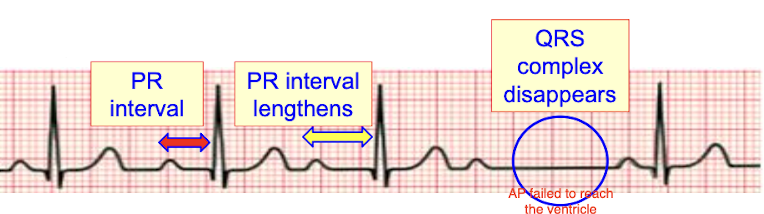
What characterizes Mobitz II second degree AV block, and why is it dangerois
The PR interval remains constant without gradual lengthening, but QRS complexes are suddenly dropped.
It is more dangerous because conduction may fail unexpectedly and progress to complete heart block

What conditions are associated with Mobitz II AV block
Septal myocardial infarction or degeneration of His-Purkinje conduciton system
What is third degree AV block, and what is its characteristic ECG finding?
Complete failure of electrical conduciton between the atria and ventricles, resulting in AV dissociation, where P waves and QRS complexes occur independently
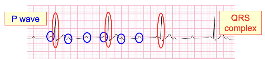
What can cause third degree AV block, and what are uts effects
Myocardial infarction or degeneration of the AV conduction system. It commonly causes bradycardia and lightheadedness.
What happens during right bundle branch block
Conduction through the right bundle branch is blocked. The left ventricle depolarizes normally, while the right ventricle depolarizes later through slower cell-to-cell conduction, producing a wide QRS complex.`
What happens during left bundle branch block (LBBB)?
Conduction through the left bundle branch is delayed or blocked. The right ventricle activates first, and depolarization spreads slowly from the right ventricle to the left ventricle, producing a wide QRS complex.
How can you distinguish first-degree, Mobitz I, Mobitz II, and third-degree AV block on an ECG?
First-degree: PR interval greater than 0.20 seconds; no dropped QRS.
Mobitz I: PR progressively lengthens until a QRS drops.
Mobitz II: Constant PR intervals with sudden dropped QRS complexes.
Third-degree: P waves and QRS complexes occur independently (AV dissociation)