Lesion Localization
 Call Kai
 Learn
 Practice Test
 Spaced Repetition
 Match
 Flashcards
 Knowt Play
1/206

There's no tags or description
Looks like no tags are added yet.
207 Terms
lesion localization
process of using the neurologic examination to determine which specific region of the nervous system is dysfunctional
The primary goal of lesion localization is to determine...
where in the nervous system the lesion is located
Why is lesion localization so important in neurology?
it determines where the problem is located, which then guides diagnostics and prevents unnecessary or inappropriate testing
Lesion localization should be done _______ diagnostic imaging. Why?
before; imaging must be targeted to the correct region
*imaging the wrong region may be useless, expensive, and potentially expose the patient to unnecessary anesthesia
The nervous system can first be divided into two major locations when localizing a neurologic lesion:
1.) central nervous system
2.) peripheral nervous system
The central nervous system is further broken down into two major strucutres:
1.) brain
2.) spinal cord
The brain is further broken down into three major regions for lesion localization:
1.) forebrain/thalamocortex
2.) brainstem
3.) cerebellum
The peripheral nervous system is further broken down into three major locations:
1.) nerves
2.) neuromuscular junction
3.) muscle
When localizing a suspected spinal cord lesion, the first major question is...
which limbs are neurologically abnormal
Spinal cord lesion localization begins with...
a gait examination
Why is gait examination important for spinal cord lesion localization?
it identifies which limbs are affected, which immediately narrows the possible lesion locations
For spinal cord lesion localization, limb involvement can initially be divided into two major patterns:
1.) all four limbs affected
2.) pelvic limbs only affected
If all four limbs are affected by a spinal cord lesion, the lesion is where?
one of the cervical regions: C1-C5 or C6-T2
If only the pelvic limbs are affected by a spinal cord lesion, the lesion is where?
in one of the more caudal regions: T3-L3 or L4-S3
The next step is then to distinguish between pelvic limb vs all four limbs is to...
use UPN vs LMN signs
Upper motor neurons (UPN)
motor pathway that originates in the brain and remains within the CNS to control movement

The upper motor neuron is considered the "_____". Why?
boss; it sends instructions and regulates movement and depends on the lower motor neuron to actually carry the signal to the muscle
Where does the upper motor neuron pathway travel?
from the brain → through the white matter of the spinal cord → toward the lower motor neurons
Functions of the upper motor neuron (3)
1.) initiate voluntary movement
2.) make postural adjustments
3.) maintain appropriate muscle tone
the combined UMN system tends to facilitate _________ muscles and inhibit ________ muscles
flexor; extensor
*UMNs exert an important inhibitory influence over extensor tone
When there is an UPM lesion, extensor tone will be ________. Why?
increased; the lesion will remove the normal inhibition of the extensor system
Lower motor neuron (LMN)
motor pathway that begins with its cell body in the gray matter of the spinal cord, leaves the CNS, and ultimately innervates skeletal muscle
Where does the lower motor neuron pathway travel?
Ventral gray column → nerve root → spinal nerve → peripheral nerve → neuromuscular junction → muscle

The lower motor neuron is considered the "________". Why?
employee; it carries out the motor command by transmitting the signal from the CNS to the muscle
Lower motor neurons are required for both:
1.) voluntary motor activity
2.) spinal reflexes
Why does LMN damage affect both voluntary movement and spinal reflexes?
the LMN provides the final motor pathway to the muscle for both voluntary movement and the local reflex arc
The LMN is aka the...
Final Common Pathway
Why are LMNs aka the Final Common Pathway?
because regardless of how well the brain and descending UMN pathways are functioning, movement cannot occur if the final connection to the muscle is disrupted
Four findings can be used to compare UMN and LMN dysfunction:
1.) motor function
2.) muscle tone
3.) spinal reflexes
4.) muscle atrophy
How can motor function be affected with either an UMN or LMN lesion?
either can cause paresis or paralysis/plegia depending on severity (so weakness alone does not distinguish the two!)
UMN dysfunction:
motor function
tone
reflexes
atrophy
motor function: paresis/ paralysis
tone: normal to increased
reflexes: normal to increased
atrophy: slower/less severe
LMN dysfunction:
motor function
tone
reflexes
atrophy
motor function: paresis/ paralysis
tone: decreased to absent
reflexes: decreased to absent
atrophy: rapid/more severe (due to denervation)
LMN signs can only occur in a limb when...
the lesion affects the LMNs that supply that limb
There are two spinal cord regions where limb LMN signs can occur:
1.) C6-T2
2.) L4-S3
C6-T2 spinal cord regions localize to _______ limbs
thoracic limbs
Why do thoracic limb LMN signs localize to C6-T2?
C6-T2 contains the LMNs that contribute to the brachial plexus and innervate the thoracic limbs
L4-S3 spinal cord regions localize to _______ limbs
pelvic limbs
Why do pelvic limb LMN signs localize to L4-S3?
L4-S3 contains the LMNs that contribute to the lumbosacral plexus and innervate the pelvic limbs
There are four major spinal cord regions used for lesion localization:
1.) C1-C5
2.) C6-T2
3.) T3-L3
4.) L4-S3

The localization process can therefore be simplified into two questions:
1. Which limbs are affected? This tells you which two regions are possible
2. Are the affected limbs showing UMN or LMN signs? This narrows those two possibilities to one region
If all four limbs are affected, the lesion can initially be localized to one of two spinal cord regions:
1.) C1-C5
2.) C6-T2
If only the pelvic limbs are affected, the lesion can initially be localized to one of two spinal cord regions:
1.) T3-L3
2.) L4-S3
Once the affected limbs have narrowed the lesion to two possible spinal cord regions, the next step is to determine...
whether the affected limbs demonstrate UMN or LMN signs
All four limbs affected: UMN vs. LMN findings distinguish between...
C1-C5 vs. C6-T2
Pelvic limbs only affected: UMN vs. LMN findings distinguish between...
T3-L3 vs. L4-S3
C1-C5 spinal cord lesions
affect the cervical spinal cord cranial to the thoracic limb LMNs
C1-C5 spinal cord lesions:
affected limbs:
thoracic limbs (UPM/LMN signs):
pelvic limbs (UPM/LMN signs):
affected limbs: all four
thoracic limbs (UPM/LMN signs): UMN signs
pelvic limbs (UPM/LMN signs): UMN signs
Why do C1-C5 spinal cord lesions affect all four limbs?
Because descending motor pathways to both the thoracic and pelvic limbs pass through this region
Clinical signs of a patient with a C1-C5 spinal cord lesion
-ataxia and tetraparesis or tetraplegia
-muscle tone normal to increased
C6-T2 spinal cord lesion
cervical spinal cord lesion that affects all four limbs and produces LMN signs in the thoracic limbs with UMN signs in the pelvic limb
C6-T2 spinal cord lesions:
affected limbs:
thoracic limbs (UPM/LMN signs):
pelvic limbs (UPM/LMN signs):
affected limbs: all four
thoracic limbs (UPM/LMN signs): LMN signs
pelvic limbs (UPM/LMN signs): UMN signs
Why does a C6-T2 lesion cause LMN signs in the thoracic limbs?
C6-T2 contains the LMNs that directly innervate the thoracic limbs
Why does a C6-T2 lesion cause UMN signs in the pelvic limbs?
the lesion interrupts descending UMN pathways to the pelvic limbs but is cranial to the pelvic limb LMNs at L4-S3, so the pelvic limb LMNs/reflex arcs remain intact
Clinical signs of a patient with a C6-T2 spinal cord lesion
-decreased to absent thoracic limb muscle tone
-normal to increased pelvic limb muscle tone
T3-L3 spinal cord lesions
thoracolumbar spinal cord lesions that leave the thoracic limbs normal and produces UMN signs in the pelvic limbs
T3-L3 spinal cord lesions:
affected limbs:
thoracic limbs (UPM/LMN signs):
pelvic limbs (UPM/LMN signs):
affected limbs: pelvic limbs only
thoracic limbs (UPM/LMN signs): remain normal
pelvic limbs (UPM/LMN signs): UMN signs
Why does a T3-L3 lesion produce UMN signs in the pelvic limbs?
it interrupts descending UMN pathways but is cranial to the pelvic limb LMNs at L4-S3, so the LMNs and local reflex arcs remain intact
Clinical signs of a patient with T3-L3 spinal cord lesions
-increased muscle tone of the pelvic limbs
-normal muscle tone of the thoracic limb
L4-S3 spinal cord lesion
caudal spinal cord lesion involving the LMNs that innervate the pelvic limbs, producing pelvic limb LMN signs
L4-S3 spinal cord lesions:
affected limbs:
thoracic limbs (UPM/LMN signs):
pelvic limbs (UPM/LMN signs):
affected limbs: pelvic limbs
thoracic limbs (UPM/LMN signs): normal
pelvic limbs (UPM/LMN signs): LMN signs
Why does an L4-S3 lesion produce LMN signs in the pelvic limbs?
the lesion directly affects the LMNs and local reflex arcs responsible for pelvic limb function
Clinical signs of a patient with a L4-S3 spinal cord lesion
-decreased to absent muscle tone of the pelvic limb
-normal muscle tone of thoracic limb
Four spinal cord regions: which limbs are affected?
C1-C5: all four limbs
C6-T2: all four limbs
T3-L3: pelvic limbs only
L4-S3: pelvic limbs only
Four spinal cord regions: UMN vs. LMN findings
C1-C5: thoracic UMN + pelvic UMN
C6-T2: thoracic LMN + pelvic UMN
T3-L3: thoracic normal + pelvic UMN
L4-S3: thoracic normal + pelvic LMN
The easiest way to understand spinal cord localization is to avoid memorizing four disconnected lists. Instead...
work through the same two questions every time (which limbs are affected? are there UMN or LMN signs?)
For a patient with a suspected spinal cord lesion, think:
1. Which limbs are abnormal?
All 4 → cervical → C1-C5 or C6-T2
Pelvic only → thoracolumbar/lumbosacral → T3-L3 or L4-S3
Then:
2. Look at muscle tone and spinal reflexes in the affected limbs.
Thoracic LMN signs? → C6-T2
No thoracic LMN signs? → C1-C5
or
Pelvic LMN signs? → L4-S3
No pelvic LMN signs / UMN signs? → T3-L3
LMN signs occur when the lesion hits the...
spinal cord region containing the LMNs that directly supply that limb
The four major spinal cord regions are useful for general localization, however...
lesions can sometimes affect only part of a spinal cord region, producing a more specific pattern of neurologic deficits
Rather than affecting the entire region, a lesion can affect particular spinal cord segments and therefore particular peripheral nerves. This can produce a mixture of...
UMN and LMN findings within the same limb!
Within the C6-T2 region, lesions can sometimes be localized more specifically to...
C6-C8
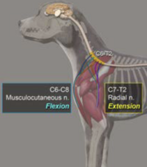
A C6-C8 lesion can produce two contradictory findings in the thoracic limbs:
1.) increased extensor tone
2.) decreased flexor reflex
Increased thoracic limb extensor tone is an ______ sign
Decreased thoracic limb flexor reflex is an ______ sign
UMN
LMN
Therefore, a C6-C8 lesion can produce both ______ and ______ signs within the same thoracic limb
UMN; LMN
Thoracic limb extension is through what nerve?
radial nerve
Thoracic limb flexion is through what nerve?
musculocutaneous nerve
Between the radial and musculoskeletal nerves, which receives more contribution from the C6-C8 spinal nerves?
musculoskeletal
Therefore, a lesion concentrated around C6-C8 can cause weakness of ________ while ________ remains increased
flexion; extension
Overall C6-C8 localization pattern
-all four limbs may be affected
-thoracic limbs have increased extensor tone but decreased flexor reflex
-pelvic limbs have UMN signs
Cervical Spondylomyelopathy Pattern
aka Wobbler syndrome; commonly produces the specific C6-C8 lesion pattern
Two anatomical locations must be distinguished when discussing spinal lesions:
1.) spinal cord segment
2.) vertebral level
spinal cord segment
functional portions of the spinal cord associated with particular spinal nerves
vertebral level
refer to the actual bones of the vertebral column surrounding the spinal cord
True or false: A spinal cord segment and its corresponding vertebra are not necessarily located at the same level
true!
Spinal cord segments and vertebral levels especially don't correspond in the ______ spinal cord
caudal
As you move caudally, spinal cord segments are located progressively more ______ than their corresponding vertebrae
cranially
Why are caudal spinal cord segments located cranial to their corresponding vertebrae?
the spinal cord ends before the vertebral column ends, so the caudal spinal cord segments become compressed into more cranial vertebral levels
Why is the knowing that spinal cord segments and vertebral levels don't always line up important?
For example, if you say there is a lesion involving the L4-S3 spinal cord segment, this does not mean it lies within the L4-S3 vertebrae
The L4-S3 spinal cord segments are actually located within which vertebral levels in most dogs?
L4-L5 vertebrae
Cauda equina
the collection of spinal nerve roots that continues caudally through the vertebral canal after the spinal cord itself has ended
At which vertebral body does the spinal cord end in the dog?
L5!!
*EXAM
At the L6-L7/L7-S1 vertebral region, there is no longer spinal cord; instead there are primarily...
nerve roots of the cauda equina
The cauda equina is part of the CNS or PNS?
PNS
Therefore, lesions affecting the caudal lumbar/lumbosacral vertebral column may affect _________ nerve roots rather than the _______ ________ itself
peripheral; spinal cord
Peripheral nerves have a greater ability to ______ following injury than the CNS
regenerate
The clinically relevant caudal spinal cord segments are primarily located around the ______ vertebral level
L5!!!
*Exam
Which spinal cord segments are particularly associated with the L5 vertebral region?
S1-S3
S1-S3 spinal cord segments are especially important for what nerve?
pudendal nerve
The pudendal nerve is important for...
urinary and fecal continence
A lesion involving the S1-S3 spinal cord segments can therefore result in...
urinary and/or fecal incontinence
The femoral nerve responsible for pelvic limb extension/weight support arises primarily from...
L4-L6
The sciatic nerve is associated primarily with...
L6-S2