GIT
 Call Kai
 Learn
Learn Practice Test
 Spaced Repetition
 Match
Match Flashcards
 Knowt Play
1/238

There's no tags or description
Looks like no tags are added yet.
Name | Mastery | Learn | Test | Matching | Spaced | Call with Kai | Chat |
|---|
No analytics yet
Send a link to your students to track their progress
239 Terms
Fill out the following table
Term | Definition |
|---|---|
Gum inflammation | |
Mouth inflammation | |
Rumenitis/reticulitis/omasitis/abomasitis/gastritis | Forestomach/stomach inflammation |
Caecum inflammation | |
Colon inflammation | |
Diarrhoea | Small bowel diarrhoea = |
Melaena | |
Dysphagia | |
Vomiting | |
Ill-thrift |
Term | Definition |
|---|---|
Gingivitis | Gum inflammation |
Stomatitis | Mouth inflammation |
Rumenitis/reticulitis/omasitis/abomasitis/gastritis | Forestomach/stomach inflammation |
Typhlitis | Caecum inflammation |
Colitis | Colon inflammation |
Diarrhoea | Excess water in faeces |
Melaena vs. Haematochezia vs. Dysentery | Digested blood in faeces from stomach/proximal GIT = Black and tarry Undigested blood in faeces from lower GIT = Bright red Severe haemorrhagic diarrhoea with flecks of mucosa (term derived from human dysentery) |
Dysphagia | Difficulty swallowing |
Vomiting vs. Regurgitation | Forceful expulsion of gastric contents through oesophagus and mouth |
Ill-thrift | Weight loss/poor BCS in production animal |

7 Diagnostic tests for GI diseases
Imaging (best for structural diseases eg. GDV)
Haematology and serum biochemistry
Serology
PCR
Faecal samples
Bacterial culture (culture ≠ disease)
Faecal egg counts
PCR
GIT samples fixed in formalin (biopsy or histology)
FNA (cytology)
When/what should PM samples of the GIT be taken and analysed? Why?
ASAP! Collect histology samples from recently dead animals OR euthanising multiple (eg. calf diarrhoea) that are unlikely to recover due to rapid autolysis of the GIT which interferes with diagnostic tests
Which image shows TRUE enteritis?

BOTH
L: Dark red discolouration = Congestion
R: Fibrin strands = Difficult to pull apart
4 Tips for PM examination of the GIT
Open multiple parts/organs of the GIT (NOT just in the abdomen (split mandible and move pluck)
Pay close attention to mucosal surfaces
Take multiple histology samples even with no gross lesions present
Handle carefully and do NOT scrape mucosa and place into formalin for histology (removed top layer of mucosa)
Sample to collect from a ruminant GIT when parasitism is a DDx
ENTIRE abomasal contents + first 10m of small intestine for worm count
Describe the bon-bon technique
Used to collect fresh/uncontaminated intestinal samples for culture or PCR
Method: Tie either end of short intestinal segment and cut → Place in STERILE pottle with NO FORMALIN to avoid contamination of contents

Which image shows TRUE AM intusussception?
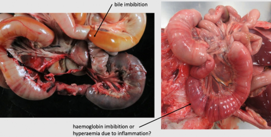
LEFT: True intussusception (dark areas = true haemorrhage) and difficult to pull apart
RIGHT: PM intussusception (Hb imbibition OR hyperaemia due to inflammation?) and easy to pull apart
Small Animal Diseases of the Oral Cavity
Congenital DDx
5 Tooth DDx
3 Inflammatory DDx
7 Neoplastic DDx
Congenital DDx: Cleft palate (palatoschisis) and cleft/hare lip (cheiloschisis) (3)
Tooth DDx:
Dentigerous cysts (3)
Enamel hypoplasia (3)
Pigmentation of teeth (3)
Dental attrition (4)
Dental plaque/calculus and periodontal disease (5)
Inflammatory DDx:
Feline indolent/eosinophilic ulcer (4)
Feline ulcerative stomatitis/glossitis (3)
Other causes of oral ulcers in dogs and cats (5)
Neoplastic DDx:
Gingival hyperplasia (3)
Epulis = Reactive, hyperplastic and benign lesions (3)
Canine oral papilloma (4)
Oral squamous cell carcinoma (SCC) (5)
Sarcomas (3)
Oral melanoma (4)
Oral lymphoma (3)
What is this?
Definition (+ 2 types)
3 Aetiologies
Pathogenesis
Sequelae
2 Other skeletal abnormalities


Cleft Palate (Palatoschisis) and Cleft/Hare Lip (Cheiloschisis) (3)
Definition:
Cleft Lip = Congenital cleft of the upper lip which may be superficial or extend into the nostrils
Cleft Palate = Congenital cleft of the hard and/or soft palate
Aetiologies: Sporadic/unknown
Teratogenic plants (hemlock, lupines)
Teratogenic drugs (griseofulvin)
Genetics (Charolais calves, some dog breeds)
Pathogenesis: During embryonic development, two halves of the face do not completely come together → Gap formation
Sequelae: Death due to aspiration pneumonia
Other Abnormalities: Crooked calf = Lupine ingestion in early gestation
Arthrogryposis = Contracted joints
Scoliosis/Kyphosis = Curved spine
What is this? (2)
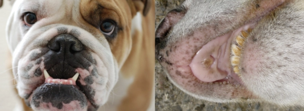
Brachygnathia Superior/Inferior (2)
LEFT = Brachygnathia superior
Definition: Shortening of the upper jaw (pugs, pekinese, boxers, Jersey and Angus cattle)
RIGHT = Brachygnathia inferior
Definition: Shortening of the lower jaw (ruminants and horses = parrot mouth)
Aetiology: Inherited
Sequelae: Severe → Difficulty prehending and chewing food → Malocclusion → Attrition and periodontal disease
What is this?
Structure and appearance
3 Signalments
Behaviour

Dentigerous Cyst/Odontogenic Cyst (3)
Appearance: Swelling in the jaw/gums which contains part/all of tooth protruding into liquid-filled lumen lined by epithelium
Signalment:
Common in NZ sheep (mandibular incisors)
Brachycephalic dogs = Bilateral cysts
Vestigial wolf teeth (horses)
Behaviour: Benign, but can destroy adjacent tissue as they enlarge
What is this?
Appearance
Pathogenesis
3 Causes

Enamel Hypoplasia (3)
Appearance: Pitted areas/irregular indentations in enamel with discolouration (vs. tartar)
Pathogenesis:
Inflammatory/metabolic disease in utero or in young animals
Systemic disturbance affects ameloblast function to make white/shiny enamel
Enamel fails to form in young animals (deciduous and permanent respectively)
Causes:
Canine distemper in dogs (permanent teeth)
BVDV in calves (in utero)
Fluorine poisoning in cattle (brown discolouration of teeth)
Pigmentation of Teeth (3)
4 Causes
Fluorosis = Brown due to enamel hypoplasia
Tetracycline in young/pregnant dam = Yellow ± enamel hypoplasia

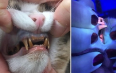
Congenital erythropoietic porphyria in calves, cats and pigs = Pink teeth/bones (fluoresce under UV) due to inherited defect in enzymes involved in heme synthesis → Porphyrin accumulation in teeth and bones
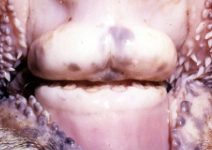
Mineral salts in ruminant cheek teeth become impregnated with chlorophyll = Black teeth (incidental finding)

Dental Attrition (4)
Sequelae
5 Causes of accelerated dental attrition
Sequelae: Ill-thrift
Causes:
Malocclusion
Abnormally soft teeth (eg. odontodystrophies)
Abnormal chewing movement (eg. due to pain)
Loss of opposing tooth
Abrasive diet
NZ sheep grazing low and chewing abrasive soil = #1 reason for culling
Horses crib biting
Dental Plaque/Calculus and Periodontal Disease (5)
Structure of plaque vs. calculus
6 Sequelae
Plaque: Non-mineralised composite mass of
Bacteria
Food particles
Tissue cells
Calculus: Mineralised plaque mainly formed mainly from calcium carbonate precipitated from saliva
Sequelae:
Gingivitis and periodontitis (carnivores)
Tooth loss
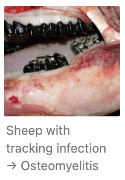
Osteomyelitis and pulpitis
→ Euthanasia due to poor penetration of AB into bone
Retrobulbar abscess
Oronasal fistula (tracking infection through sinuses, nasal cavity and face)
Caries (infundibular necrosis) = Cavities/erosive lesions of the teeth
Signalment: Horses and sheep
Pathogenesis: Organic acids produced by bacteria in plaque → Decalcification of enamel
Location: Occlusal surface of maxillary 1st molar


What is this? (cat lip)
3 Locations
Signalment
Appearance
Cytology
2 Aetiologies
Treatment
Prognosis
2 DDx


Feline Indolent/Eosinophilic Ulcer (4)
Location: Chronic ulcerative lesion of the mucocutaneous junctions of the
Lips (esp. upper lip either side of midline)
Oral mucosa (tongue, palate or gums)
Skin
Signalment: Cats of ALL ages/sex/breed
Appearance: Well-demarcated, reddish-brown shallow ulcers which may progress rapidly
Indolent/rodent ulcers = Upper lip
Cytology: Eosinophils and neutrophils (also plasma cells and mast cells)
Aetiologies:
Immune-mediated allergic disease
Part of the eosinophilic granuloma complex
Primary dysfunction of eosinophils
Treatment: DIFFICULT (steroids, cryosurgery, cyclosporine, antibiotics???)
Prognosis: Recurrence common unless 1˚ allergen identified
DDx: Differentiate with impression smear/skin scraping for cytology
Oral mast cell tumours
Oral SCC
What is this?
Signalment
Pathogenesis
4 Clinical signs
4 Predisposing factors
3 Locations

Feline Ulcerative Stomatitis/Glossitis (3)
Signalment: Older cats
Aetiology: Multifactorial → Imbalance in oral microflora (spirochetes and G- anaerobes #1)
Clinical Signs: Chronic disease often in conjunction with periodontal disease
Dysphagia
Hypersalivation
Halitosis (necrotic tissue)
± Weight loss
Risks: Viral infection
Calicivirus
Feline herpesvirus-1
Feline leukaemia virus
Feline immunodeficiency virus
Location:
Fauces #1 = Junction of mouth and pharynx
Gingiva
Tongue
DDx:
Feline plasma cell gingivitis/pharyngitis
Catarral stomatitis
4 Other causes of oral ulcers in dogs and cats
Renal failure and uraemia
Excess urea in blood → saliva
Urea broken down by urease-producing bacteria normally present in mouth
Ammonia production which is caustic to the oral mucosa resulting in oral ulcers
Ingestion of caustic chemicals
Biting electrical cords
Feline calicivirus (more virulent strains form vesicles → ulcers esp. tongue)

What is this?
Signalment
Location
Pathogenesis
Sequelae
Treatment
Prognosis
DDx
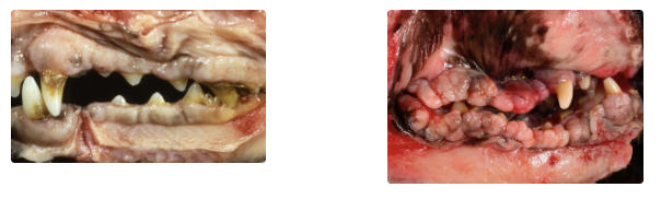
Gingival Hyperplasia (4)
Signalment: Cats and brachycephalic dogs (esp. Boxers)
Location: Upper canines most obvious
Pathogenesis: Chronic inflammation (eg. periodontal disease) → Locally extensive and proliferative reaction → Increased cell number
Potentially caused by cyclosporine, Ca channel blockers and anticonvulsants?
Sequelae: Extra tissue creates pockets which accumulate food and plaque
Treatment: Surgical removal
Prognosis: Recurrence
DDx: Peripheral odontogenic fibroma (solitary lesion NOT proliferative as with gingival hyperplasia)
What is this?
Definition
Diagnosis
4 Types


Epulis
Definition: Group of reactive, hyperplastic and benign lesions within the oral cavity
Diagnosis: Histology with curative biopsy
Types:
Peripheral odontogenic fibroma (fibromatous epulis) = POFs
Pyogenic granuloma
Peripheral giant cell granuloma
Other odontogenic tumours which originate from the dental germinal cells (eg. ameloblastoma, odontoma, cementoma)
What is this?
Appearance
Location
Agent
Prognosis
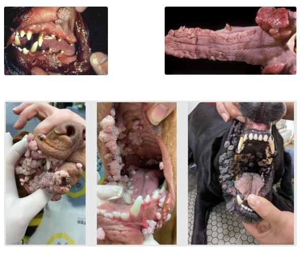
Canine Oral Papilloma (4)
Appearance: Benign epithelial proliferations (warts) which appears as smooth and pale nodules → White-grey, cauliflower-like
Location: Lips, hard palate, gums, tonsil and buccal mucosa
Agent: Canine papillomavirus 1 infection (highly infectious often involving several puppies in a kennel)
Prognosis: GOOD
Regression and recovery in 2 - 3 months due to strong immune response (dogs at puberty)
Older dogs = Immunosuppression due to underlying disease process/immunosuppressive therapy
What is this? (cat)
Location
Appearance
Behaviour
DDx


FELINE Oral Squamous Cell Carcinoma (SCC) (5)
Location: Ventral tongue
Appearance: Red-grey friable mass with ulcerated centre that bleeds easily
Behaviour: Highly malignant with extensive local invasion into soft tissue and bone
Metastasis to lymph nodes late in disease process (early resection → Good prognosis)
Rarely lung metastasis
Neoplastic epithelial cells with neutrophils due to ulceration and inflammation
DDx: Early gingival SCC similar to gingivitis
What is this? (dog tongue and liver)
Location
3 Aetiologies
Behaviour
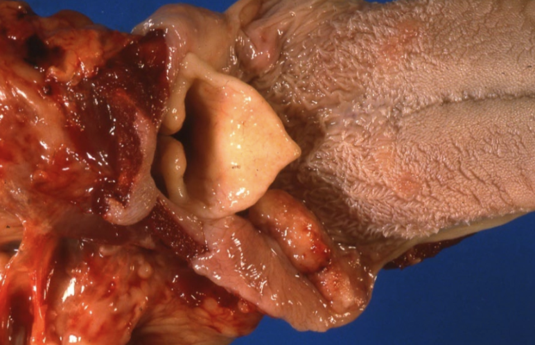

Canine Oral Squamous Cell Carcinoma (SCC) (5)
Location: Tonsil and occasionally gums (usually unilateral)
Aetiology: Multifactorial??? More common in older animals:
Gingivitis
Carcinogens in smoke
Canine papillomavirus
Behaviour: Tonsillar SCC = Fast to metastasis to regional lymph nodes (retropharyngeal), thyroids, lungs and bones vs. gingival = Similar behaviour as cats
What is this?
Behaviour
Signalment
Appearance
Prognosis

Oral Fibrosarcoma (3)
Behaviour: Highly malignant tumours with local invasion into bone and rapid metastasis (rapid growth)
Signalment: Younger dogs (25% in dogs < 5yr)
Large breeds (esp. Golden Retriever)
Appearance: Large, nodular fleshy masses arising from the gums and attached to underlying bone
Prognosis: Local recurrence common after surgical removal
Metastasis to regional lymph nodes and lungs likely present at time of diagnosis
What is this?
Signalment
Location
Diagnosis

Oral Lymphoma (3)
Signalment: Reported in dogs, cats and horses (less common oral tumour)
Location: Tonsils, tongue, gums, mucocutaneous junctions or pharynx ± other widespread organ involvement (multicentric lymphoma)
Diagnosis: FNA and biopsy (differentiate from inflammation)
What is this?
Signalment
Location
Appearance
Behaviour
Prognosis

Oral Melanoma (4)
Signalment: #1 oral malignant tumour of dogs (rare in cats)
Location: Gums and lips
Appearance: Grey to black (sometimes no pigment = Amelanotic melanoma)
Behaviour: Rapid grow with ulceration, bone invasion and metastasis
Prognosis: ~2 - 3m survival (even with treatment)
Metastasis to regional lymph nodes and lungs likely present at time of diagnosis (also spinal cord and brain)
2 Most common malignant oral tumours in CATS vs. DOGS
CATS:
Oral SCC
Oral fibrosarcoma
DOGS:
Oral melanoma
Oral SCC
Oral fibrosarcoma
Large Animal Oral Cavity Diseases
4 Viral vesicular DDx
6 Erosive and ulcerative DDx
3 Papular DDx
4 Bacterial/fungal DDx
Viral Vesicular DDx:
Foot-and-mouth disease (FMD) (5)
Vesicular stomatitis (VS) (3)
Swine vesicular disease (SVD) (3)
Senecavirus A (SVA) (3)
Erosive and Ulcerative DDx:
Bovine viral diarrhoea (mucosal disease) (5)
Malignant catarrhal fever (MCF) (4)
Calf ulcerative stomatitis (3)
Acorn poisoning (3)
Glossitis in photosensitisation (3)
Bluetongue (3)
Papular DDx:
Bovine papular stomatitis (3)
Contagious ecthyma (“scabby mouth”, contagious pustular dermatitis) (5)
Bacterial/Fungal DDx:
Oral necrobacillosis (necrotic stomatitis/laryngitis, calf diphtheria) (3)
Actinobacillosis (”woody tongue”) (4)
Actinomycosis (”lumpy jaw”) (3)
Thrush (4)
Vesicular vs. ulcerative viral diseases
Vesicular diseases usually present as ulcers → Vesicular and ulcerative viral diseases should be on the SAME DDx list for oral ulcers
Cannot differentiate between viral vesicular diseases → Assess which species are infected
Describe the ONLY endemic VESICULAR disease similar in appearance for FMD in NZ
Species and aetiology
Pathogenesis
Appearance of lesions
Species/Aetiology: Pigs grazing furanocoumarin-containing plants (eg. celery/parsnips)
Pathogenesis: Furanocoumarin compounds react with UV light to form vesicles
Appearance: ≤5cm vesicles/bullae on the lips, snout and legs
Foot-and-Mouth Disease (FMD) (5)
Agent
Signalment
Pathogenesis
Clinical signs
Cattle (6)
Pigs (3)
Sheep/goat (3)
Prognosis
Prevention
Agent: Family = Picornaviridae
Aphthovirus (aphtho- = ulcer)
7 serotypes with numerous subtypes (vaccination against SPECIFIC serotype in the area)
Signalment: Highly contagious disease of cloven-footed animals
Cattle = Indicator host (severe lesions → Easy detection)
Sheep, goats, deer = Maintenance host (mild lesions, primarily lameness)
Pigs = Amplifier hosts (shed lots of virus with milder lesions that predominate on hooves)
Camelids = Reasonably resistant (no transmission or carrier status) Horse NOT affected by FMD
Pathogenesis:
Viraemia
Degeneration of middle layers in stratified squamous epithelium
Potential space created fills with serous fluid → Vesicles
Vesicles coalesce → Bullae
Bullae rupture within 24hr → Erosions/ulcers
Clinical Signs:
Cattle
Generalised: Fever (>40˚C), anorexia and depression
Reduced milk production
Drooling and thick saliva (vesicles in mouth = painful)
Lameness (vesicles in interdigital skin and coronet)
Vesicles on teats, vulva and rumen
Neonates = Death from myocarditis (Tiger heart = streaks of inflammation)
Pigs
Small vesicles on snout ± mouth
Lameness (hoof lesions)
Mortality in piglets due to GI, pancreas and heart lesions
Sheep/Goats = Mild
± 2˚ bacterial infection (eg. footrot)
Abortion/foetal mummification
Sudden death in young lambs (lesions in heart/muscle)
Prognosis: Low mortality (1 - 5%)
High morbidity (massive outbreaks of highly contagious disease) → Consider lameness and loss of production
Prevention: Prohibited to feed pigs uncooked food waste from ships or aircrafts
Pigs have highest risk for virus entering NZ (spread via direct contact/wind)

Vesicular Stomatitis (VS) (3)
Agent
Species affected
Appearance
Geographical distribution
Transmission
Importance
Agent: Rhabdoviridae family (genus = vesiculovirus)
Species: Cattle, pigs and HORSES/equids (ONLY vesicular disease of horses)
Sheep and goats relatively resistant
Appearance: As for FMD
Geographic Distribution: Endemic in some USA states, Central and South America
Transmission: Direct contact and insects
Importance: Production loss in cattle + resembles FMD

Swine Vesicular Disease (SVD) (3)
Agent
Species affected
Geographical distribution
Location of lesions
Prognosis
Prevention
Importance
Agent: Picornaviridae family (enterovirus)
Species: Pigs ONLY
Geographical Distribution: Europe and Far East
Location: Feet (between heel and coronary band) ± oral lesions (10%)
Prognosis: Often mild/subclinical (more significant loss in piglets)
Prevention: Do NOT feed pigs food waste from ships/aircrafts
Importance: Clinical indistinguishable from all other vesicular diseases

Senecavirus A (SVA) Infection of Pigs (3)
Agent
Species affected
Geographical distribution
Agent: Picornaviridae family (emerging enterovirus)
Species: Pigs ONLY
Geographical Distribution: USA, Canada, Brazil and China Location and appearance as for other swine vesicular diseases
Bovine Viral Diarrhoea Virus (BVDV) (5)
Agent
Signalment
8 Clinical signs (+ stage of gestation)
Agent: Family = Flaviviridae → Genus = Pestivirus 2 genotypes = BVD-1 and BVD-2 2 biotypes = Non-cytopathic and cytopathic
Signalment: Cattle, deer and camelids
Clinical Signs:
Embryonal death (0 - 40 days)
Abortion (40 - 120 days)
Premature birth
Stillbirth (120 - 280 days)
Foetal mummification
Dummy calves = Weak and non-viable (120 - 280 days)
Teratogenesis (75 - 150 days)
Cerebellar hypoplasia
Hydranencephaly
Hypomyelinogenesis
Alopecia
Retinal pathology
Cataracts
Microphthalmia
Brachygnathism
Enamel hypoplasia
Subclinical immunosuppression (post-natal infection)
Pregnant cows may give birth to clinically normal/small/weak calves that are Ab+ and Ag-
Acute BVDV
Agent
Transmission
4 Clinical signs
Signalment
Prognosis
4 Methods of diagnosis
Agent: Non-cytopathic strain of BVDV (BVDV-2 may cause more severe lesions resembling MD)
Transmission: Direct/indirect contact with bodily fluids
Clinical Signs: Subclinical or mild
Lethargy
Mild occulonasal discharge
Occasional mild oral erosions
Occasional diarrhoea
Signalment: Acute BVD = 3 - 6m calves (waning maternal immunity)
MD = 6 - 24m cattle
Prognosis: High morbidity and low mortality (clears within 2 - 3w)
Diagnosis:
Ag ELISA
Ab ELISA = Pooled milk/serum
PCR = Pooled milk/serum
SNAP test on serum/ear-notch


Mucosal Disease (5)
Signalment
Pathogenesis
5 Clinical signs
Prognosis
5 PM findings
Signalment: 6 - 24m cattle
Pathogenesis:
PI calf = Infection of dam at 40 - 120 days gestation with non-cytopathic strain of BVDV in utero
Foetus becomes infected → Immunological tolerance
Ab- and Ag+
Superinfection OR mutation of non-cytopathic BVDV in PI animals
Cytopathic BVDV infection
Lack of immune response in PI calves
Death
Clinical Signs:
Lethargy and anorexia
Dehydration
Diarrhoea
Nasal discharge
Rough coat
PM:
Erosions and ulcers around muzzle, in mouth, oesophagus (linear and irregular ulcers), reticulorumen and omasal leaves
Small, punctate ulcers and areas of haemorrhage in abomasal mucosa
Blood and fibrin overlying Peyer's patches (BVD attacks rapidly dividing cells eg. crypts of intestine and lymphoid cells in germinal centres)
± Congestion, necrosis, ulceration and fibrinous inflammation of large bowel
± Exudative skin lesions around perineum, base of horns, interdigital skin and heels of feet

How to differentiate between Mucosal Disease and MCF? (2)
Necrotic/haemorrhagic in Peyer’s patches is a feature of MD and NOT MCF (lymphoid hyperplasia)
Brain, kidney and adrenal glands = Vasculitis with MCF ONLY
What is this? (calf)
Signalment
3 Clinical signs
Appearance
Location
Histology
4 DDx
Aetiology
Prognosis

Calf Ulcerative Stomatitis (CUS) (3)
Signalment: 3 - 6m dairy calves (call MPI)
Clinical Signs:
Ill-thrift
Oral ulceration (± oesophagus and forestomach)
Diarrhoea
Appearance: Mild/solitary to multifocal/severe ulcers
Location: Mouth → oesophagus and forestomach
Histology: Inflammation of intestinal crypts and enteritis
DDx:
BVDV (rule out with BVDV test)
MCF
Uraemia
Caustic ingestion
Aetiology: Multifactorial? Similar to summer scour in Australia and UK
Prognosis: Variable morbidity and mortality
Recovery in 4 - 6 weeks
Outbreak may last 3 - 8 weeks on farm
What is this? (cow oesophagus and kidney)
Pathogenesis
2 PM findings
Diagnosis

Acorn Poisoning (3)
Pathogenesis:
Cattle ingests acorns/oaks leaves
Tannins are toxic to the kidneys → AKI
Uraemia
Uraemic toxins cause oral ulceration and haemorrhage
PM:
Shallow oral ulceration and haemorrhage (also oesophagus, rumen, intestines)
Swollen and pale kidneys surrounded by haemorrhage and oedema
Diagnosis: Serum biochemistry with urinalysis = Renal azotaemia and isosthenuria
Cannot use faecal sampling for tannins (already passed out by the time of diagnosis)
What is this?
Agent
Transmission
Geographical distribution
Signalment
Pathogenesis
3 Clinical signs
2 Sequelae
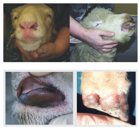
Bluetongue (3)
Agent: Family = Reoviridae → Genus = Orbivirus
25 serotypes with variable pathogenicity (10 serotypes in Australia)
Transmission: Culicoides spp. (midges/gnats)
Distribution: Africa, Middle East, parts of Asia, Australia, USA, parts of Europe
Exotic to NZ
Signalment: Sheep = Clinical disease
Goats and cattle = Subclinical/mild disease
Pathogenesis:
Endothelial damage
Microthrombi formation
Ischaemic necrosis
Ulceration of oral cavity
Oedema and haemorrhages
PM:
Cyanosis of tissues → Blue tongue and mouth (rare)
Petechiae of coronets and bulbs of sheep claws
Ecchymoses in pulmonary artery wall
Sequelae:
Torticollis (wry neck) in recovering sheep due to scarring after muscle necrosis of neck
Foetal malformations (eg. hydranencephaly) after infection during pregnancy
What is this?
Species affected
Location and appearance
Pathogenesis

Glossitis in Photosensitisation (3)
Species: Cattle with photosensitisation of ANY cause (eg. FE)
Location/Appearance: Erosions of ventral midline of tongue
Pathogenesis: Light of certain wavelengths excite photodynamic compounds in the tongue when animal licks its nares and muzzle
What is this?
Agent
Signalment
4 Causes
Appearance
Location
Diagnosis

Oral Necrobacillosis (Necrotic Stomatitis/Laryngitis, Calf Diphtheria) (3)
Agent: Fusobacterium necrophorum (anaerobic G+ bacteria that likes necrotic tissue)
Signalment: Cattle
Aetiology: 2˚ to previous mucosal damage in caudal pharynx
Trauma (eg. drenching, bolus)
Infectious bovine rhinotracheitis (herpesvirus)
BVD
Papular stomatitis infection
Appearance: Large, well-demarcated, yellow-grey, dry areas of coagulative necrosis surrounded by a hyperaemic zone
Location:
Adults = Oral cavity ONLY
Young animals = Spread to trachea (→ aspiration pneumonia), oesophagus or systemically
Diagnosis: Gross lesions ± organism in smears
Difficult to culture as strict anaerobes
What is this?
Agent
Signalment
Aetiology
Inflammatory response

Actinobacillosis (“Woody Tongue”) (4)
Agent: Actinobacillus lignieresi (G- coccobacilli part of NORMAL oral flora)
Signalment: Cattle
Aetiology: Trauma allows bacteria to invade
Inflammatory Response: Pyogranulomatous inflammation and fibrosis centred on club colonies
Club colonies = Immune complexes surrounding the bacteria often seen grossly as sulphur granules
Associated lymphangitis and regional lymphadenitis common
What is this?
Agent
Aetiology
Inflammatory response
Location
Appearance
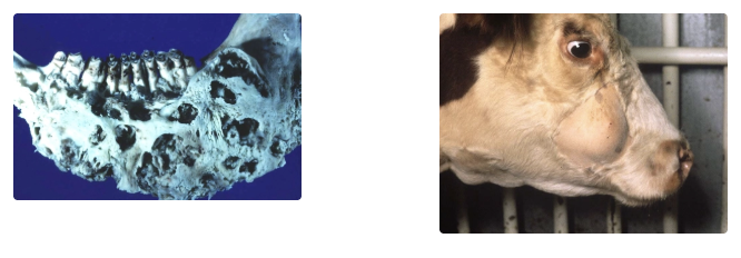
Actinomycosis (“Lumpy Jaw”) (3)
Agent: Actinomyces bovis (G+ filamentous bacteria that is normal flora in the GIT)
Aetiology: Trauma (eg. tooth eruption or rough feed) → Invasion of oral submucosa and mandible/maxilla
Inflammatory Response: Pyogranulomatous osteomyelitis
Location: Mandible and maxilla ± soft tissue and lymph node involvement
Appearance: Similar lesions to actinobacillosis (including sulphur granules)
Prognosis: Lesions develop slowly over months with bone involvement = Difficult to treat
What is this?
Agent
Inflammatory response
Location
Risk factor
Signalment
Appearance

Thrush (4)
Agent: Candida albicans (yeast)
NOT likely primary pathogen
Inflammatory Response: Superficial inflammation of stratified squamous epithelium
Location: Mouth, oesophagus, stomach or rumen) → Accumulation of excess epithelial debris on surface
Risk: Concurrent mild debilitating disease or antibiotic therapy resulting in altered microbial flora
Signalment: Young animals (worse in piglets)
Appearance: White layer of excessive, partly keratinised epithelium and exudate ± hyphae
Oesophagus and Stomach Diseases
5 Oesophageal DDx
6 Rumen DDx
4 Displacement DDx
3 Circulatory DDx
Oesophageal DDx:
Oesophageal stenosis (3)
Oesophageal obstruction (3)
Megaoesophagus (3)
Oesophagitis (3)
Parasitic diseases (3)
Rumen DDx:
Primary (frothy) bloat (4)
Secondary (free gas) bloat (4)
Rumen acidosis (5)
Milk rumenitis (4)
Mycotic rumenitis/reticulitis/omasitis (zygomycosis) (5)
Traumatic reticuloperitonitis (5)
Displacement DDx:
Diaphragmatic hernia (4)
Gastric dilation/volvulus (GDV) in dogs (5)
Abomasal displacement and/or volvulus in cattle (4)
Dilation of the stomach/abomasum in other species (4)
Circulatory DDx:
Gastrorrhagia and hyperaemia (3)
Oedema of the stomach/abomasal wall (3)
Infarction of the stomach in pigs (3)
Overview of the Oesophagus
Structure
3 OTHER types of oesophageal diseases
Structure: Lined by stratified squamous epithelium with mucus glands
Inner circular + outer longitudinal layer of skeletal muscle
Other DDx:
Viral diseases causing oral ulceration → Erosion/ulceration of mucosa (eg. BVD and MCF)
Skeletal muscle diseases (eg. nutritional myopathy)
Neuromuscular junction diseases (eg. myasthenia gravis)
What is this? (horse and dog oesophagus)
4 Causes (+ DDx)
Appearance
2 Clinical signs

Oesophageal Stenosis (3)
Causes:
Cicatrisation (scar tissue formation) due to previous injury/reflux
External pressure (eg. tumour, lymph node as for TB, thyroids)
Developmental defect (eg. persistent right aortic arch in puppies)
Spirocerca lupi round worm granuloma
Appearance: Often mucosal necrosis at stenosis with dilation of proximal oesophagus
Clinical Signs:
Difficulty swallowing
Regurgitation of food
What is this? (horse oesophagus and dog thorax)
3 Causes
4 Sequelae

Oesophageal Obstruction (3)
Causes:
2˚ to stenosis
Foreign bodies (eg. turnips, apples, bones)
Choke = Oesophageal obstruction in horses (or ruminants) caused by impaction of inadequately chewed feed → Stretched oesophagus
Associated with dental disease
Gastro-oesophageal intussusception in dogs (stomach telescopes into oesophagus)
Sequelae:
Pressure necrosis and ulcerated mucosa
→ Oesophageal rupture/perforation → Pleuritis (esp. grass awns)
Ruminant oesophageal obstruction → Free gas bloat
Healing of large ulcers → Scarring and stenosis
6 Causes of oesophagitis (+ DDx)
Viral disease
Papular stomatitis → Circular mucosal erosions more common at the proximal end
BVD → Superficial longitudinal erosions more common at the distal end
Mucosal disease → Deeper + more haemorrhagic erosions BVD
MCF → Deeper + more haemorrhagic erosions than BVD
Necrobacillosis
Thrush
Traumatic injury (eg. doxycycline pill in small animals)
Chemical injury (eg. acids/alkalis, batteries, oak)
Reflux oesophagitis = Gastric acid and bile salts reflux into distal oesophagus → Erosion and ulceration due to:
Dysfunction of the lower oesophageal sphincter due to anaesthesia (eg. not fasting for Caesarean section)
Increased abdominal pressure
Airway occlusion
Hiatal abnormalities

What is this?
Pathogenesis
3 Sequelae
2 Aetiologies (+ DDx)
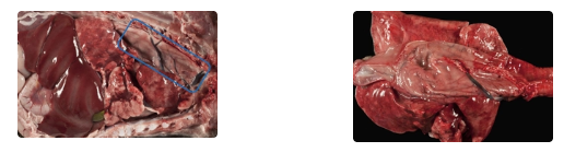
Megaoesophagus (3)
Pathogenesis:
Motor dysfunction causes the entire oesophagus to become flaccid and dilated
Failure of normal peristaltic waves to develop and pass food down (achalasia)
Ingesta accumulates in dilated lumen (may become larger than stomach)
Undigested food is regurgitated
Sequelae:
Oesophagitis
Malnutrition
2˚ aspiration pneumonia
Aetiologies:
Congenital megaoesophagus = Defect in afferent autonomic part of reflex
Signalment: Great dane, GSD, Irish setter
Secondary to diseases affecting NMJ
Myasthenia gravis
Hypoadrenocorticism (Addison's disease)
Neuropathies/myopathies
Lead poisoning
Canine distemper
What is this?
Appearance
Host
Importance

Sarcocystis gigantea
Appearance: Giant protozoal cysts = White nodules ≤1cm long resembling rice grains beneath the oesophageal serosa
Host: IH = Sheep
DH: Cat
Importance: Incidental finding
3 Parasitic diseases of the oesophagus (+ which are exotic to NZ + species affected)
Gasterophilus in horses (occasional)
Sarcocystis gigantea in sheep
Spirocerca lupi in dogs (EXOTIC)
5 Features to examine in rumen contents at PM (+ most likely DDx)
Dry = Dehydration
Ammonia smell = Urea toxicity
Low rumen pH = Acidosis
Flakes of lead paint = Toxicity
Toxic plants (eg. oleander, yew, tutu, weeds, sump oil)
Yellow pollen from pine tress = Incidental finding

What is this?

Gastric Foreign Bodies (1)
Trichobezoars = Hair balls
Phytobezoars = Plant fibre balls
Predisposing factors:
Very young animals deprived of dietary fibre
Lice infestation and excessive licking
Importance: Incidental finding
What is this?
2 Causes
6 PM findings

Ruminal Tympany (Bloat) (4)
Causes:
Primary (frothy) bloat
Secondary (free gas) bloat
PM:
Distended rumen with foamy contents
Dark blood which does not clot due to anoxia from impaired respiration (deoxygenated blood and congestion)
Bloat-line in the oesophagus = Congested/dark cervical (cranial) oesophagus with pale/blanched thoracic (caudal) mucosa → Distinguish from PM bloat
Pale lungs compressed into cranial thorax by bulging diaphragm
± Haemorrhage, congestion and oedema of cervical muscles
± SC oedema of inguinal and perineal regions (decreased venous return)
Primary (Frothy) Bloat
Cause
Pathogenesis
Cause: Cattle grazing pastures with high legume content
Pathogenesis:
Cattle graze lots of legumes which contain cytoplasmic proteins
Proteins stabilises gas bubbles produced by rumen fermentation
Layer of foam created (vs. bubbles normally popping)
Prevents formation of free gas cap in the dorsal sac of rumen which is required to stimulate eructation
Accumulated foam blocks cardia
Rumen pressure increases → Increased intra-abdominal pressure on diaphragm, abdominal organs and vena cava
Impaired respiration + Reduced venous return + Rumen mobility ceases → Sudden death
Secondary (Free Gas) Bloat
3 Causes
Less common
Cause: Physical/functional defects in eructation of gas
Foreign body obstruction (eg. turnip, potato, plastic bag)
Oesophageal stenosis
Vagal nerve damage
Normal rumen pH (+ 4 regulators of rumen pH)
Normal pH: > 5.5 and regulated by
Saliva (bicarbonate)
Epithelial absorption of VFAs
Microbial metabolism of VFAs
Movement of fluid into distal GIT
What is this?
Aetiology (+ 3 example causes)
3 Clinical signs
2 Types (+ characteristics)

Rumen Acidosis (5)
Aetiology: Ingestion of lots of readily fermentable carbohydrates which animal has not been accustomed
Grey and watery rumen contents with rumenitis
Examples:
Grain introduction (eg. barley, maize and wheat)
Accidental breakout → Access to grain, apples, kiwifruit, turnips, swedes, potatoes, kumara, fodder beet
Break-feeding/drought-feeding → Hungry/dominant animals get more feed
Recently calved cows transitioned to lactation rotation too quickly (high CHO and insufficient fibre for saliva production) → SARA
Clinical Signs: 3Ds
Diarrhoea
Dehydration
Decreased milk production
Types:
Subacute Rumen Acidosis (SARA)
Rumen pH = 5 - 5.5
Increased concentration of VFAs in rumen
Relatively low concentrations lactic acid (< 10 mmol/L)
Dominance of G- flora
Acute Clinical Rumen Acidosis (ACRA)
Rumen pH < 5
Dramatic increase in lactic acid concentrations (mainly D-lactate)
Defaunation = Absent rumen protozoa
Dominance of G+ flora (Streptococcus bovis and Lactobacillus spp.)
Rumen Acidosis (5)
Pathogenesis
5 Methods of diagnosis
6 Sequelae
Pathogenesis: Increased fatty acids in both SARA and ACRA (which buffering capacity cannot keep up with) leads to:
Absorption of fatty acids and acidosis (profound uncompensated metabolic acidosis → Collapse and death in ARCA)
Ruminal atony (fatty acids act on receptor that inhibit reticulorumen motility)
Increased intraruminal osmotic pressure → Fluid moves from blood into rumen → Dehydration + haemoconcentration + diarrhoea
± 2˚ infection (diagnosis depends on amount of fermentable grain relative to amount expected)
Fusobacterium necrophorum
Trueperella pyogenes
Fungal
Diagnosis:
History and diet
Clinical signs (ACRA → Dehydration, colic, rumen stasis, increased HR, ± CNS signs when severe)
CBC/biochemistry (metabolic acidosis with reduced BE but similar to other metabolic disorders: hypocalcaemia, toxic mastitis, metritis)
Measure rumen pH with electronic pH meter/narrow range indicator paper (immediately after sample collection)
Microscopy: No motile protozoa
Histology: Epithelial necrosis and inflammation (red)
Sequelae:
Frothy bloat
Zygomycosis of forestomach wall
Ruminal/abomasal necrobacillosis → Superficial foci of mucosal coagulative necrosis → Liver abscesses → Caudal vena cava thrombosis → Pulmonary embolic aneurysm (PEA) = Rupture of pulmonary vasculature → Fatal bilateral epistaxis
Abomasal/duodenal ulcers
Polioencephalomalacia (reduced thiamine-producing bacteria)
Laminitis (release of histamine and endotoxin → Peripheral vasoconstriction)

What is this? (calf rumen)
Signalment
Pathogenesis
2 Clinical signs
Sequelae
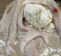
Milk Rumenitis (4)
Signalment: Calves < 4 - 6w
Pathogenesis:
Calves fed milk from bucket/stomach tube (no suckling reflex)
Failure of oesophageal groove reflex
Milk spills into immature rumen and reticulum
Milk putrefaction → Rumen distension and rumenitis (milder than grain overload)
Damaged rumen mucosa
Clinical Signs:
Diarrhoea
Bruxism
Sequelae: Mycotic rumenitis

What is this?
Agents
Pathogenesis
3 PM findings

Mycotic Rumenitis (Zygomycosis) (5)
Agent: Zygomycete fungi of the genera: Mucor, Rhizopus and Absidia = Saprophytic fungi found in environment
Pathogenesis:
Damage to mucosa of the rumen/reticulum/omasum
Fungi invade venules in submucosa
→ Thrombosis and infarction of the forestomach wall
Lesions spread through ALL layers of the forestomach wall
Fungi may spread through blood to liver
PM:
Large, circular, red-black areas of thrombosis and necrosis which extend throughout the forestomach wall
± Localised fibrinous peritonitis
± Spleen and liver affected
Importance of paramphistomes (Calicophoron calicophorum) (2)
Incidental finding when adult stages in reticulum

What it this?
TWO aetiologies
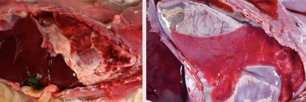
Diaphragmatic Hernia (4)
Stomach displaced into pleural cavity through:
Traumatic diaphragmatic hernia (eg. HBC) = Left image
Congenital diaphragmatic hernia
Incomplete development of lungs due to presence of intestines in chest = Right image
What is this?
Signalment
Pathogenesis
3 PM findings
5 Clinical features

Gastric Dilation Volvulus (GDV) (5)
Signalment: Deep-chested dogs after large meal
Pathogenesis:
Food, fluid and gas accumulate in stomach
Impaired eructation and pyloric outflow
Stomach rotates around oesophagus
Spleen move into a right ventral position and oesophagus becomes occluded →
Obstruction of gastric and splenic veins →
Infarction of gastric mucosa and congestion/infarction of spleen
PM:
Stomach contents is blood-stained
Congested stomach wall
± Ruptured stomach due to ischaemic necrosis
Clinical Features: Requires emergency treatment to prevent death
Circulatory shock (decreased venous return and CO)
Electrolyte imbalance
Acid-base imbalance
± Cardiac arrhythmia
± DIC
Abomasal Displacement (4)
Timing
2 Types
Diagnosis
Timing: Common around calving (underlying metabolic disease, ketosis, metritis → Atony) and seldom fatal unless complicated by volvulus
Types:
LDA
RDA → Volvulus
Diagnosis: Ping heard on percussion of abdomen somewhere between olecranon and tuber coxae ± blood work
6 Clinical pathology findings of displaced abomasum
PCV, Hb, RBC
WBC
Creatinine and urea
Electrolytes
Muscle enzymes
BOH
Haemoconcentration due to dehydration
± Inflammatory/stress leukogram (neutrophilia with left shift)
Pre-renal azotaemia due to dehydration (increase urea and creatinine with concentrated urine)
Mild hyponatraemia and hypokalaemia with marked hypochloraemia due to loss of HCl and electrolytes into lumen of abomasum
Elevated CK and AST if cow was down
Increased BOH = Common in any ruminant off-feed and indicate ketosis
What is this> (horse stomach)
Cause
Pathogenesis
Sequelae

Stomach Dilation in Horses (4)
Cause: Consumption of excess carbohydrates or excessive feeding
Pathogenesis:
Bacteria fermentation of carbohydrates
Production of gas and acid → Increased osmotic pressure
Water moves into stomach down osmotic gradient → ± Gastric rupture along greater curvature, and death due to shock and peritonitis
Sequelae: Survival → Laminitis
What is this?
Pathogenesis
Diagnosis

Vagal Indigestion
Pathogenesis:
Sequel to traumatic damage and scarring of the vagus nerve (eg. traumatic reticuloperitonitis) which assists with digestion
Abomasum and rumen become greatly distended with lots of dry ingesta
→ Visible distension of abdominal wall (papple-shape: LEFT = Apple, RIGHT = Pear)
Diagnosis: Clinical appearance of papple + exclusion of other DDx
What is this?
TWO causes (+ pathogenesis)
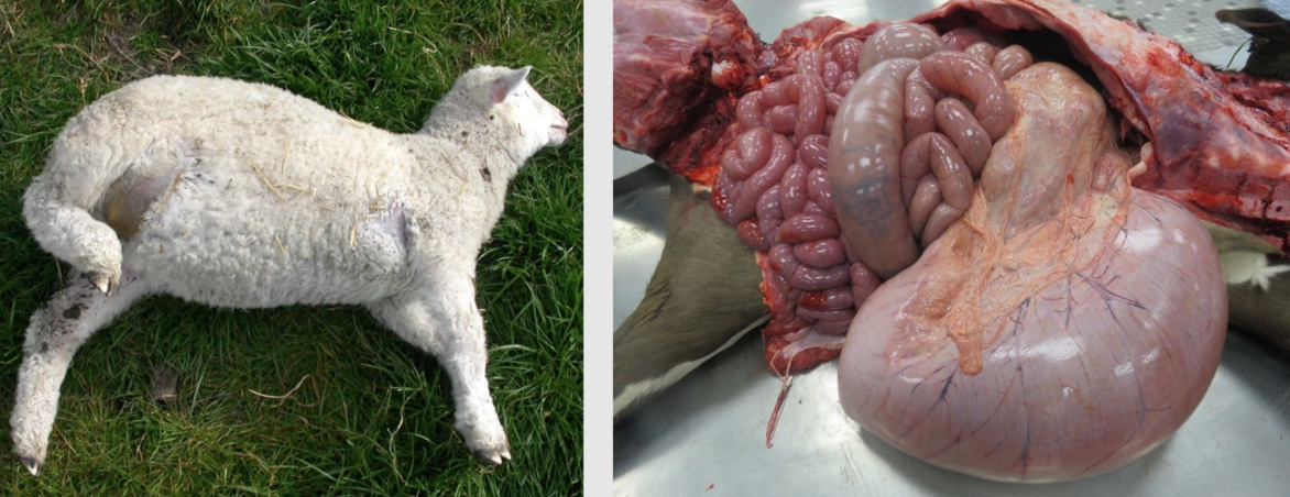
Lamb/Calf Abomasal Distension (4)
Causes:
2˚ to pyloric outflow obstruction (eg. straw impaction)
Abomasal bloat associated with Sarcina or Clostridia bacteria
Bottle-fed animals given large volumes of milk in a single feed
→ Bacteria ferment lactose → ∆abomasal pH
→ Lots of gas production (Sarcina and Clostridia) → ± Abomasal rupture
2 Causes of active gastric hyperaemia
Physiological after ingestion of food
Pathological due to acute gastritis
4 Causes of gastrorrhagia (gastric haemorrhage)
Uraemia in dogs (damaged submucosal arteries ± mineralisation of gastric wall)
Gastric ulceration
Haemonchosis in sheep (attachment of worms)
Trauma and coagulopathies
What is this? (pig stomach)
Agent
6 Causes in ruminants

Oedema of Stomach Wall (3)
Agent: Oedema disease caused by E. coli infection
Ruminants: oedema of abomasal folds due to
Chronic wasting diseases (cachexia)
Haemonchus
Ostertagia/Teladorsagia infections
Arsenic poisoning
MCF
What is this? (pig stomach)
Pathogenesis
5 Aetiologies
Location
Appearance

Infarction of the Stomach in Pigs (3)
Pathogenesis:
Severe acute infectious disease causes endotoxaemia/septicaemia
→ DIC resulting in widespread coagulation
Venous infarction of fundic mucosa of the stomach
Aetiologies:
Salmonellosis
Coliform enteritis
Erysipelas
Swine fever
Swine dysentery
Location: Mucosal surface of greater curvature
Appearance: Large, dark red-black areas of infarction ± fibrin/excessive mucus
Describe 6 causes of gastritis/abomasitis
Acute enteric infection
eg. Abomasitis in sheep = Salmonella Typhimurium OR Hindmarsh
Irritant/caustic poison
eg. Arsenic and fluoride in superphosphate fertiliser → Fluid intestinal contents and nephrosis
Fusobacterium necrophorum
2˚ to ulcerative disease (eg. BVDV)
Mycotic haemorrhagic abomasitis
2˚ to ulcerative disease (eg. BVDV)
Nematodes
Braxy = Sporadic abomasitis in lambs and calves caused by Clostridium septicum
Pathogenesis:
Associated with ingestion of frozen food in cold weather
Creates local tissue damage which allows bacterial invasion
Exotoxin produced by Cl. septicum causes rapid death

Ulceration of the Stomach/Abomasum (5)
2 Normal mucosal protective mechanisms of the abomasum
2 Mechanisms causing stomach ulceration
Normal Mucosal Protection:
Bicarbonate (secreted by saliva) covers the mucosa of the cardia, fundus and pylorus
Prostaglandins stimulate bicarbonate and mucus secretion AND enhance gastric mucosal blood flow
Stomach Ulceration:
Compromised mucosal protective mechanisms #1 (eg. loss of mucus, prostaglandins or bicarbonate)
Hypersecretion of acid
What is this?
Appearance
4 Clinical signs
2 Sequelae

Ulceration of the Stomach/Abomasum (5)
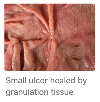
Appearance:
Small ulcers = Red/haemorrhagic OR black due to blood digested by acid → Heal by granulation
Large ulcers = Haemorrhage ± death (esp. pigs)
Clinical Signs:
Melaena = Dark and tarry faeces due to digestion of blood
Haematemesis = Coffee ground appearance due to digested blood
Anaemia
Sudden death due to haemorrhage
Sequelae:
2˚ fungal infection (Aspergillus or Mucor) in ruminants
Peritonitis due to perforation into peritoneal cavity
5 Causes of gastric ulceration in cattle (+ pathogenesis)
Acute stress → Hyperacidity → Small punctate (1 - 2mm)/linear areas of haemorrhage and ulceration in abomasum
Undeveloped rumen → Roughage enter abomasum → Irritation → Ulcerated abomasum of young calves
Traumatic reticuloperitonitis/displaced abomasum → Abomasal atony in adults
Malignant lymphoma → Grossly thickened folds susceptible to ulceration and haemorrhage
Viral infection: BVD, MCF, FMD
What is this? (pig stomach)
Pathogenesis
Other location

Ulceration of the Pars Oesophagea
Pathogenesis: Young growing pigs
Finely ground rations (also associated with high dietary copper, whey, starchy diets low in protein, high dietary unsaturated fatty acids)
Increased water in stomach and loss gastric content partitioning
Pars oesophagus = Square area of tissue at the entry of the stomach = Squamous epithelium with no buffering capacity becomes susceptible to attack by gastric acid
→ Fissures in hyperplastic parakeratotic epithelium ± Candida albicans hyphae
Progression to erosion and ulceration
Other Location: Ulceration of fundus in older pigs due to chronic infection with Hyostrongylus rubidus
± Fatal haemorrhage
What is this? (horse stomach)
Location
4 Causes

Ulceration of the Margo Plicatus
Location: Margo plicatus
Causes: May perforate and bleed (esp. foals)
Bots
Phenylbutazone (also small intestine and right dorsal colon #1)
Stress (eg. horse in training)
Surgery
3 Causes of gastric ulceration in dogs
Uncommon
Uraemia
NSAIDs
Foreign bodies
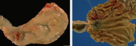
Forestomach Parasites
2 Horse parasites
3 Ruminant parasites
Horse:
Bots = Larval stage of Gasterophilus flies
Draschia (Habronema) megastoma
Ruminant:
Haemonchus contortus
Ostertagia/Teladorsagia
Trichostrongylus axei
What is this?
Lifecycle
2 Sites
Clinical signs
Appearance

Horse Bots (3)
Lifecycle:
Gasterophilus flies deposit eggs on face and legs
Horses ingest eggs through licking
L1 penetrates oral mucosa and moults to L2
L2 migrate down the GIT
Larvae pass into faeces to pupate
Sites in GIT:
G. interstinalis = Attached to gastric mucosa of pars oesophagea in clumps
G. nasalis = Pylorus and proximal duodenum
Clinical Signs: Incidental
PM: ± Small areas of ulceration and granulomatous inflammation
What is this?
Length of parasite
Location in host
Appearance of lesions
Clinical signs

Draschia (Habronema) megastoma (3)
Length: 20mm nematode
Location: Burrows into stomach wall of horses
Appearance: Granulomatous inflammation 20 - 40mm diameter ± suppuration with 2˚ infection by pyogenic bacteria
Clinical signs: NONE!
What is this?
Signalment
Pathogenesis
Diagnosis
3 Clinical signs
5 PM findings
Haematology

Haemonchus contortus (Barber’s Pole Worm)
Signalment: Sheep and goats of ALL ages (lambs more susceptible to clinical disease = 2000 - 3000 significant burden)
Insignificant in cattle
Pathogenesis: L4 and adult suck blood in abomasum
Diagnosis: High FEC (may be low due to acute death before PPP of 15 days or recent drench)
Clinical Signs:
Sudden death
Exercise intolerance
Submandibular oedema (panhypoproteinaemia) No diarrhoea
PM:
Pale carcass (MM)
± Ventral SC oedema, hydrothorax and ascites due to hypoproteinaemia
Grossly visible worms in abomasum
Brown fluid in abomasum (formation of acid haematin from Hb)
Foam up trachea due to pulmonary oedema
Haematology: Iron-deficiency anaemia = Regenerative → Non-regenerative (keratocytes, schistocytes, hypochromasia and microcytosis)

Trichostrongylus axei
Importance
Appearance of lesions
Unimportant in ruminants of ALL ages in NZ
Appearance: Catarrhal gastroenteritis with small ulcers after mucosal emergence
What is this?
Signalment
Pathogenesis
Appearance
2 Types
2 Clinical pathology findings
# Larvae needed to produce disease

Ostertagia/Teladorsagia
Signalment:
Type I = Sheep and cattle < 1yr (± adults)
Type II Ostertagiosis = Cattle 1 - 2yr
Pathogenesis:
Larvae burrow into abomasal mucus glands
Gland hyperplasia and metaplasia of epithelium (unspecialised cuboidal cells replace chief and parietal cells)
Loss of chief cells → Reduced HCl production → Neutral abomasal pH
→ Pepsinogen cannot be activated to pepsin → Reduced protein digestion → Diarrhoea
Bacteria are NOT killed
Pepsinogen absorbed into bloodstream
Hypoalbuminaemia → Ventral SC oedema (abomasal oedema when severe in lambs/calves)
Appearance: Small, pale, umbilicated nodules 1 - 2mm grossly on mucosal surface within a few days of infection → Morocco leather appearance with heavy infection → Superficial necrosis and inflammation with emergence of immature adults
Types:
Type I = Lambs and calves ingest lots of larvae over short period
Type II infection = Larvae retard development in mucosa to have mass emergence months later in winter/spring
Laboratory:
Hypoalbuminaemia (protein-losing gastroenteropathy)
Increased serum/plasma pepsinogen which increases from ingestion, then again with emergence (no pepsinogen increase when worms encysted)
Disease: 10,000+ larvae needed to produce clinical disease
What is this?
Signalment
2 Aetiologies
Pathogenesis


Atresia (4) = No patency in normal lumen
eg. atresia jejuni, ilei, coli, ani
Signalment: Holstein calves and foals → Spiral colon empty and flaccid
Aetiologies:
Recessive inherited trait (carrier bulls removed from AI serve in NZ)
Potential association with early amniotic vesicle palpation for pregnancy diagnosis???
Atresia ani = Heritable trait occurring in most species (may be associated with spinal/genitourinary malformations
Progression: Normal at birth → Faecal accumulation → Abdominal distension → Death within few days
List 3 types of intestinal obstructions (+ definitions)
Intraluminal/Intrinsic obstructions = Obstructed LUMEN of intestine
Extrinsic obstructions = External factor compressing intestinal lumen
Functional obstructions = No physical occlusion, but food cannot pass through intestine due to lack of peristalsis
What is this?
Type of intestinal obstruction
7 Examples

Intraluminal/Intrinsic Intestinal Obstruction
Foreign body (eg. corn cob, strip of cloth, plastic bags, string, fruit sone, ball)
Bezoar
Ascarids in pigs and foals
Chronic constipation and colon impacted with faecal matter
Enterolith = Spherical mineral concretations occasionally found in horse colons (usually do NOT cause obstruction)
Congenital atresia
Scarring/stenosis
What is this?
Type of intestinal obstruction
5 Examples

Extraluminal/Extrinsic Intestinal Obstruction
Tumour (eg. sheep adenocarcinoma or horse lipoma)
Strangulation (eg. herniation or lipoma)
Fibrous adhesion
Fat necrosis in cattle (lipomatous proliferation of fat surrounding intestine)
Abscess
What is this?
Type of intestinal obstruction
2 Examples
Definition
Causes

Functional Obstruction
Grass sickness (equine dysautonomia) = Subacute partial paralysis of intestines in HORSES
Cause: Unidentified neurotoxin → Necrotic degeneration of autonomic ganglia in abdominal cavity
Diagnosis: Histology intestinal nerves
Paralytic ileus = Diffuse absence of normal intestinal tone and peristalsis → Flaccid intestines distended by fluid caused by:
Post-abdominal surgery involving intestines, peritonitis
Spina bifida
Spinal injury
What 2 things does severity/consequences of intestinal obstruction depend on?
Location of intestine obstructed (upper vs. lower GIT): Obstructed intestines interfere with propagation of peristaltic contractions distally
Simple or strangulated
UPPER Small Intestinal Obstruction
Pathogenesis
Clinical pathology findings
RBC and TP
WBC
Urea, creatinine and USG
Electrolytes
Acid-base balance
Phosphorus
Pathogenesis: ACUTE AND MORE SEVERE
Lots of fluid normally produced by the upper GIT accumulates proximal to blockage
→ Loss of water and electrolytes into lumen
Increased local peristalsis initially proximal AND distal to obstruction → Pain
Reverse peristalsis above point of obstruction → Intense vomiting
More fluid and gas accumulates → Intestinal ileus
Clinical Pathology:
Relative erythrocytosis and hyperproteinaemia (dehydration)
Inflammatory leukogram = Neutrophilia with left shift (damaged intestine)
Pre-renal azotaemia (dehydration) and optimally concentrated urine
Reduced serum Na, Cl and K (loss of electrolytes into gut/vomiting)
Metabolic alkalosis (loss of gastric acid) ± Hypercapnoea (compensatory respiratory acidosis)
± Increased serum phosphorus (pre-renal azotaemia and reduced excretion of P in saliva of ruminants)
Pathogenesis of lower intestinal obstruction
LESS ACUTE AND OFTEN MORE CHRONIC AETIOLOGY
Lots of fluid normally produced by the proximal GIT accumulates proximal to blockage
Less significant loss of fluid and electrolytes into lumen (larger absorptive surface area)
Gas and fluid pressure build → Altered mucosal permeability
→ Readily absorbed bacterial toxins
→ Toxaemia