622 11/9 L Streptococcus Intro
 Call Kai
 Learn
 Practice Test
 Spaced Repetition
 Match
 Flashcards
 Knowt Play
1/89

There's no tags or description
Looks like no tags are added yet.
90 Terms
Objectives

Skin Characteristics that Prevent Bacterial Growth (first line of defense)
antimicrobial peptides (b-defensins, cathelicidins)
anitmicrobial lipids (sebum)
leukocytes (intraepidermal T cells and epidermal langerhands cells)
dead keratinocytes (barrier) ad living keratinocytes (cytokine production)
pH, salt, temperature
physical barrier (tight junctions)
Skin Characteristics that Promote Bacterial Growth (commensals and some pathogens)
pH, salt, temp, moisture
estimated 100-1000 cfu/cm² colonizing skin, mostly aerobic and facultative anaerobes
Factors that Influence distribution, Composition, and Density of Bacterial Growth on Skin
moisture, pH, salt, temp, exfoliation, bathing habits, antibacterial products
Commensals
resident bacteria of the skin
7 Resident Skin Bacteria (Commensals)
staphylococcus epidermis
cutibacterium acnes
micrococcis spp
corynebacteria spp
lactobacillus spp
anaerobic gram + cocci
aerobic gram - rods
Transient Skin Bacteria (from mucous membranes or environment, pathogens)
staphylococcus aureus (gram + cocci)
streptococcus pyrogens (gram + cocci)
Manifestations of Direct Skin Entry
impetigo on the epidermis
erysipelas and cellulitis in the dermis
necrotizing fasciitis all the way to the fascia and muscle
folliculitis, furuncle, and carbuncle in hair follicles

Direct Entry Infections Diagram
from cut, scrape, wound, or injection

5 Infections from Direct Entry
impetigo
erysipelas
folloculitis
cellulitis
necrotizing fascitis
Impetigo
infection from direct entry, honey-colored crusts, very contagious, most common in kids around the mouth
bullous impetigo is crusts with bullae, usually caused by staph aureus
Impetigo Image

Impetigo Bacteria
strep pyrogens
staph aureus
Erysipelas
infection from direct entry, clear demarcation from unaffected skin
involved dermis and lymphatics (can cause swelling of lymph nodes)
Erysipelas Image

Erysipelas Bacteria
s pyogenes (usually)
Folliculitis
infection of direct entry, infection of hair follicle, carbuncles and boils
hot tub folliculits (special case assocaited with pseudomonas aeruginoas)
Folliculitis Image

Folliculitis Bacteria
s aureus
Cellulitis
infecion from direct entry, deeper dermal or subcutaneous infection that is spreading, painful, not well-demarcated
usually begins from an abrasion, wound, or mild trauma
Cellulitis Bacteria
s pyrogenes or s aureus
Cellulitis Image

Necrotizing Fasciitis
infection from direct entry, frequently fatal, requires quick attention with antibiotics or surgical debridement
media has linked it to strep pyrogenes (flesh eating bacteria) but it can be caused by many bacterial infections, MRSA
Necrotizing Fasciitis Bacteria
anaerobes and facultative anerobes
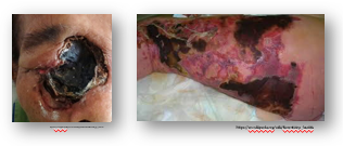
Necrotizing Fasciitis Image
Gram + Cocci Flow Chart

Streptococci spp Characteristics
cocci commonly arranged into chains or pairs, gram +, catalase negative
non-motile, non-spore forming, facultative anaeboes

SSTI
skin and soft tissue infections
Lancefield Classification of Strep
s pyogenes is strep A (GAS)
s agalactiae is strep B (GBS)

Streptococcus pyogenes: Epidemiology
lancefield classification is group a strep (GAS), 600 million infections annually
normal flora of skin and mucous membranes, skin and soft tissue infections after introduction through break in skin/deep tissue
Location of S pyogenes Infections
skin in the summer
respiratory in winter, and school aged children
S pyogenes Complications/Sequelae
acute rheumatic fever, scarlet fever, glomerulonephritis
S pyogenes Transmission
person-to-person through respiratory droplets, and skin to skin
we like our own bacteria but not that of other people
S pyogenes Patient Risk Factors
age 5-15 yo
influenza infection in elderly
trauma, burns, and surgery
immunodeficiency
crowded living situations (dorms, daycare)
S pyogenes Clincal Diseases
superficial skin infections like impetigo and erysipeals
deep skin infections like cellulitis and necrotizing fascitis
pharyngotonislitis (strep throat)
S pyogenes Toxin-Mediated (Supperative, happens during infection) Disease
scarlet fever
streptococcal toxic shock syndrome (STSS)
S pyogenes Post-Streptococcal (Nonsupperative) Disease
rheumatic fever
glomerulonephritis
S pyogenes Other Infections
bacteremia
pneumonia
septic arthritis
S pyogenes: Pharyngotonsilitis
strep throat, can extend to otitis media/ear infection in children
characterized by intense acute pharyngitis, tonsilitis, redness and edema of mucus membranes, purulent exudate
S pyogenes: Impetigo
superficial bacterial infection, highly contagious
can be primary (direct bacterial invasion of previously normal skin) or secondary (infection at sites of minor skin trauma)
2 Forms of Impetigo
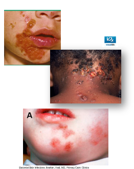
nonbullous (crusting, strep pyogenes)
bullous (blistering, staph aureus)
Nonbullous Impetigo
most common form, from strep, honey-colored crusts
lesions begins as papules surrounded by erythmea, then progess to pustules that enlarge and break down to form crusts with a characteristic golden apperance
usually involved the face and extremities
Bullous Impetigo
from staph, more common in 2-4 yo children
vesicles enlarge to form flaccid bullae (blisters) with clear yellow fuild that later becomes darker and more turbid

S pyogenes: Erysipelas
superficial skin infection (upper dermis) with lymph node enlargement and systemic symptoms
lesions distict from uninvolved skin with clear demarcation

S pyogenes: Erysipelas Predisposing Factors
impaired venous/lymphatic drainage
immunocompromised
S pyogenes: Cellulitis
a rapidly spreading infection of the dermis and subcutaneous fat layer with poorly defined borders
severe cases may include vesicle and bulla formation, pustules, and necrosis
S pyogenes: Cellulitis Clinical Presentation
fevers, chills, malaise may precede onset of skin manifestations
erythema, pain, warmth, and swelling
patchy areas may be present

S pyogenes: Cellulitis Risk Factors
breaks in skin, chronic skin disease, edema caused by venous insufficiency or lymphatic obstruction, IV drug use
S pyogenes: Necrotizing Fasciitis
streptococcal gangrene, bacteria infect the fascia planes
progress rapidly over several days
S pyogenes: Necrotizing Fasciitis Clinical Presentation
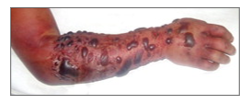
erythema, warmth, swelling, significant pain
signs of systemic illness: fever, lethargy, hypotension, tachycardia
high mortality
Rapid Progression of Necrotizing Fasciitis Over Several Days
skin changes color from red-purple to blue-gray patches
skin breakdown with bullae containing thick pink or purple fluid
cutaneous gangrene may be seen, destruction of blood vessels and skin necrosis
S pyogenes: Necrotizing Fasciitis Risk Factors
penetrating wounds, surgical wounds, immunosuppression, diabetes, obesity
Scarlet Fever
scarlatina, possible complication of untreated or persistent streptococcal infections during infection

Scarlet Fever Clincal Presentation
high fever > 38.8 C, sandpaper rash, strawberry tongue (fungiform papillae on surface of tongue), circumoral palor
enlarged cervical lymph nodes

Rheumatic Fever
possible complication of untreated or persistent streptococcal infections, occur after infection
mediated by M protein (strep virulence factor) is cross-reactive with human cardiac myosin leading to rheumatic fever where antibodies develop against M protein then can attack the heart
Rheumatic Fever Clinical Presentation
fever, polyarthritis, carditis, erythema marginatum, chorea, subcutaneous nodules

What triggers toxic shock-like syndrome from s. pyrogenes infection?
erythrogenic exotoxin A
triggers massive inflammatory response with IL-1, IL-2, TNF-a, and IFN-y
Toxic Shock-Like Syndrome Clinical Presentation
fever, rash, shock
from s pyrogenes
Post-Streptococcal Glomerulonephritis
usually develops 1-5 weeks after acute pharyngitis or streptococcal skin infection
deposition of immune complexes in the kidneys
Post-Streptococcal Glomerulonephritis Clincal Presentation
blood and protein in urine, edema, hypertension, renal insufficiency, oliguria (nonsuppurative)
majority of patients recover but it can be fatal or develop into chronic disease in rare cases (1-2% of pt have renal failure)
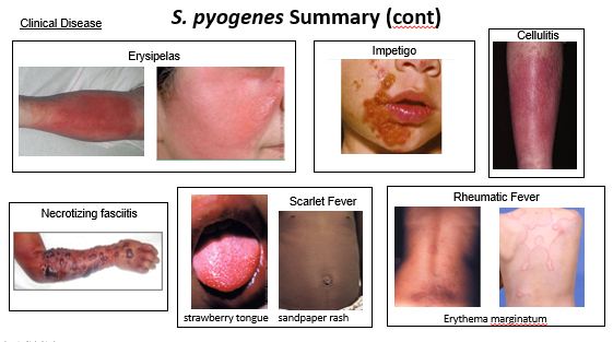
S. pyogenes Clinical Diseases Summary

Virulence Factor
a trait of a pathogen that mediates pathogenesis, virulence, or destruction of host cells/tissues
6 Bacterial Virulence Factors for Strep and Staph
structural virulence
adherence
enzymes
toxins
immune evasion
antibiotic resistance
4 S pyogenes Structural Virulence Factors
m protein (major)
hyaluronic acid capsule (anti-phonetic/slippery, poorly immunogneic)
fibronectin-binding proteins (f protein causes adherence to cell surface)
lipotechoic acid (adherence to epithelical cells and ecm)
M Protein
major structural virulence factor of s pyogenes, adherence, blocks complement activity making it anti-phagotic
determinant of long lasting type-specific immunity
can lead to cross-reactive with cardiac myosin (type ii hypersensitivity), resulting in rheumatic fever
3 S pyogenes Enzyme Virulence Factors
C5a peptidase (immune evasion that inactivates C5a)
streptokinase A and B
DNase A, B, C, D (dissemination, degrades free DNA in pus)
Streptokinase A and B
enzyme virulence factor of s pyogenes, dissemination
cleaves plasminogen to form the proteaseplasmic, plasmic cleaves fibrinogen and fibrin, escape from blood clots and fibrin deposits (dissemination)
3 S pyogenes Cytotoxins Virulence Factors
streptolysin S (SLS)
streptolysin O (SLO)
supertantigens (streptococcal pyrogenic exotoxins types A/B/C, superantigens overactivate the immune system and cause host damage)
Streptolysin S (SLS)
cytocixin virulence factor of s pyogenes, lyse rbcs/leukocytes/platelets
s = serum stable, oxygen stable, responsible for b-hemolysis on blood agar
stimulates release of lysosomal contents when engulfed, leading to phagocyte death
Streptolysin O (SLO)
cytocixin virulence factor of s pyogenes, lyse rbcs/leukocytes/platelets
oxygen-labile
lab titers (anti-streptolysin-o antibodies) are an indication of past infection
inhibited by cholesterol in skin lipids, so no anti-streptolysin O antibodies in skin infection
4 S pyogenes Immune Evasion Virulence Factors
C5a peptidase (inactivates C5a a soluble chemoattractat that recruits PMNs)
anti-phagotic (hyaluronic acid capsule, M protein)
streptococcal inhibitor of complement (SIC, inactivates complement MAC)
SLO and SLS (apoptosis of PMNs and macrophage)
Getting to S. pyogenes on Gram + Cocci Flow Chart

6 Lab Diagnositcs of S pyogenes (Used for all Staph and Strep Infections)
gram stain
catalase test
hemolysis
antibiotic sensitivity test
lacefield classification
PYR test
Gram Stain
looking at bacteria under microscope after differential staining
S pyogenes Gram Stain
gram + cocci

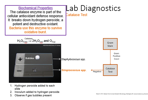
Catalse Test of S pyogens
catalase negative (no bubbling, cannot break down hydrogen peroxide to survive oxidative burst of immune reaction)
Hemolysis
growth on blood agar to observe if it breaks down the blood, can be beta, gamma, or alpha
S pyogens Hemolysis
beta hemolysis
beta Hemolysis
complete (full) lysis of erythrocytes via hemolysin, clearing around and/or under colony, transparent when help up to light
ex: group A strep S pyogenes, group B S agalactiae
gamma Hemolysis
no hemolysis
ex: enterococcus (E. faecalis), nonenterococcus (S. bovis)
alpha Hemolysis
partial lysis of erythrocytes, greenish tinge around and/or under colony
ex: s pneumoniae, viridans streptococci (s mutans)
Hemolysis Summary

Antibiotic Sensitivity
kirby-baur or disk diffusion, different zones of inhibition, bacteria can be sensitive (S) or resistant (R)
Antibiotic Sensitivity of S pyogenes
sensitive to bacitracin
Lancefield Classification
classification based on cell wall carb antigens (most commonly used for group A, B, C, F, and G)
latex agglutination: add latex bead solution to flat surface, mix bacterial strain in solution, and evaluate consistency

Lancefield Classification of S pyogenes
group A (GAS)
PYR Test
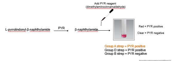
commonly used for different groups of strep, instant biochemical test
red = PYR positive
clear = PYR negative
S pyogens PYR Test
group A (s pyogenes) and D strep are PYR positive and will be red
group B strep is PYR negative and will be clear
S. pyogenes Summary

S pyogenes Clinical Disease Summary