Week 5: Respiratory System, case study 6+2.5
 Call Kai
 Learn
 Practice Test
 Spaced Repetition
 Match
 Flashcards
 Knowt Play
1/66

There's no tags or description
Looks like no tags are added yet.
67 Terms
What is the main job of the conducting zone, and what structures are in it?
Job: air pipeline that warms, moistens, and filters incoming air.
structures: nose, nasal cavity, pharynx, larynx, trachea, bronchi, ad bronchioles.
what is the main job of the respiratory zone, and what structures are in it?
Job: actual site of gas exchange (swapping oxygen and CO2)
Structures: respiratory bronchioles, alveolar ducts, alveolar sacs, and alveoli (~300 million air sacs)
How do cartilage, tissue lining, and muscle change moving down the airways?
Cartilage: C-shaped rings in trachea→ plates in bronchi→ completely disappears in small bronchioles.
Lining: Psuedostratified ciliated columnar ( nose/trachea)→ protective stratified squamous (throat friction) → simple cuboidal ( bronchioles).
Smooth Muscle: increases in relative amount as cartilage decreases.
what are bronchopulmonary segments and trachealis muscle?
Segments: pyramid-shaped sections of a lung lobe with their own separate air supply (bronchus) and blood supply.
Trachealis muscle: smooth muscle on the back of the trachea that squeezes during coughing to blast out mucus.
How does a newborn’s throat (larynx and pharynx) differ from an adult’s?
High position: sits much higher up in the neck.
Epiglottis: touches the soft palate, creating separate paths for Ian and milk.
benefit: allows babies to suckle milk and breath though their nose at the same time.
shape: newborn epiglottis haves a curved (omega) shape.
How do the right and left lungs differ in lobes, fissures, and shapes?
Right lung: larger/wider; has 3 lobes (superior, middle, inferior) separated by 2 fissures (horizontal and oblique).
left lung: smaller (fits heart); has 2 lobes ( superior, inferior) separated by 1 fissure (oblique). features a cardiac notch and lingual.
what are the apex, base, and hilum of the lung?
Apex: pointed top tip extending up near the first rib.
base: flat bottom resting on the diaphragm.
Hilum: doorway slot on the inner side where bronchi, blood vessels, and nerves enter/exit.
what are the two layers of the pleura?
Visceral pleura: inner protective layers stuck directly to the lung surface.
Parietal Pleura: outer layer lining the inside of the chest wall and top of the diaphragm.
Where do the upper and lower respiratory tracts originate in the embryo??
Divider: the larynx marks the boundary between upper and lower development.
Upper tract: Develops in the head from surface ectoderm (nasal places) and endoderm (pharynx).
Lower tract: sprouts in the body during week 4 as a bud (respiratory diverticulum) off the forregut endoderm, wrapped in splanchnic mesoderm.
where are key development timepoints and why does lung function happen so late?
Wk 4-5: lung bud forms and splits into main bronchi.
Month 6-7: primitive air sacs and surfactant production start; gas exchange becomes possible by month 7.
Why function is late: breathing requires 3 things that only mature late in pregnancy: ultra-thin alveolar walls, dense surrounding blood capillaries, and surfactant (which prevents air sacs from collapsing).
What are Esophageal Atresia (EA) and Tracheoesophageal Fistula (TEF)?
Cause: failure of the respiratory bud to separate cleanly from the foregut esophagus during week 4-5.
Esophageal Atresia(EA): the esophagus ends in a closed/blind pouch instead of reaching the stomach.
tracheoesophageal fistula (TEF): an abnormal connecting tunnel between the trachea and esophagus.
Most common type: EA with lower/distal TEF (makes up 90% of cases).
Nose
external facial structure that provides an airway; warms, moistens and filters incoming air.

Nasal Cavity
Internal space behind the nose divided by a septum; lined with ciliated mucus membrane to clean air.

Nasal conchae
Three bony ridges ( superior, middle, inferior) on the side walls of the nasal cavity that swirl air to warm and humidify it.

Nasopharynx
upper section of the throat behind the nasal cavity; air-only passage lined with pseudo stratified ciliated columnar tissue.

Oropharynx
Middle section of the throat behind the mouth; shared pathway for both food and air lined with protective stratified squamous tissue.

Epiglottis
Flexible, leaf-shaped flap of elastic cartilage at the top of the larynx folds down to block food/water from entering the trachea during swallowing.

vocal folds
true vocal cords inside the larynx that vibrate as air moves past them to produce sound.

Trachea
flexible windpipe carrying air from the larynx to the lungs, help open by rigid hyaline cartilage rings.

Thyroid cartilage
shield-shaped hyaline cartilage forming the front wall of the larynx; features the laryngeal prominence (“Adam’s apple”)
Cricoid cartilage
full ring of hyaline cartilage located directly below the thyroid cartilage at the base of the larynx.

tracheal ring
C-shaped band of hyaline cartilage embedded in the tracheal wall to prevent the air way from collapsing inward.

Carina
Sensitive ridge of cartilage at the bottom base of the trachea where it branches into the left and right main bronchi.

Primary bronchi
First two main branches off the trachea entering each lung at the hilum (right primary bronchus is shorter and more vertical).
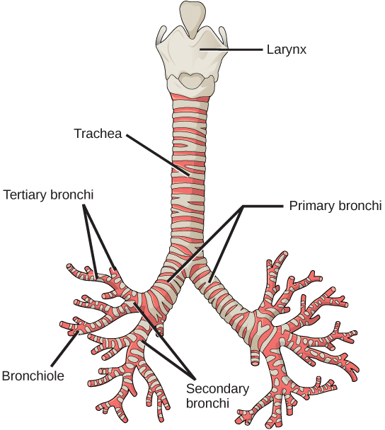
(secondary) lobar bronchi
secondary branches of the bronchial tree supplying specific lung lobes ( 3 branches in the right lung, 2 in the left).

(tertiary) segmental bronchi
Smaller tertiary branches supplying individual bronchopulmonary segments inside each lobe.

Pulmonary arteries
Blood vessels carrying deoxygenated blood away from right ventricle of the heart into the lungs.

Pulmonary veins
Blood vessels carrying freshly oxygenated blood from the lungs back into the left atrium of the heart.

Diaphragm
Main dome shaped muscle of respiration separating the chest cavity from the abdomen.

Lung: apex
Pointed, rounded top tip of each lung extending up near the first rib.

Lung: base
Broad, concave bottom edge of each lung that sits directly on the diaphragm.

Lung: anterior border
sharp front edge where the costal and mediastinal surfaces meet at the front of the chest.

Lung: Posterior border
smooth, rounded back edge of the lung alongside the vertebral column.
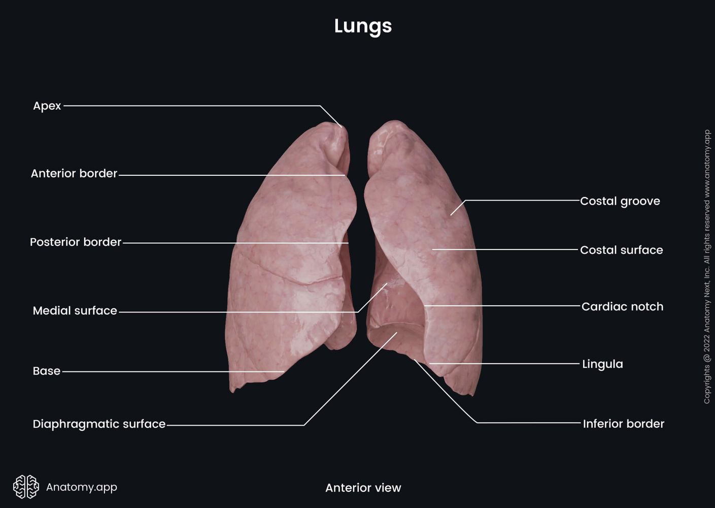
Lung: inferior border
Bottom rim of the lung encircling the base.

lung: costal surface
Large outer surface of the lung pressed against the ribs.

Lung: mediastinal surface
Inner surface of the lung facing middle chest organs (heart, trachea, esophagus)

Lung: Diaphragmatic surface
Bottom surface of the lung resting directly on top of the diaphragm.

Lung Hilum
Intend region on the inner mediastinal surface where bronchi, blood vessels, and nerves enter/exit the lung.

Left lung features
Divided into 2 lobes (superior, inferior) by 1 fissure (oblique fissure); features a cardiac notch (cutout for the heart) and a lingula.

Right lung features
Divided into 3 lobes (superior, middle, inferior) by 2 fissures (horizontal and oblique fissures).

slide: alveoli
Microscopic air sacs with ultra-thin walls surrounded by capillaries; site of gas exchange.

Slide: Bronchiole
Small airway (<1 mm) identified under a microscope by its thick ring of smooth muscle and complete lack of cartilage.

slide: Alveolar duct
Elongated microscopic passageway lined with alveoli leading into alveolar sac.

Slide: ciliated pseudo stratified columnar epithelium
airway tissue lining with cilia on top and goblet cells scattered throughout to trap and move mucus.

slide: tracheal rings (hyaline cartilage)
clear/glassy cartilage tissue providing rigid support in the tracheal wall.

slide: Seromucous glands
glands located in the submucosa layer of the trachea that secrete fluid and mucus.

Case study: 6+2.4 questions
What was the first sign that caught the doctors attention that the newborn boy might have esophageal atresia?
Excessive drooling and spitting up clear fluid. Within his first 12 hours of life, clear liquid kept coming out of his mouth and nose. When doctors tried passing a feeding tube down into his stomach, it got stuck and twisted up in his upper esophagus.
what sign alerted doctors that the newborn boy might also have a tracheoesophageal fistula (TEF)?
A swollen, bloated stomach (distended abdomen). An extra tunnel (fistula) connected his windpipe to his food pipe, letting breathed air go straight into his stomach.
what is Meconium?
A newborns baby’s first poop. it is dark, sticky, green-black bowel content formed while the baby is still inside the womb.
Why was passing meconium and having an open (patent) anal canal important to note?
to rule out other birth defects in the lower digestive tract. Babies born with food pipe defects often have related birth defects (VACTERL association), including a blocked or missing anus. Passing meconium proved his bottom opening was normal and clear.
Why was the baby put on antibiotics?
To prevent a severe lung infection (aspiration pneumonia). because of the extra opening between the windpipe and food pipe, stomach acid and spit could spill over into his lungs and case dangerous lung inflammation.
What germ layers do the lungs, trachea, smooth muscle, and cartilage come from?
Inner lining (epithelium of trachea, bronchi, lungs): Endoderm layer.
Smooth muscle and cartilage: mesoderm layer (splanchnic Mesoderm).
How does a tracheoesophageal fistula (TEF) form during pregnancy?
During week 4 and 5 of pregnancy, the walls (tracheoesophageal folds) that divide the main tube into a windpipe and food pipe fail to join (fuse) together completely, leaving an open connection between them.
how does esophageal atresia form during pregnancy?
The dividing wall (tracheoesophageal folds) grow in the worn position, pinching off the food pipe into two separate, unconnected pouches.
What is polyhydramnios?
An abnormally large buildup of amniotic fluid the womb. Normally, a fetus swallows amniotic fluid. because this bays food pipe was blocked, he couldn’t swallow it, so extra fluid built up around him.
what if the two ends of the esophagus were too far apart tot connect during surgery?
Surgeons would tie off the windpipe connection (fistula) and feed the baby through an IV line. They would wait for the top pouch to grow longer. if it still didn’t reach, they would make a new food pipe (Neo-esophagus) using a piece of the baby’s stomach or intestine.
what important nerve must surgeons be very careful not to damage during this surgery, and why?
The right vagus nerve ( and its recurrent laryngeal brach). It carries vital nerve signals controlling stomach digestion and voice box muscles needed for breathing and vocal sounds.
What happens ethically/legally if a baby has a fistula and a mental disability and the parent refuses surgery?
under medical ethics and child protection rules (like baby Doe regulations), parents cannot withhold life-saving surgery for a fixable physical defect just because a child has a disability. The hospital would get an ethics committee and court involved to protect the child and allow the surgery.
Hankin Case 2.4 (Pneumothorax)
What nerves supply feeling to the pleura (lung linings)?
Outer lining (parietal Pleura): Intercostal nerves (along the ribs) and phrenic nerves (across the top/center), which carry sharp pain signals.
Inner lining (visceral pleura): sympathetic nerves.
what clues allowed doctors to rule out pneumonia for this patient?
He had no fever (was non-febrile).
Chest x-rays showed no fluid spots or infected lung tissue ( no infiltrates)
His chest pain started suddenly without a cough or illness.
what is atelectasis?
A collapsed lung (or a portion of lung that has deflated and cannot fill with air).
what risk factors (predisposing factors) did this patient have for a pneumothorax?
young adults male (26 years old).
Tall and thin build (6 feet 4 inches, 168 lbs)
Heavy cigarette smoker (15 cigarettes per day).
why is a tension pneumothorax much more dangerous than a regular pneumothorax?
Air gets trapped in the chest cavity like a one-way valve air goes in when breathing in, but cannot get out. The rising pressure crushes the healthy lung, pushes heart vessels out of place, blocks blood return to the heart, and can be fatal.
what causes the sharp, one-sided (unilateral) pain in a pneumothorax?
a small air blister (bled) on the lung surface pops, spilling air and a small amount of blood into the chest cavity. Blood irritates the sensitive outer lung lining (parietal Pluera), causing sharp pain on that side.
what blood test can rule out a heart attack (myocardial infraction)?
serum Troponin test (troponin T and I). if heart muscle cells are damaged, troponin leaks into the blood and registers high.