BIPN 105 Midterm 1
 Call Kai
 Learn
 Practice Test
 Spaced Repetition
 Match
 Flashcards
 Knowt Play
1/82

Earn XP
Description and Tags
covers LE 1 and 3-6 (not LE 2), Labs 1 and 3-6 (not Lab 2), problem sets 1-3 (excluding some problems in problem set 2), ALSO complete names of Rs/their transduction pathways/cell types they're on/its effects, equations, setups of each Lab & why, any drugs used, physiology behind expected results
83 Terms
what is the instrument used in Lab 1: Bioinstrumentation?
oscilloscope
what does oscilloscope measure/graph (aka name the x and y axes & what each measures)
how would you adjust each (2 for each “axes”)
^ what does the “gain” mean?
what is the units for timebase?
graphs voltage vs. time
y-axis gain of range, which tells the sensitivity of the data
decrease range to get more sensitive data
(ex: from 100 mV→ 10 mV to get more sensitive data b/c can get and observe more detailed changes)
increase range to be get less sensitive data
(ex: from 10 mV→ 100 mV to get less sensitive data b/c get less data points)
x-axis gain of timebase, which tells the sweep speed
decrease timebase = graph is more compressed
(ex: 10 ms/div → 100 ms/div will mean data is crammed in same screen space)
increase timebase = graph is stretched out
(ex: 100 ms/div → 10 ms/div will mean you are more zoomed in on data)
—
^ “gain” is the amplification factor that is adjusted by the Gain control of the preamplifier
timebase units are time/division (← 1 “division” is 1 grid square)
functions of oscilloscope (3) & basic on how each works (1 each)
^ what always must be done with a transducer?
detect electromagnetic fields (EMFs)
plug a wire into the oscilloscope & place that wire near an electrical source to record voltage v. time
measure electrical activity in a nerve
stimulating electrodes cause AP down a nerve to the recording electrodes, is connected to a preamplifier (which amplifies electrical signals from stimulating electrodes before reaching the oscilloscope) & then to the oscilloscope (where electrical activity is measured)
measure skeletal muscle contractions (← mechanical events)
HAVE TO USE A TRANSDUCER, which converts the mechanical event (aka change in muscle length) into a voltage
^ (aka change mechanical event to an electrical event that can be measured by the oscilloscope)
^ the transducer must always be calibrated, which is used to get a calibration equation that will convert volts into the appropriate units

what are the 4 preamp’s settings that can be controlled?
input control switch
(positive monopolar, ← compare 1 recording electrode to reference point
bipolar, ← uses 2 recording electrodes & cancels out any background noise
or ground ← for calibration to stop noise from influencing data)
low cutoff filter (← set min voltage of recorded data)
high cutoff filter (← set max voltage of recorded data)
gain (← input voltage is multiplied by the gain)
explain analog to digital mismatch
how do you solve this problem/mismatch?
computers take discrete data/samples, while life takes analog data of continuous events
SO you solve this by taking more OR as many data points possible
(ex. of this mismatch is if the recorded data started to increase before the stimulation actually occurs)
name the 3 types of filters (of an oscilloscope)
& describe how each works
__
when do we use AC vs DC couple? (1 general & 1 specific for each)
band pass
use to remove noise that has frequencies above/high cutoff or below/low cutoff the frequency of the signal we want to record (aka allows a certain “band” of frequencies to pass)
AC couple
works by:
setting non-changing or slowly changing parts of the signal to 0
lets fast changing parts of the signal go through unchanged
DC couple
works by:
letting all parts of the signal go through unchanged
__
use AC couple to set the baseline to 0 & used for fast events, like APs
use DC couple when the AC couple is off & used for slow events, like transducers measuring MECHANICAL events

def. grounding
function of grounding electrodes (1)
_
def. stimulus artifact
importance of it being a “field”
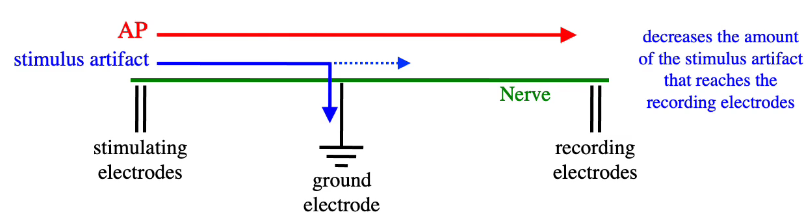
stimulus artifact is when a nerve is zapped with a certain amount of voltage, the voltage will __ spread down the nerve b/c the nerve is surrounded by __ __
uses a low resistance circuit where passively spreading electricity goes into the ground
grounding electrodes decrease the amount of the stimulus artifact that reaches the recording electrodes
__
stimulus artifact - is the recorded deflection (aka unwanted electrical field/signal recorded when the stimulus pulse is delivered)
“field” means that the stimulus artifact can hit 2 r.e. (1 +, 1 -) at the same time & subtract them to get a bipolar recording
stimulus artifact is when a nerve is zapped with a certain amount of voltage, the voltage will passively spread down the nerve b/c the nerve is surrounded by salty water/solution
remember:
stimulus artifact is different from stimulus pulse
in the Scope application in Stimulator box, you can change (4 ← and def. delay and duration)
delay (← from (0,0) of axes to onset)
duration (← time from onset to nothing happening anymore)
range
amplitude
why may this cause damage to the Powerlab: if range for Channel 1 is not changed from 20 mV to 5 V?
the preamp’s sensitivity is very high at 20 mV, so any large electrical signal from the stimulating electrode will overwhelm the preamp’s input circuit,
WHILE increasing the range will give you less sensitive data BUT the less sensitive data does include those within the larger voltage range of 5 V
if you adjust the preamp gains & some of the data goes off the screen, what could you do to fit it all on again? (2)
you can either:
decrease the sensitivity by increasing the range
decrease the gain
what are the 4 Mode settings of the stimulator? (def. briefly)
off
pulse (1 pulse in 1 sweep)
double (2 pulses in 1 sweep)
multiple (multiple pulses in 1 sweep)
__
sweep MB refers to filling the screen(?)
how to measure latency of the stimulus artifact
is the time delay between the start of the stimulus & onset of the stimulus artifact
5 parts of the neuron/nerve cell & briefly def. each
in lab 3: frog sciatic nerve lab, what specific part of the neuron are we looking at?
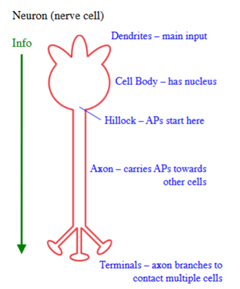
dendrites - main input
cell body - has nucleus
hillock - where APs start
axon - carries APs towards other cells
terminals - axon branches out to contact multiple cells
_
the sciatic nerve lab looks at the axon function (b/c we are cutting out the cell body & terminals)
membrane potential (Vm) is deter. by what 2 things?
& what are each of those 2 things based on?
__ potential balances the __ potential
_
give normal concentrations of Na+ and K+ inside and outside of a normal human/animal cell when at rest
_
give ex. of how both potentials work together
deter. by
chemical potential - based on concentration gradients
electrical potential - based on charge
(ex: if K+ exits the cell due to moving from its high→low concentration gradient, then the inside of the cell is now more “-” charged SO that attracts cations to enter the cell, such as K+)
electrical potential balances the chemical potential
_
inside:
[K+] = 150 mM
[Na+] = 15 mM
(separated by cell membrane)
outside:
[K+] = 5.5 mM
[Na+] = 150 mM

what does Nernst equation calculate for?
give formula & what does the variable stand for
is applied under what condition (1)?
what 3 assumptions are made related to this equation?
calculates the equilibrium potential (Eion) ← where Eion is the ELECTRICAL potential difference of an ion across the cell membrane that balances the chemical potential of that ion
when temp is at 37 degrees C
z = the ion’s charge
3 assumptions:
there is a voltage across the membrane
there is NO net flow of the ion across the membrane (b/c chemical and electrical potentials balance e/o out / aka are equal in strength but going in opposite directions)
if an ion crosses the membrane, Vm → Eion

give E_ion for Na+, K+, and Cl-, using the known concentrations
EK = - 90 mV
ENa = +61 mV
ECl = -70 mV
_
where chemical potential pulls Na+ into the cell, making the inside largely positive (aka “+” electrical potential) WHICH would repel the “+” charge of Na+, SO Na+ is pushed back out of the cell at the same rate

what does Goldman equation calculate for?
give formula & what does the variable stand for
is applied under what condition (1)?
__
give Vm at rest
at rest, compare permeabilities of Na+ and K+ (& explain why)
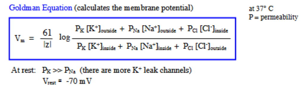
calculates the membrane potential of the cell
when temp is at 37 degrees C
P= = permeability
__
at rest:
Vm = -70 mV
PK » PNa
b/c at rest, there are more open K+ leak channels (than open Na+ leak channels) B/C the RMP is closer to the EK of -90 mV
for INTRAcellular recording
explain
when is it used?
insert a microelectrode into the neuron to measure voltage (aka membrane potential) of inside the cell & compare it to the reference electrode located outside the neuron → to get the true membrane potential (aka charge/potential across the cell membrane)
is good for recording the true Vm of ONE neuron
for APs and VT
t/f: APs are the rapid temporary rise and fall of electrical voltage across cell membrane that allows cells like neurons and muscle cells to communicate, and is activate by depolarizing above the VT
def. VT
t/f: threshold is not fixed, but depends on (what 2 variables? and explain ← 2)
explain: APs are considered to be “all-or-nothing” (2)
__
VT acts as a __ __
neurons use __ __ to code information (this means…)
true
__
VT - membrane potential at which an AP is fired/initiated
threshold is not fixed, but depends on amplitude/strength & duration of the stimulus
if have short stim. duration, then need higher stim. amp
if have longer stim. duration, then can have shorter stim. amp
__
APs are considered to be “all-or-nothing”
if de- makes Vm subthreshold → no AP fired
if de- makes Vm suprathreshold → AP is fired
__
VT acts as a noise filter
neurons use frequency modulation to code information
b/c neurons can’t change the signals by changing the size of the AP, so they can increase or decrease the frequency of APs firing, which affects which signals are sent (i.e. pressing harder on your finger to receptor potentials, so more likely to reach VT, so fire APs at a higher frequency)
AP phases are generated by (1), whose activity depends on the (1)
_
explain steps for each (including how they connect in the next stage): de-, re-, hyperpolarization, and returning to RMP/Vrest
__
match inactivation/deactivation aka closed state with time-dependent/voltage-dependent
AP phases are generated by voltage-gated (VG) ion channels, whose activity depends on the membrane potential (Vm)
_
depolarization:
a stimulus brings the Vm to or past VT
the VG-Na+ channels are activated → get large FNa (aka large electrochemical driving force of Na+)
Na+ influx → depolarization
repolarization:
VG-Na+ channels inactivate
b/c are time-dependent & the inactivation gate of these channels open and close very fast
VG-K+ channels activate
K+ efflux → rapid repolarization
hyperpolarization:
get excess K+ efflux
(b/c VG-K+ channels are slow to close)
return to Vrest:
VG-K+ channels deactivate
b/c are voltage-dependent, NOT time-dependent
VG-Na+ channels deactivate
b/c are voltage-dependent
& VG-Na+ channel deactivation is needed to removed inactivation of these channels
Vm (membrane potential) → returns to Vrest WHERE leak channels are the only activated channels
………..
inactivation is time-dependent
deactivation is voltage-dependent (aka channel closes due to change in Vm, specifically repolarization)
(image as extra reference)

APs are __ events that spread along the membrane
as APs spread, they __ so they don’t get smaller
very __ # of ions actually cross during an AP, so it __ affect the gradients
repolarization is NOT due to __ __ b/c …
IF THE MEMBRANE STAYS DEPOLARIZED, then (2)
APs are local events that spread along the membrane
as APs spread, they regenerate so they don’t get smaller
very small # of ions actually cross during an AP, so it doesn’t affect the gradients
repolarization is NOT due to ATPase pumps b/c ATPase pumps are too slow
(remember ATPase pumps 3 Na+ out, 2 K+ in)
IF THE MEMBRANE STAYS DEPOLARIZED, then:
Na+ channels stay inactivated
and you have to must repolarize in order to remove Na+ channel inactivation
def. refractory periods
name & def. the 2 types
what part of an AP is the duration of each refractory period type?
…
why do you need a “big” stimulus for that specific refractory period?
__
in lab 3: frog sciatic nerve lab, what are the 2/3 assumptions made above refractory periods?
how do you test these assumptions?
__
if you get RRP graph but want to get ARP graph, what do you do?
refractory period - how long a neuron must wait before it can fire a 2nd AP
absolute refractory period (ARP)
a 2nd stimulus will NOT fire a 2nd AP regardless of the stimulus strength b/c VG-Na+ channels are still inactivated
is duration of de- & re-
relative refractory period (RRP)
a BIG 2nd stimulus will fire a 2nd AP b/c there has been enough time to remove inactivation of VG-Na+ channels & there is still high K+ permeability bc/ some VG-K+ channels haven’t deactivated yet
is duration of hyper- & returning to Vrest
…
^ you need a “big” stimulus for RRP in order to fire a 2nd AP b/c the Vm is further away from the VT (b/c RRP is during hyper- and returning to RMP), so a bigger stimulus can cause bigger increase in Vm more increase likelihood of being suprathreshold and firing another AP
__
2 assumptions made above refractory periods:
we use very big stimuli, so any neuron in its relative refractory period (RRP) will fire a 2nd AP
if a neuron doesn’t fire a 2nd AP, it is in its absolute refractory period (ARP)
…
^ test by doing a small 1st stimulus pulse & very big 2nd stimulus pulse, where:
is ARP if there is no 2nd AP
is RRP if there is a 2nd AP due to the big second pulse
__
if you get RRP graph but want to get ARP graph, then make the 2nd pulse occur closer to the 1st pulse

explain the spread of AP along an axon
at rest, the inside Vm of the cell is “-” charged & outside is “+” charged
when there is a stimulus causes an AP to fire, there is local reversal of charge where at that local region of the axon, the inside of the cell is “+” charged & outside is “-” charged, while the non-local areas are the opposite/same as before
as AP propagates down axon, the previous segments return to same as before

2 things that affect conduction velocity & how
^ def. myelination
def. saltatory conduction (aka 3 parts)
_
name the 2 types of fibers (1 of them has 4 types ← differentiate b/w them), def. the 2 types, and give the conduction velocity of the 2 types
^ which of the 4 types of A fibers are most sensitive vs. least sensitive
also def. Type B fibers
_
the sciatic nerve is mostly made of type __ fibers
diameter
larger diameter = higher velocity
myelination (aka lipid bilayer wrapped around an axon that acts as insulation so the passively spreading depolarization/electricity down the myelin sheaths doesn’t fall below the threshold)
increased myelination = higher velocity
_
saltatory conduction:
AP jumps from node to node, while depolarization passively spreads down myelin
APs are regenerated at the nodes of Ranvier, so they don’t get smaller as they spread down the axon
APs spread much faster when myelinated
__
A fibers (Aalpha, Abeta, Agamma, Adelta)
large, myelinated
20-100 m/s
^ Aalpha fibers are the largest and most myelinated, while Adelta are the smallest and least myelinated
^ Aalpha fibers are the most sensitive (aka first to detect a CAP), while Adelta are the least sensitive
^ b/c larger diameter axons have lower thresholds
…
C fibers
small, unmyelinated
1-2 m/s
…
B fibers
myelinated, but have smaller diameters than Type A fibers
_
the sciatic nerve is mostly made of type A fibers
def. compound nerves
def. CAP (compound action potential)
^ __ diameter axons have __ thresholds
CAP is the __ of the individual __-__-__ APs of __ neurons; CAP is NOT an __-__-__ AP
what happens to CAP if you apply a very small stim, small stim, and as you increase the stim amplitude?
what does max CAP mean?
__
if there are 2 stimulating electrodes, what happens in the cell at the + vs. - stimulating electrode?
compound nerves - the bundles of different types of axons
CAP - the summation of individual APs
larger diameter axons have lower thresholds (Aalpha > Adelta)
CAP is the summation of the individual all-or-nothing APs of multiple neurons; CAP is NOT an all-or-nothing AP
__
if you apply a
very small stim → no CAP (b/c is below the threshold of all neurons)
small stim → very small CAP (b/c is only above the threshold of large diameter/low threshold Aalpha neurons)
as you increase the stim amplitude → CAP increases (b/c you start to recruit higher threshold neurons)
max CAP = all neurons have been recruited
__
in the cell @ the - stimulating electrode:
attracts cations to that local region → depolarization
& if suprathreshold, can fire an AP that spreads down either side of the axon
in the cell @ the + stimulating electrode:
attracts anions → hyperpolarization

for EXTRAcellular recording
explain
if there is 1 “+” and 1 “-” recording electrode, what type of recording do you get? (& how do you calculate it aka formula)
what does the voltage vs. time graph look like if local depolarization of the AP hits the negative r.e. only, then in b/w the 2 r.e., and then hits the positive r.e. only?
^ so at (-) r.e., depolarization is a __ deflection
^ so at (+) r.e., depolarization is a __ deflection
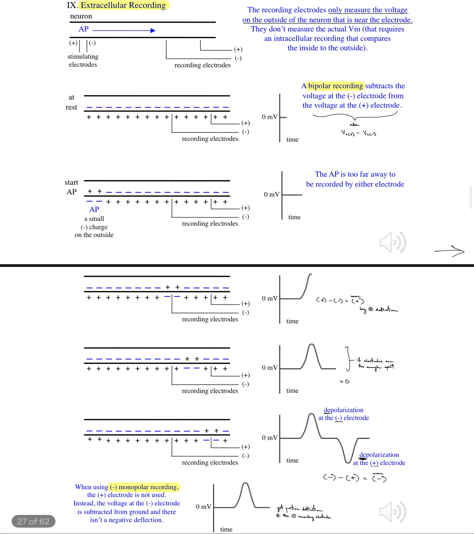
2 stimulating electrodes (+ and -) initiate AP that propagates down the neuron’s axon towards the 2 recording electrodes (+ and -), where the r.e. only measure the voltage on the outside of the neuron that is near/local to the electrode (meaning they don’t measure the actual Vm which requires intracellular recording)
_
get bipolar recording
Vr(+) - Vr(-) ← aka voltage at (+) r.e. minus the voltage at (-) r.e.
__
hits the negative r.e. only → get big positive deflection
(+) - (-) = more +
then in b/w the 2 r.e. → back to 0 mV
0-0 = 0
then hits the positive r.e. only → get a big negative deflection
(-) - (+) = more negative
…
^ so at (-) r.e., depolarization is a positive deflection
^ so at (+) r.e., depolarization is a negative deflection
cont. for EXTRAcellular recording
explain using a (-) monopolar recording (earlier was about bipolar recording)
how do you calculate the voltage
so you would get a __ deflection
only the (-) r.e. is used
Vground - Vr(-)
so you would get a positive deflection (at the negative r.e.)
MB don’t need to know
for lab 3: frog sciatic nerve, what was the setup of electrodes for:
the bipolar recording of CAP
finding threshold recruitment
looking at effects of stimulus polarity (2)
looking at conduction velocity (2)
refractory periods
strength-duration curve
bipolar recording of CAP:
(A) + s.e., (B) - s.e., (C) ground electrode, (D) - r.e., (E) + r.e.
threshold recruitment
(A) + s.e., (B) - s.e., (C) ground electrode, (D) - r.e.
looking at effects of stimulus polarity
from (A) + s.e., (B) - s.e., (C) ground electrode, (D) - r.e.
to switching positions of “+ and -” stimulating electrodes
looking at conduction velocity
from (A) + s.e., (B) - s.e., (C) ground electrode, (D) - r.e.
to moving the “-” r.e. 10 mm to (E)
refractory periods ← same as threshold recruitment
strength-duration curve ← same as threshold recruitment
aka starting from threshold, a (-) monopolar setup was used

how to calculate:
CAP latency
__
def. maximal vs supramaximal
_
2c) why does the CAP stop increasing even though the stimulus voltage is increased?
CAP latency is
time difference between onset of stimulus artifact & peak of CAP
__
maximal - stimulus voltage that first results in the largest response
supramaximal - any stimulus voltage above the maximal
_
even tho the stimulus voltage still increases, the CAP stops increasing in size b/c all the neurons have been recruited & brought to/above threshold (← seen as a plateau)
why does CAP have a longer latency & smaller amplitude when the (+) and (-) s.e. are switched to B and A positions respectively, instead of A and B, on the nerve chamber setup?
when (+) s.e. is first and (-) s.e. is second, the (-) s.e. attracts cations that can depolarize to be suprathreshold → AP, which has to travel less length from the (-) s.e. to the (-) r.e.
when (-) s.e. is first and (+) s.e. is second, the (-) s.e. attracts cations that can depolarize to be suprathreshold → AP BUT it has to travel a longer length towards the r.e. → resulting in longer latency
__
when (+) s.e. is first and (-) s.e. is second, AP is directly from (-) s.e. to r.e. so the amplitude is normal
when (-) s.e. is first and (+) s.e. is second, hyperpolarization from anions attracted to (+) s.e. is right after depolarization, SO some of the A fibers won’t reach threshold to fire → resulting in smaller CAP amplitudes

give formula for:
conduction velocity
diff. b/w “difference in latencies” vs. “_AP latency”
_
what is stimulus interval?
as intervals decrease b/w 2 CAPs, why does CAP 2 amplitude decrease?
conduction velocity = (distance b/w electrodes) / (difference in latencies)
where “difference in latencies” is the time difference b/w 2 CAP or MAP peaks
vs. “CAP/MAP latency” is the time from the onset of the stimulus artifact to either the CAP peak or the onset of MAP
__
stimulus interval is the time b/w offset of stimulus 1 & onset of stimulus 2 (aka time between 2 stimuli)
…
b/c some axons are still in their RRP (relative refractory period) from the first stimulus, which means that less axons have fully recovered SO less axons can fire again in response to the second stimulus → lower CAP 2 amp
explain why as CAP duration increases, the CAP latencies increase as well (include how threshold is involved)
(CAP latency is measured as the time b/w onset of stimulus artifact & peak of CAP)
b/c:
latency is the time it takes for the cations to accumulate at the (-) stimulating electrode, and in order to fire an AP, it takes time to accumulate cations to depolarize and reach VT
a longer CAP duration allows for more time for membrane to reach threshold to make up for the weak stimulus → so get longer CAP latency
def. rheobase & chronaxie (← MB have to know?)
__
why does stimulus with amp. below the rheobase not initiate an AP, even if it has a very long duration?
rheobase - the minimum voltage needed to get CAP when applied for an infinitely long duration
(so any stimulus with a voltage below the rheobase will never fire a CAP, regardless of the stimulus duration)
chronaxie - the duration of the stimulus needed when the amplitude is 2x the rheobase
(a lower chronaxie = tissue is very excitable, so acts like a fast nerve fiber)
(^^ both rheobase and chronaxie refer to the nerve, not the muscle, b/c of CAP)
__
in the case of 69 mV and threshold of 70 mV, an AP won’t be fired regardless of how long the duration of the stimulus is
b/c as cations gather at the (-) stimulating electrode, cations will repel e/o & also diffuse away → SO it never reaches that threshold
…
however, when an AP does fire, it is b/c cations gather fast enough to cause an AP to form (and they gather fast enough b/c of the strong stimulus from the “-” stimulating electrode)
NMJ (neuromuscular junction) is located b/w (2)
describe the anatomy of the NMJ, including all involved parts
^ vesicles are __ __ bags
there are ~300,000 vesicles/terminal & ~10,000 Ach/vesicle
synaptic cleft (def. it) is filled with what type of fluid?
for receptors:
def. cholinergic & nicotinic
t/f: nmAChRs are only located on skeletal muscle
nmAChRs are __-dependent __ channels that require __ ACh to activate
is a non-specific __ cation channel (meaning?)
t/f: b/c RMP is close to EK+, it means there is a larger force for Na+ influx and smaller force for K+ efflux, which is why nmAChR activation causes Na+ influx into depolarization
NMJ is b/w motor neuron terminals & skeletal muscle fibers
(smooth muscles don’t involve the NMJ)
anatomy:
motor neuron has synaptic vesicles filled w/ ACh & P/Q type Ca2+ channels (← which allow Ca2+ to enter the pre- terminal to cause vesicle exocytosis of ACh into the synaptic cleft)
synaptic cleft
endplate - is the specialized part of the postsynaptic membrane of a skeletal muscle that has a high density of nmAChRs
__
vesicles are lipid bilayer bags
there are ~300,000 vesicles/terminal & ~10,000 Ach/vesicle
synaptic cleft - is the space b/w pre- membrane and post- membrane
is filled w/ basal lamina
for Rs:
cholinergic - respond to ACh
nicotinic - sensitive to the drug nicotine
true (nmAChRs are only located on skeletal muscle)
nmAChRs are ligand-dependent ion channels that require 2 ACh to activate
is a non-specific mixed cation channel (meaning that it allows for Na+ influx & K+ efflux)
true
describe the mechanism: nerve AP → muscle AP
endplate potential (EPP) is __
how is it affected by small amount, enough, and large amount of ACh in the synaptic cleft?
for this lab we assume:
there is __ NMJ per skeletal muscle cell, and the NMJ is located where in the muscle cell?
Aalpha motor neuron is depolarized suprathreshold & fires an AP that propagates towards the pre- terminal
the de- activates the P/Q Ca2+ channels to allow Ca2+ influx
cause vesicle exocytosis of ACh into the cleft
ACh diffuses across the cleft
2 ACh bind to the nmAChR → to get nmAChR activation → causes post- skeletal muscle de- aka the EPP (endplate potential)
the EPP will spread to either side along the membrane to activate adjacent VG-Na+ channels → cause de- that will fire a MAP if it is suprathreshold
__
EPP is graded (a graded potential), so:
small amount of ACh → get small EPP that will not reach VT → so no MAP
enough ACh → get an EPP that is > VT → get MAP
large amount of ACh → get very large EPP → get MAP
(remember EPP is graded potential so EPP amp is dependent on stimulus strength, while MAP is all-or-nothing)
__
for this lab we assume:
there is 1 NMJ per skeletal muscle cell, and the NMJ is located in the middle of the muscle cell

describe 3 mechanisms of terminating depolarization across the endplate (include 1 enzyme)
is AChE breakdown of ACh fast? (give time)
stop vesicle exocytosis of ACh by BOTH:
Ca2+ diffusing away from vesicles
use ATPase pumps to pump Ca2+ from cytosol (inside) to outside the cell/motor neuron
ACh dissociates from nmAChRs → so nmAChRs deactivate → EPP fades away
ACh is removed from the synaptic cleft by:
ACh diffusing away
the enzyme acetylcholinesterase (AChE) breaks down ACh in the cleft → into acetate + choline, which happens very fast aka <1 ms
describe mech. of recycling ACh (include 3 “things”)
choline uptake into the pre- terminal via the Na+-choline cotransporter (← transports both Na+ and choline into the pre- terminal)
choline acetyltransferase (ChAT) will take choline + acetyl-CoA → into ACh + CoA
ACh will be packaged into vesicles via the vesicle ACh transporter (VAChT)
(note: the vesicles are formed from part of the pre- terminal membrane)

what causes fatigue? (aka 2 connecting parts)
why do you get depletion of ACh/vesicle?
what happens during recovery? how long does it take?
__
def. synaptic delay
it depends on (3 ← give the time each takes & the total synaptic delay)
BUT in lab, what do we measure as the synaptic delay?
why is this longer?
so the normal range of synaptic delay in lab is?
fatigue is caused by high frequency nerve stimulation → that depletes the ACh from vesicles
b/c vesicles are depleted faster than you can recycle them
recovery - is the recycling of ACh, recycling of vesicles, and filling up new vesicles w/ ACh
takes seconds to minutes (long time)
__
synaptic delay - delay b/w pre- depolarization & post- depolarization
depends on:
release of NTs (0.3 ms)
diffusion across cleft (0.05 ms)
nmAChR activation (0.15 ms)
total synaptic delay is ~0.5 ms
BUT in lab, we measure as the synaptic delay from pre- depolarization to when the MAP reaches the recording electrodes
this is longer b/c it includes the time for EPP to reach threshold & the time for MAP to spread to the recording electrodes
SO normal range of lab’s synaptic delay is 1-3 ms
def. facilitation
describe mech. of facilitation
facilitation - when the stimulus occurs at shorter intervals, the EPP amplitudes are increasing for a short-term
short intervals of stimuli (aka fast occurring stimuli) result in residual intracellular Ca2+ near the pre- terminal membrane
b/c not enough time for Ca2+ to diffuse away or be removed by the ATPase pump
→ which increases the activation of P/Q Ca2+ channels
get increased Ca2+ influx into pre- terminal → get increased ACh release → activate nmAChRs → get short-term growing EPPs


diff. b/w Aalpha motor neuron & skeletal muscle in terms of:
RMP aka Vrest
VT
absolute refractory period (ARP)
conduction velocity
__
what are other NTs in the body (4 incl. ACh), the Rs they act on aka “-nergic” (4), and what systems do they act on & the function?
what drug blocks mAChRs?
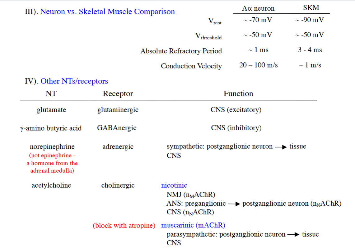
for Aalpha motor neurons: (← have faster ARP & conduction velocity)
RMP is -70 mV
VT is -50 mV
ARP is 1 ms
conduction velocity is 20-100 m/s
for SKM:
RMP is -90 mV
(remember that RMP is closer to EK+ b/c there are more K+ leak channels open at rest)
VT is -50 mV
(SKM need larger depolarization to reach threshold compared to Aalpha motor neurons)
ARP is 3-4 ms
conduction velocity is 1 m/s
__
glutamate — glutaminergic Rs
CNS (excitatory)
gamma-amino butyric acid — GABAnergic Rs
CNS (inhibitory)
norepinephrine (from adrenal medulla) — adrenergic Rs
CNS
sympathetic, specifically where postganglionic neuron meets the tissue
acetylcholine (ACh) — cholinergic Rs
for nicotonic (nAChR):
act on NMJ (nmAChR)
act on autonomic NS: where preganglionic neuron meets postganglionic neuron (nnAChR)
CNS (nnAChR)
for muscarinic (mAChR):
parasympathetic NS: where postganglionic neuron meets tissue
CNS
for myasthenia gravis syndrome
symptoms (1)
usually starts in (1 w/ 2) & progresses to (1 w/ 3)
cause (1)
how do you diagnose it? (2 ← 1 of them has 2)
(^ incl. name of drug w/ its def.)
treatment (2)
symptom:
muscle weakness/fatigue w/ repetitive use
usually starts in small cranial muscles, like extraocular & throat (so affects vision and swallowing)
& progresses to larger muscles, like arms, legs, and can affect breathing in some cases
diagnose it by:
having person do repetitive contractions until fatigue starts & then, inject edrophonium (← a short acting AChE inhibitor)
at 30 seconds → fatigue decreases
at 10 minutes → fatigue returns (b/c AChE returns/is no longer inhibited)
treatment:
long-acting AChE inhibitors
anti-immune therapy
should CAP and MAP thresholds be the same or different? why?
different b/c
for MAP, ACh has to travel across the cleft, bind to receptors and cause EPP that then has to active VG Na channels for depolarization suprathreshold to fire MAP
while CAP just has to travel down the axon w/in a nerve
in general, why do the CAP and MAP amplitudes increase w/ higher stimulus amplitudes?
b/c they involve more axons
for CAP: recruit more axons, so more APs contribute to the compound AP which increase CAP amp.
for MAP: recruit more axons, which so overall get more ACh released into the NMJ to activate more muscle cells → more MAPs can come together to make a larger MAP amp. (but there is a limit of the max MAP amp.)
why does CAP amp continue to increase after the max MAP has been achieved?
for CAP, more axons recruited just means getting a bigger compound AP amplitude
for MAP, once all the axons of the muscle have been recruited, then the MAP reaches its max MAP amp.
the time interval b/w the nerve AP (CAP) and the muscle AP (MAP) is composed of what 3 major factors/phases?
give formulas for:
conduction time
MAP latency
synaptic delay
this means that the conduction time for the MAP to spread along the sarcolemma and reach the muscle recording electrodes is negligible/insignificant
the conduction time of the CAP traveling along/propagating down the nerve
synaptic delay
the conduction time of the MAP to spread along the sarcolemma to the muscle recording electrodes
__
conduction time = (distance b/w “-” stimulating electrode & “-” muscle recording electrode) / conduction velocity
MAP latency is the time from the onset of the stimulus artifact to the ONSET of the MAP
synaptic delay = MAP latency - conduction time

if the muscle r.e.’s weren’t near the center of the muscle, would the calculated synaptic delay be increased, decreased, or the same? why?
increased
b/c MAP would need to travel a longer distance to the muscle r.e.’s, which takes more time → so have longer MAP latency
in the synaptic delay formula, a longer MAP latency will give you a longer synaptic delay
is there another way to accurately measure synaptic delay if you could use other equipment?
add more recording electrodes at different locations along the nerve and muscle (can look specifically at when APs end at a nerve & start on a muscle)
why is it important to know what the CAPs are doing when assessing IF facilitation is occurring?
if you are not at the max CAP, then facilitation isn’t happening
b/c if CAP amp. increases, then it means more axons are still being recruited, which will mean more axons are recruited to active more muscle cells/fibers to get a larger MAP amp. ← which isn’t facilitation
b/c at max CAP, the same number of axons in the nerve are being activated, so the increased MAP can only be due to the facilitation at the NMJ
if the data at a small time interval shows 5 CAPs but 3 MAPs, why is that?
b/c there is not enough time for the muscles to relax before contracting again (the interval is too short)
if there is a lot of ACh in the NMJ or that ACh stays in the NMJ for a long time, what happens to the MAP? name & explain this mech.
the MAP can decrease
b/c of of the depolarization blockade - where continued or high ACh presence in the NMJ causes the muscle fiber to get hyperexcitable at first, but becomes unresponsive to further stimulation
b/c the adjacent VG-Na+ channels that usually cause depolarization will be in a constant inactivated state (b/c constant depolarization means there is no chance to repolarize, so stay in inactivated state) → so you get less depolarization and lower MAP amp. over time
how do you know where fatigue occurs based on the graphs giving the CAP and MAP?
3 possible locations of fatigue: nerve, NMJ, muscle
fatigue at nerve: if CAP amp. decreases, which causes MAP amp. to decrease too
fatigue at NMJ: if CAP amp. is constant & there’s a big MAP aka big muscle contraction still
(aka conclude that fatigue is at the NMJ by eliminating that it’s not at nerve or muscle)
fatigue at muscle: little to no contraction as seen w/ decreased MAP amp.
basic anatomy of skeletal muscle
organize from big to small: muscle, myofibril, myofiber
describe myofibril and myofiber
__
for a myofibril:
def. sarcomere, myofilaments, Z-line
name the 2 myofilaments & diff. b/w them
name the 2 types of bands & diff. b/w them
skeletal muscle is __ muscle b/c of the (2)
muscle → myofiber → myofibril (helps to think of “e” before “r” in alphabet so myofibEr is bigger than myofibRil)
myofiber
is 1 muscle cell
is multinucleated (aka has multiple nuclei)
there is 1 NMJ/myofiber, where NMJ is always located in the center of a myofiber
diameter: 10-80 microns
spans length of the entire muscle
myofibril
have NO nuclei
diameter: 1 micron
__
for a myofibril:
sarcomere - the contractile part of myofibril, where myofilaments pull their Z-lines closer together to get tension & shorten the muscle
myofilaments - the individual protein strands that make up myofibrils
actin (thin filament)
myosin (thick filament)
Z-line - anchors actin in place (aka helps actin stay in place)
A band - the width of myosin, which includes “myosin + myosin/actin overlap”
I band - is only actin (is not the full length of actin b/c doesn’t include the myosin/actin overlap)
…
skeletal muscle is striated (striped) muscle b/c of the A and I bands

def. sarcoplasmic reticulum (SR)
def. sarcolemma (& describe ← 2)
def. T-tubule/transverse tubule (& describe ← 1)
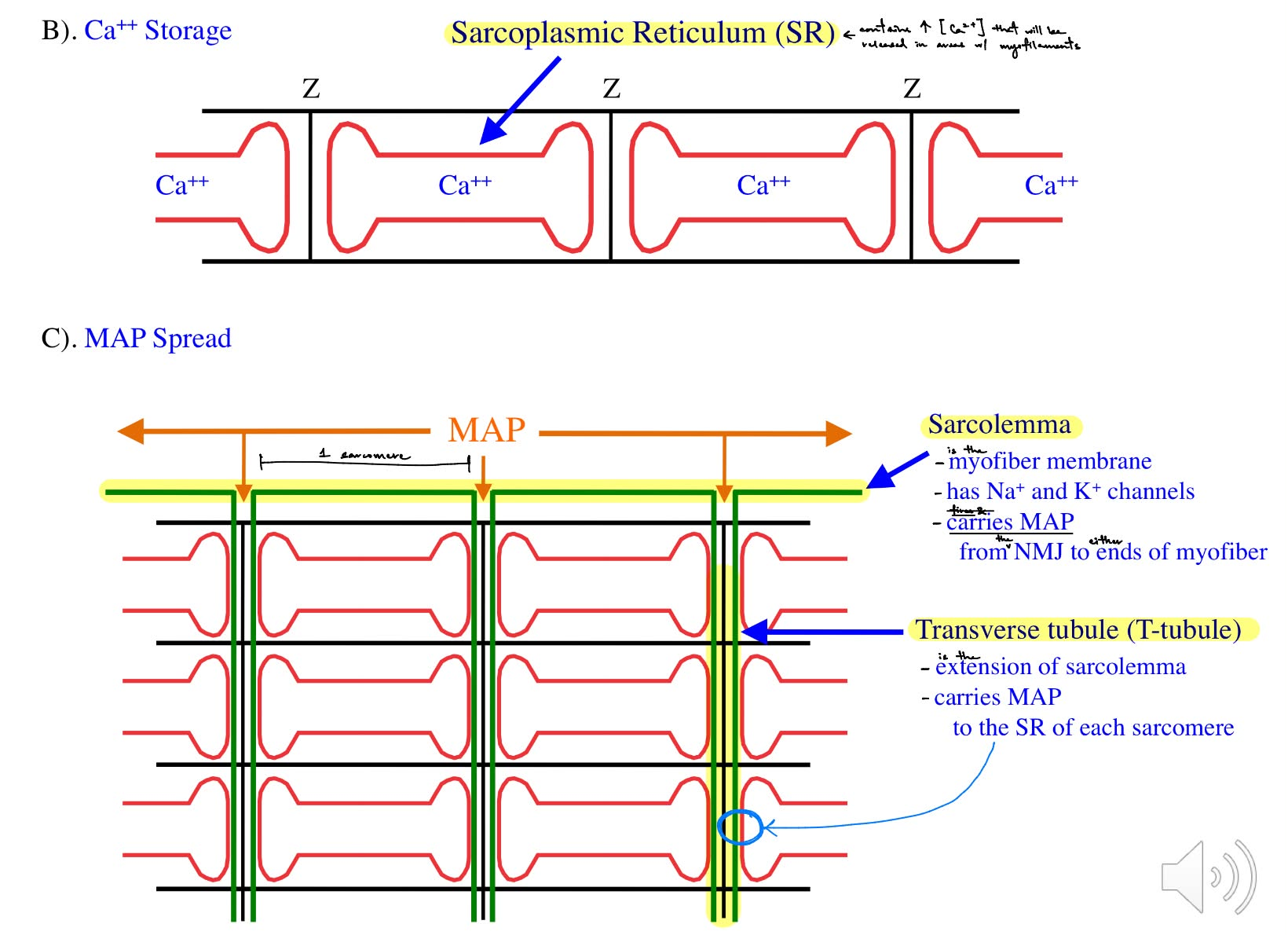
sarcoplasmic reticulum (SR) - contains high concentrations of Ca2+ that will be released in areas w/ myofilaments
sarcolemma - the myofiber membrane
has Na+ and K+ channels
carries MAP from the NMJ to either ends of the myofiber
transverse tubules (T-tubules) - the extension of the sarcolemma
carries MAP to the SR of each sarcomere
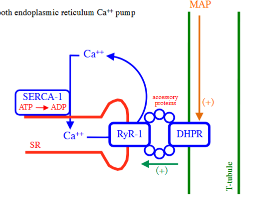
what 3 Rs/pumps are involved in Ca2+ release of skeletal muscles?
for 2 of them, state what type of Ca2+ channel they are & if they are voltage or ligand-dependent
for the last one, describe it (1)
__
explain the mech. of Ca2+ release in skeletal muscles
_
how do you stop a contraction in terms of SERCA pump?
RyR-1 (ryanodine receptors), which are SR Ca2+ channels (sarcoplasmic reticulum Ca2+ channels)
are ligand-dependent b/c mechanically coupled to DHPRs
DHPR (dihydropyridine receptors), which are LTCCs (L-type Ca2+ channels)
are voltage-dependent & long-activated/L-type
SERCA-1 (smooth endoplasmic reticulum Ca2+ pump)
uses ATP to pump Ca2+ back into the SR
__
mech. of Ca2+ release in skeletal muscles:
MAP spreads along the sarcolemma & down T-tubules towards the SR
depolarization of MAP activates DHPRs (a type of LTCCs that are V-dependent) located along the T-tubules
DHPR undergoes conformational change, involves connected accessory proteins → which trigger activation of RyR-1 channels
so get Ca2+ release from SR into the cytosol
myofilaments are now exposed to the Ca2+
where Ca2+ in the cytosol will bind to troponin of the complex, which causes conformation change of the complex → to move tropomyosin away from the myosin-binding sites of actin
myosin heads can now bind to actin → form crossbridges & powerstroke USING myosin ATPase uses converts ATP/energy
can trigger a contraction
__
to stop a contraction, the SERCA pump uses ATP to pump Ca2+ back into the SR (ATP → ADP)
describe mech. of a skeletal muscle contraction, in terms of the mechanical (start w/ myosin and actin) vs. electrical mechanisms (start at the Aalpha motor neuron)
def. fatigue
mechanically, what happens when the muscle is at rest aka not contracting?
__
skeletal muscle is __, meaning …
t/f: Abeta and Agamma motor neurons are NOT associated w/ MAP
t/f: you need to increase the permeability of Ca2+ across the SR membrane in order for Ca2+ to diffuse out of the SR into the cytosol for contractions
t/f: myosin ATPase is always active in SKELETAL muscle
mechanical part:
myosin binds to actin using energy/ATP, where myosin ATPase converts ATP → ADP
crossbridge forms → powerstroke
__
electrical part:
Aalpha motor neuron has an AP that travels to the pre- terminal
activate P/Q Ca2+ channels (which increases permeability of Ca2+ across the pre- membrane) → Ca2+ influx into the pre- terminal
ACh is released → diffuse across the cleft
nmAChR activation → (Na+ influx and K+ efflux b/c nmAChR is a non-specific mixed cation channel) → EPP
get MAP if EPP > VT
MAP spreads along the sarcolemma & down T-tubules
once MAP reaches the DHPRs (a type of LTCC) → (involves accessory proteins)
→ activate RyR-1 channels (a type of SR Ca2+ channel) (which increases the SR membrane’s permeability to Ca2+) → Ca2+ moves from the SR to the cytosol
Ca2+ in the cytosol causes:
Ca2+ to bind to the troponin/tropomyosin complex → complex undergoes conformational change → uncovers myosin-binding sites on actin → crossbridges → powerstroke
muscle contracts → increases tension and/or shortens the muscle
__
fatigue - ATP depletion (b/c not enough ATP for crossbridges to form & for powerstroke to occur)
…
mechanically, when the muscle is at rest aka not contracting:
the troponin/tropomyosin complex covers actin, which blocks the myosin-binding sites on actin
__
skeletal muscle is neurogenic, meaning it requires nerve input to trigger an AP/contraction
…
true: Abeta and Agamma motor neurons are NOT associated w/ MAP
true: you need to increase the permeability of Ca2+ across the SR membrane in order for Ca2+ to diffuse out of the SR into the cytosol for contractions
true: myosin ATPase is always active in SKELETAL muscle
describe mech. for skeletal muscle relaxation
if there is Ca2+ still present, what does this mean?
SERCA-1 pumps Ca2+ from the cytosol back into the SR
myosin unbinds from actin, so have no crossbridges
troponin/tropomyosin complex covers the myosin-binding sites on actin
__
if there is Ca2+ still present, then myosin will bind to actin to form crossbridges & have a powerstroke b/c Ca2+ binds to troponin, causing conformational change of the complex WHICH will pull away to expose the myosin-binding sites on actin

name the 3 types of muscle contractions/phases (not specific to any muscle)
def. each
~2 conditions needed for isotonic contraction/phase to occur
__
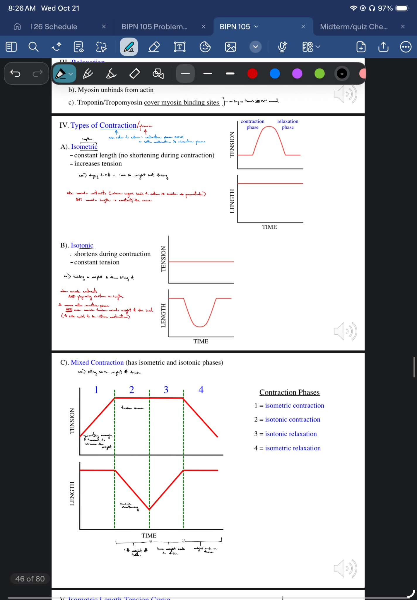
for the mixed contraction pic, state which contraction is which # 1-4
isometric
during an isometric contraction, the muscle has a constant length (doesn’t shorten during contraction), but tension increases
isotonic
during an isotonic contraction, the muscle shortens in length, but has constant tension
mixed contraction
has both isometric & isotonic phases
__
isotonic phase/contraction occurs:
after isometric phase/contraction has finished
AND once active tension overcomes the afterload weight
(for the pic, also look at the tension graphs and length graphs for the isometric and isotonic contractions/phases)

describe the isometric length-tension curve (aka what it shows)
def. passive tension, active tension, and total tension
why does the passive tension curve increase as muscle length increases?
t/f: passive tension does NOT involve actin/myosin interactions
why does the active tension curve increase and then decrease as muscle length increases?
__
(pic) explain what happens past E & b/w D to E, C to D & b/w C to B, B to A
show tension (y) vs. muscle length (x), where there are 3 curves representing passive, active, and total tension
…
passive tension - the force generated by stretching a relaxed muscle without any contraction, due to the elasticity of the muscle
active tension - the force generated by actin and myosin binding to form crossbridges during a contraction
total tension = passive tension + active tension
__
passive tension increases as muscle length increases b/c stretching a muscle (aka increasing muscle length) will pull on elastic non-contractile muscle structures, WHICH increases the amount of passive tension
true: passive tension does NOT involve actin/myosin interactions
__
active tension curve increases b/c you always start the the preload muscle length (L)
shortening the muscle (aka decreasing muscle length / going to the left of L) means that the muscle has shortened isometrically where active tension is trying to overcome the afterload, THEN shortens isotonically where the muscle shortens due to the now constant high active tension
active tension curve decreases b/c you always start the the preload muscle length (L)
if you stretch the muscle, the myofilaments will move too far apart & have no myofilament overlap, which means there is no crossbridge formation which decreases active tension
____
C to B: actins overlap block each other’s binding sites, fewer crossbridges possible
A to B: more actins overlap & myosins hit the Z lines more blocked binding sites, damage


give “formulas” for total tension
before a contraction
during a contraction
def. the total tension curve
actual tension developed during a contraction depends on the __
before a contraction:
total tension = passive tension
during a contraction:
total tension = passive tension + active tension
__
total tension curve - the max tension possible at a given length during an isometric contraction
….
actual tension developed during a contraction depends on the load
def. preload, afterload, total load
__
for preload:
what happens to muscle length if you increase the preload?
the preload sets (2)
for afterload:
afterload is felt by the muscle __ contractions start
what conditions will cause muscle to shorten vs. not shorten during a contraction, in terms of the afterload?
for total load:
what conditions will cause muscle to shorten vs. not shorten during a contraction, in terms of the total load?
__
For isometric length-tension curve, the afterload ONLY affects (1) and does NOT affect (1)
explain why
preload - the maximum stretch/load before a contraction (aka is the weight supported by the muscle that causes the muscle to stretch)
increasing the preload will increase the muscle length aka stretch the muscle more
preload sets the passive tension & muscle length before a contraction starts
afterload - the resistance/load that the muscle must overcome in order for the muscle to shorten & lift the load (aka is the weight NOT supported by the muscle so the muscle does NOT stretch)
afterload is felt by the muscle after contractions start
if active tension < afterload → no muscle shortening during the contraction (isometric contraction only)
if active tension > afterload → muscle shortens during the contraction (both isometric and isotonic contractions)
total load = preload + afterload
if total tension < total load → no muscle shortening during the contraction (isometric contraction only)
if total tension > total load → → muscle shortens during the contraction (both isometric and isotonic contractions)
__
For isometric length-tension curve, the afterload ONLY affects muscle length and does NOT affect total tension
b/c the muscle length is constant, meaning that there will be the same amount of myosin/actin binding and same amount of crossbridges formed

explain the pic
__
what happens if the afterload = 0?
knowing the 3 top right pictures,
during a contraction,
the muscle will first shorten isometrically in order to increase the active tension to overcome the afterload, while still maintaining constant length
THEN, the muscle will shorten isotonically as seen by moving left (towards decreased length) towards the total tension curve
^^ some only go through one of these phases, and are called pure isometric and pure isotonic contractions
(^^ LOOK at the red, blue, and green shapes that depict this)
notice that relaxation occurs in the opposite direction, where there is first isotonic relaxation phase & then isometric relaxation phase
(look at the arrows)
__
if the afterload = 0, then will only get isotonic contraction (pure isotonic contraction)
b/c start at the passive tension curve at L (the length of the muscle after being stretched by the preload), it can shorten isotonically b/c active tension > 0 (afterload)

for multiple muscle twitches (referring to MAPs)
state the 2 things that can happen at low frequency (← include the term & its def. for 1 of them)
what can happen at medium frequency (aka name the term, define it, and state the condition in which this occurs)
^ include how long skeletal MAP durations & twitch durations are
__ events are much shorter than __ events
summation is of __ events (aka __, not __)
what always occurs at high frequency (aka name the term & define it)
muscles with high frequency stimulus will __ the fastest b/c …
at lower frequency:
get MAPs of the same amp. repeatedly, or
get treppe - get increasing MAP amplitudes with every stimulus b/c there is increasing SR Ca2+ release for every contraction UNTIL it maxes out
at medium frequency:
summation - where previous MAPs add onto newly firing MAPs to get high MAP amplitudes
occurs b/c the skeletal MAP duration (<5 ms) is much shorter than the twitch duration (10’s of ms), SO another twitch can be triggered when the previous twitch hasn’t completely relaxed SO the twitch tensions summate
aka 1 MAP causes 1 twitch (mechanical event) → before that twitch is done, another MAP is fired that causes another twitch → twitches start summating
^^ electrical events are much shorter than mechanical events
^^ summation is of mechanical events (aka twitches, NOT MAPs)
at high frequency:
tetanus - when the muscle has generated its max tension, so has a sustained contraction
muscles with high frequency stimulus will fatigue the fastest b/c they use ATP the fastest

def. motor unit
__
Usually in humans, an AP in the Aalpha motorneuron will generate a MAP/contraction in __ of the motor unit’s muscle fibers because __ ACh is released at each NMJ to generate EPPs > VT of __ muscle fiber.
In frogs, some of the presynaptic terminals may __ release enough ACh for the EPP to reach VT, so a MAP/contraction will __ be initiated in some of the muscle fibers.
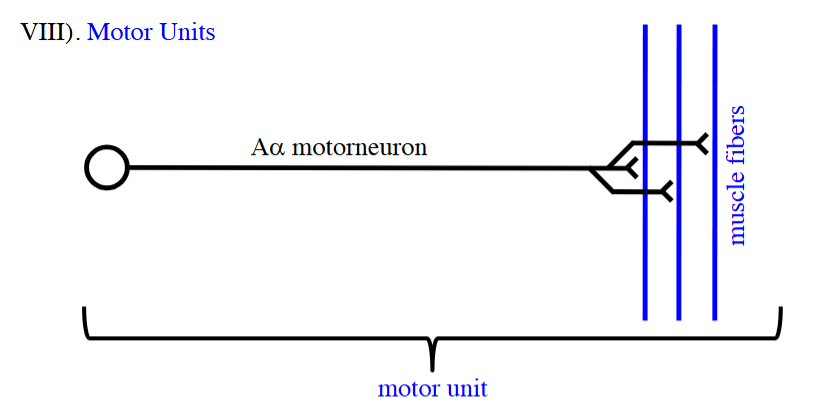
motor unit - the Aalpha motor neuron & all the skeletal muscle fibers/myofibers that it innervates
__
Usually in humans, an AP in the Aalpha motorneuron will generate a MAP/contraction in all of the motor unit’s muscle fibers because enough ACh is released at each NMJ to generate EPPs > VT of each muscle fiber.
In frogs, some of the presynaptic terminals may NOT release enough ACh for the EPP to reach VT, so a MAP/contraction will NOT be initiated in some of the muscle fibers.
when you make a calibration curve, which variable is the x vs. y axis?
purpose of calibration equation
weight (g) is the x-axis
transducer output voltage (mV) is the y-axis
…
purpose of calibration equation is to convert volts into the appropriate units
for lab 5: frog skeletal muscle
what is the solution used to fill up the muscle bath? & its purpose
what was used to stimulate the sciatic nerve?
__
how do you find & solve for the active tension when looking at the graph?
the sciatic nerve is in a muscle bath filled with Ringer’s solution (← keep isolated muscle alive so that we can see contractions occurring outside of the body)
use a suction electrode to stimulate the sciatic nerve
__
how to find & solve for the active tension when looking at the graph:
find the contraction that has the largest amplitude (“peak mV - baseline mV”) ← aka this is the largest contraction
then, use the calibration equation to convert the transducer output voltage from mV to grams of active tension
~ review:
what causes summation vs. tetanus?
summation is caused by medium frequency stimulation,
where skeletal MAP durations being a lot shorter than twitch durations, so rapid nerve pulses/MAPs arrive before a skeletal muscle fiber can fully relax SO leftover Ca2+ ions will build up & increase tension (of the muscle
twitches)
tetanus is caused by very high frequency stimulation,
where muscles do not relax at all (no relaxation phase), which locks the muscles in a sustained contraction
cont. for lab 5: frog skeletal muscle
purpose of using Grass stimulator
what factors affect how long a muscle fiber takes to fatigue? (2)
__
which muscle fiber type is most likely to fatigue first?
which muscle fiber type is resistant to being fatigued & is most likely maintaining the low amount of tension during the plateau?
the Grass stimulator delivers controlled electrical pulses for nerve or muscle stimulation
__
how long muscle fiber takes to fatigue depends on:
the muscle fiber type
the stimulus frequency b/c that affects how frequently the muscle fiber is being stimulated/contracted & using up ATP
__
fast-twitch muscle fiber type is most likely to fatigue first
slow-twitch muscle fiber type is resistant to being fatigued & is most likely maintaining the low amount of tension during the plateau
for SMOOTH muscle
for a myofiber:
in broad terms, how does the diameter & length of smooth muscle myofiber compare to skeletal muscle myofiber?
1 myofiber has __ nucleus/nuclei
the myofilaments in smooth muscle:
are (2)
do they have sarcomeres?
this means that there is __ __ of myofilaments throughout the cell → so the myofiber is NOT __ (aka is __)
myofiber of a smooth muscle:
smooth muscle myofiber is a lot smaller in diameter and length compared to a skeletal muscle myofiber
1 myofiber has 1 nucleus/nuclei
the myofilaments in smooth muscle:
are actin & myosin
NO sarcomeres
this means that there is random distribution of myofilaments throughout the cell → so the myofiber is NOT striated (aka is smooth)
for smooth muscle
first, what are the 3 phases of AP? (this relates to below Q’s answer)
describe the mech. of APs, in terms of:
slow wave depolarization at rest
depolarization
repolarization
spread of AP
_
slow wave depolarization at rest acts as a __, which shows that smooth muscle is __ (← def. term)
slow wave depolarization at rest:
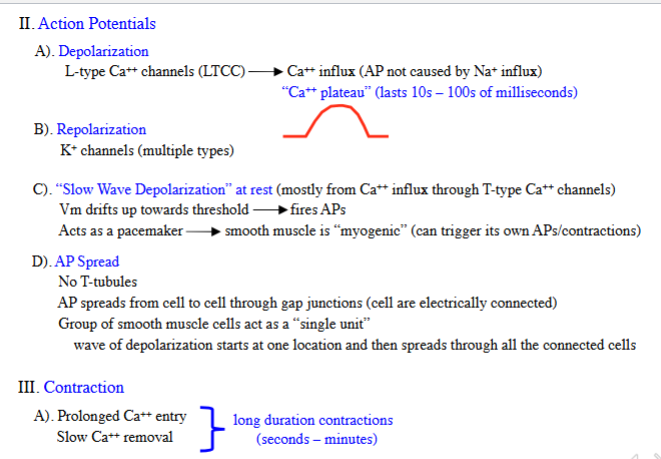
at rest, there some Ca2+ influx through T-type Ca2+ channels (← are low-voltage-dependent/activated)
so the membrane potential drifts up towards VT → fires AP if suprathreshold
depolarization:
L-type Ca2+ channels (LTCCs) are activated → Ca2+ influx
get Ca2+ plateau of 10s-100s of ms
(^ AP is not caused by VG-Na+ channels and Na+ influx like it is in SKM)
repolarization:
multiple types of K+ channels open → K+ efflux
(^ unlike the 1 type: VG-K+ channels in SKM)
spread of AP:
smooth muscles do not have any T-tubules, so AP spreads from cell-to-cell via gap junctions b/c cells are electrically connected
a group of smooth muscle cells act as a single unit
so, a wave of depolarization will start at one location & spread through all connected cells
_
slow wave depolarization at rest acts as a pacemaker, which shows that smooth muscle is myogenic (← the muscle can trigger its own APs and contractions)
for smooth muscle
describe the mech. of contraction (2 parts ← electrical and mechanical part)
smooth muscle has __ duration contractions (lasting …)
smooth muscle contractions have a __ increase in tension & __ ATP use, meaning the muscle __ fatigue
have prolonged Ca2+ influx
& slow Ca2+ removal
so
have slow myosin-actin interactions
__
as a result:
smooth muscle has long duration contractions (lasting seconds to minutes)
(due to longer Ca+ influx and slower Ca2+ removal)
smooth muscle contractions have a slow increase in tension & low ATP use, meaning the muscle doesn’t fatigue
(due to slow crossbridge formation & powerstroke)
describe the mech. of triggering a smooth muscle fiber/myofiber contraction
t/f: unlike skeletal muscle, myosin ATPase is NOT always active in smooth muscle
both skeletal and smooth muscle have the same trigger for contractions, which is __
Ca2+ influx through LTCCs, and sometimes, also get Ca2+ from the SR into the cytosol
(main source of Ca2+ is extracellularly from LTCCs)
4 Ca2+ bind to CAM (calmodulin) to form the Ca2+-calmodulin complex
the Ca2+:CAM complex binds to & activates the MLCK
the activated MLCK will use ATP to add a phosphate group from ATP to MLC (myosin light chain) & will phosphorylate the inactive myosin ATPase into active myosin ATPase w/ an attached phosphate group
now that myosin ATPase is active (so can use ATP to do crossbridge and powerstroke) & have phosphorylated MLC (so myosin head can bind to bind) → then, contraction occurs
__
true: unlike skeletal muscle, myosin ATPase is NOT always active in smooth muscle
both skeletal and smooth muscle have the same trigger for contractions, which is Ca2+
know the full names of: calmodulin (CAM) & myosin light chain kinase (MLCK)

the goal is to (1) in order for smooth muscle fibers to relax
describe the mech. of relaxation for a smooth muscle fiber/myofiber (incl. both electrical and mechanical parts)
goal: to remove Ca2_ from the cytosol
1 Ca2+ is pumped out & 3 Na+ are pumped into the cytosol via the Na+-Ca2+ exchanger (NCX) ← which uses/is driven by the Na+ gradient
Ca2+ is also pumped back in the SR via SERCA
-1Ca2+ also inhibits LTCCs in order to prevent more Ca2+ influx
Ca2+ also activates K+ channels, which cause repolarization & further inhibits LTCCs
ALSO, the myosin light chain phosphatase (MLCP) will remove the phosphate group from MLC, which turns active myosin ATPase into inactive myosin ATPase → so get reduced crossbridge cycling & powerstroke → which causes relaxation
know the full names of: myosin light chain phosphatase (MLCP) & Na+-Ca2+ exchanger (NCX)

smooth muscle contractility is regulated in the autonomic nervous system, specifically the parasympathetic & sympathetic NS
for parasympathetic NS:
what is the involved NT (1)?
__ __ receptor (1) and its G protein?
its effect
for sympathetic NS:
what is the involved NT (1) and hormone (1) from where?
__ receptor (2) and its G protein?
which NT and/or hormones affect each receptor?
its effect (1 each)
for parasympathetic NS:
NT: ACh
muscarinic cholinergic receptor (m3AChR) ← Gq transduction pathway
contraction
for sympathetic NS:
NT: norepinephrine (NE)
hormone: epinephrine (E) from the adrenal medulla
adrenergic receptors:
α1R ← Gq transduction pathway
activated by both NE & E
contraction
β2R ← Gs transduction pathway
activated by only E
relaxation

name the 2 local effects of smooth muscle contractility
^ describe mech. of stretch-induced contractions
^ describe effect on metabolic state
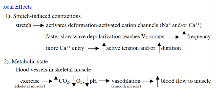
cause stretch-induced contractions
stretch activates deformation-activated cation channels of Na+ and Ca2+
for Na+:
get faster “slow wave depolarization” → so reach VT faster → which increases the frequency of smooth muscle contractions (aka higher frequency of smooth muscle contractions)
for Ca2+:
get more Ca2+ influx → (increased crossbridge cycling & powerstroke) → increases active tension and duration of smooth muscle contraction
affects the metabolic state
exercise of skeletal muscles → (higher CO2/lower O2 & pH) → vasodilation aka relaxation of smooth muscles → increased blood flow to the exercising skeletal muscle
how do these hormones affect smooth muscle contractility (aka state the involve G protein transduction pathways & its effect):
angiotensin I and II
oxytocin
epinephrine
angiotensin I and II
can act on Gq AND Gi transduction pathways
contraction
oxytocin
Gq transduction pathway
contraction
epinephrine
Gq OR Gs transduction pathway
either contraction or relaxation depending on the type of transduction pathway

def. resting tone
def. tone
give description/formula for resting tone aka passive tension for SKM vs. smooth muscle
def. plasticity
describe 2 effects w/ its specific G protein
__
in smooth muscle, why is the increase in passive tension higher than what would be predicted by the passive tension curve?
what about what causes the decrease in passive tension (that occurred after the increase)?
resting tone - the baseline tension maintained by a muscle at rest (aka passive tension ← but is calculated differently in SKM vs. SM)
tone - a state of partial sustained/prolonged smooth muscle contraction (due to maintaining a state of long-term crossbridge formation)
__
for SKM: resting tone = passive tension
for SM: passive tension = elastic elements + AP-independent crossbridges
__
plasticity - the passive tension curve can change as AP-independent crossbridges reset due to hormones, stretch, time, etc.
where Gq pathway can increase passive tension curve
where Gs pathway can decrease passive tension curve
__
increase in passive tension is higher than what would be predicted by the passive tension curve b/c of AP-independent crossbridges
the decrease in passive tension is b/c of plasticity, where AP-independent crossbridges detach to release active tension

4 things to measure in order to look at changes in smooth muscle contractility
active tension
duration
frequency
resting tone
for smooth muscle
connect the 3 types of G proteins to if they contract or relax
name 5 receptors for smooth muscles that were in LE & state what G protein transduction pathway(s) each has
Gq - contraction
Gs- relaxation
Gi - contraction
__
α1R — Gq
β2R — Gs
m3AChR — Gq
oxytocin R — Gq
angiotensin II receptor (type 1) — Gq and Gi
describe the mech. of Gq transduction pathway
the Gq receptor is activated, which activates the Gq protein → beta and gamma subunits split away from the alpha subunit
alpha subunits drops off GDP & adds GTP → have activated Gαq subunit
… (^ above happens for all transduction pathways) …
Gαq activates the membrane enzyme PLC (phospholipase C)
PLC will break down PIP2 into DAG and IP3
DAG stays in the membrane & activates PKC (protein kinase C)
→ PKC activates LTCCs, which increases Ca2+ influx → increases contractibility
→ PKC inhibits K+ channels, which delays repolarization → so LTCCs stay open longer → increases contractibility
IP3 go the cytosol → binds to IP3Rs on the SR → Ca2+ influx from SR to the cytosol → increases contractibility

describe the mech. of Gs transduction pathway
what does PDE stand for?
the Gs receptor is activated, which activates the Gs protein → beta and gamma subunits split away from the alpha subunit
alpha subunits drops off GDP & adds GTP → have activated Gαs subunit
… (^ above happens for all transduction pathways) …
Gαs activates adenylyl cyclase (AC) → AC converts ATP into cAMP
cAMP activates PKA, and then, cAMP is broken down by phosphodiesterase (PDE) into AMP
PKA activates K+ channels → get early repolarization so close LTCCs earlier and get less Ca2+ influx → decrease contractibility/increase relaxibility
PKA inhibits MLCK & activates MLCP → get less active myosin ATPase → decrease contractibility/increase relaxibility

describe the mech. of Gi transduction pathway
the Gi receptor is activated, which activates the Gi protein → beta and gamma subunits split away from the alpha subunit
alpha subunits drops off GDP & adds GTP → have activated Gαi subunit
… (^ above happens for all transduction pathways) …
Gαi inhibits adenylyl cyclase (AC) → less ATP is turned into cAMP aka less cAMP → (so less PKA is activated → have more active myosin ATPase compared to inactivate myosin ATPase) → increase contractibility
cAMP is broken down by phosphodiesterase (PDE) into AMP, which means you have no to very little cAMP → (so less PKA is activated → have more active myosin ATPase compared to inactivate myosin ATPase) → increase contractibility

review:
name the different types of GqRs, GsRs, and GiRs mentioned in LE regarding smooth muscle (not skeletal muscle)
for smooth muscle
GqRs:
m3AChR
oxytocin R
angiotensin II R
α1R
GsRs:
β2R
GiRs:
angiotensin II R