Rad II: Thoracic Radiographs
 Call Kai
 Learn
Learn Practice Test
 Spaced Repetition
 Match
Match Flashcards
 Knowt Play
1/23

There's no tags or description
Looks like no tags are added yet.
Name | Mastery | Learn | Test | Matching | Spaced | Call with Kai | Chat |
|---|
No analytics yet
Send a link to your students to track their progress
24 Terms
List indications for thoracic radiographs:
Coughing
Respiratory distress
Cardiac evaluation
Metstasis check
Where do you measure for thoracic radiographs?
Caudal border of the thoracic scapula
What is the cranial landmark for a thoracic radiograph?
Manubrium (thoracic inlet)
What is the caudal landmark for a thoracic radiograph?
Halfway between the xiphoid and the last rib.
Which view would be best in diagnosing lung disease?
RL
LL
VD
Which view would be best in diagnosing heart disease?
RL
DV
VD
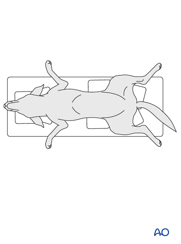
When should you never use a VD view on a patient?
Extreme respiratory distress.
How many ribs do dogs and cats have?
26 total- 13 pairs.
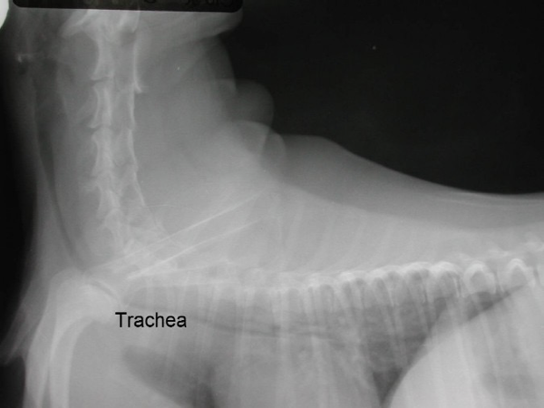
What is tracheal collapse?
The tracheal rings are weak or have a decreased amount of cartilage, causing the rings to collapse.
What animals are more prone to tracheal collapse?
Older
Obese
Toy breeds
What are the c/s of tracheal collapse?
“Goose honk” cough
Exercise intolerance
What are the radiographic signs of tracheal collapse?
Narrowing of the trachea.
What is the difference pulmonary edema and pleural effusion?
Edema- fluid in the lung tissue
Effusion- fluid in the thoracic cavity around the lung tissue
Define pneumothorax:
Entry of air into the pleural cavity in sufficient quantity to cause collapse of the lung consequent respiratory embarrassment.
Where do we measure the patient?
Thickest part of the thorax
Behind the caudal border of the scapula
Where should the center beam be?
Caudal aspect of the scapula.
Where do we place the markers?
Axillary region. (armpit)
Exposure should be taken at peak ____________.
Inspiration.
During expiration, the diaphragm is more __________ than during inspiration.
Cranial.
What is the alpha-2-agonist that we administered in lab?
Dexmedetomidine.
When would a DV view be preferred over a VD view for thoracic films?
For cardiac rads.
You are doing a thoracic metastatic check on a patient. What views/positions would you take?
RL
LL
VD
The sternum and vertebral column should be superimposed on a VD thoracic view.
True or false?
True.
One important positional parameter for the thorax is to position the forelimbs cranially and hold/sandbag them.
True or false?
True.