Anatomy and Physiology - EXAM 1
 Call Kai
 Learn
 Practice Test
 Spaced Repetition
 Match
 Flashcards
 Knowt Play
1/507

There's no tags or description
Looks like no tags are added yet.
508 Terms
Endocrine System
Composed of ductless glands that synthesize and secrete hormones, which are released into the blood and transported throughout the body
Exocrine System
Secreting substances to go outside of the body; includes the:
Salivary/merocrine gland: secretion through exocytosis
Mammary/apocrine gland: secretion through budding
Sebaceous/holocrine gland: secretion through cell disintegration
Target Cells
Have the specific receptors for a hormone to bind to it and respond
Hormone transport to target cells
Hormones released into interstitial fluid and then enter blood
Transported within blood
Randomly leave blood and enter interstitial fluid
Hormone binds to target cells’ receptors
Similarities between nervous and endocrine
Two control systems of the body
Release ligands - chemical messengers which bind to cellular receptors on particular target cells
Differences between nervous and endocrine
The endocrine system:
Transmits hormones through the blood
Targets any cells in the body with correct receptors (can be very widespread)
Exhibits longer reaction times
Has longer-lasting effects (minutes to days and weeks)
General functions of endocrine
Regulating development, growth, and metabolism
Maintaining homeostasis of blood composition and volume
Controlling digestive processes
Controlling reproductive activities
Endocrine glands
Contain epithelial tissue, which makes and releases hormones within a connective tissue framework
Endocrine organs (solely endocrine function)
Pituitary, pineal, thyroid, parathyroid, and adrenal glands
Organs with clusters of endocrine cells
Hypothalamus, skin, thymus, heart, liver, stomach, pancreas, small intestine, adipose connective tissue, kidneys, and gonads
Endocrine reflexes
Hormonal stimulation, humoral stimulation, nervous system stimulation
Hormonal stimulation (endocrine reflex)
Release of another hormone triggers hormone release
Anterior pituitary releases thyroid-stimulating hormone (TSH)
TSH stimulates thyroid gland to release thyroid hormone (TH)
Humoral stimulation (endocrine reflex)
Changes in level of nutrient or ion in the blood triggers hormone release
Blood glucose levels increase
Increased blood glucose stimulates pancreas to release insulin
Nervous system stimulation (endocrine reflex)
Stimulation of the nervous system triggers release of the hormone
Sympathetic division is activated
Sympathetic preganglionic axons stimulate the adrenal medulla to release epinephrine and norepinephrine
Steroids (circulating hormone)
Lipid-soluble molecules synthesized from cholesterol
Includes gonadal steroids (for example, estrogen)
Includes steroid synthesized by adrenal cortex (for example, cortisol)
Calcitriol sometimes classified in this group, but more accurately called a sterol
Examples of steroids
Estrogen, progesterone, testosterone, cortisol, aldosterone
Biogenic amines/monoamines (circulating hormone)
Modified amino acids
Water-soluble EXCEPT for thyroid hormone (TH)
TH is nonpolar (made from a pair of tyrosines) and lipid soluble
Examples of biogenic amines/monoamines
Catecholamines (norepinephrine, epinephrine), thyroid hormone, melatonin
Proteins (circulating hormone)
• Most hormones are in this category
• Water-soluble chains of amino acids
Examples of proteins (circulating hormone)
Antidiuretic hormone, insulin, glucagon, growth hormone, erythropoietin
Local hormones
Signaling molecules that don’t circulate in blood
Autocrine stimulation
When local hormones bind to the cells that release them
Paracrine stimulation
When local hormones bind to cells neighboring the cells that release them
Eicosanoids (local hormone)
Formed from fatty acids within phospholipid bilayer of membrane (water-soluble)
Synthesized through an enzymatic cascade
Eicosanoid formation
• Phospholipase A2 removes 20-carbon fatty/arachidonic acid from phospholipid
• Other enzymes convert arachidonic acid to a subtype of eicosanoid
Examples of eicosanoids
Prostaglandins, thromboxanes, leukotrienes
Can act as local vasoactive substances (causing vasoconstriction/-dilation)
Prostaglandin (eicosanoid)
• Stimulate pain and inflammatory responses
• Aspirin and other nonsteroidal anti-inflammatory drugs block prostaglandin formation
What blocks phospholipase A2 before it can remove arachidonic acid from the plasma membrane?
Steroid drugs
What blocks cyclooxygenase before it synthesizes prostaglandins and thromboxanes?
NSAIDs (ex: aspirin)
What blocks lipoxygenase before it synthesizes leukotrienes?
St John’s wort
Carrier proteins
Lipid-soluble hormones require these to be transported in the blood because lipid-soluble hormones do not dissolve readily in the blood
Are water-soluble proteins made by the liver
Carriers protect hormones from early destruction
Binding between hormone and carrier is:
Temporary; attachment, detachment, reattachment are common
Bound hormone
Most of the hormone (90% or more) is bound to the carrier
Unbound (free) hormone
Able to exit blood and bind to target cell receptors; only 10%> of the hormone
Most water-soluble hormones travel freely through blood, but…
A few use carrier proteins to prolong their life
A hormone’s blood concentration depends on
How fast it is synthesized and eliminated
Hormone release and blood concentration are
Positively correlated
Hormone elimination
Enzymatic degradation in liver cells
Removal from blood via kidney excretion or target cell uptake
The faster the elimination rate, the lower the blood concentration and vice versa
Half-life
Time necessary to reduce a hormone’s concentration to half of its original level; depends on how efficiently it is eliminated
Hormones with short half-life must be secreted:
Frequently to maintain normal concentration
Half-life of water-soluble hormones
Generally short; ex: small peptide hormones have a half-life of a few minutes
Half-life of steroid hormones
Generally long because carrier proteins protect them; ex: testosterone half-life is 12 days
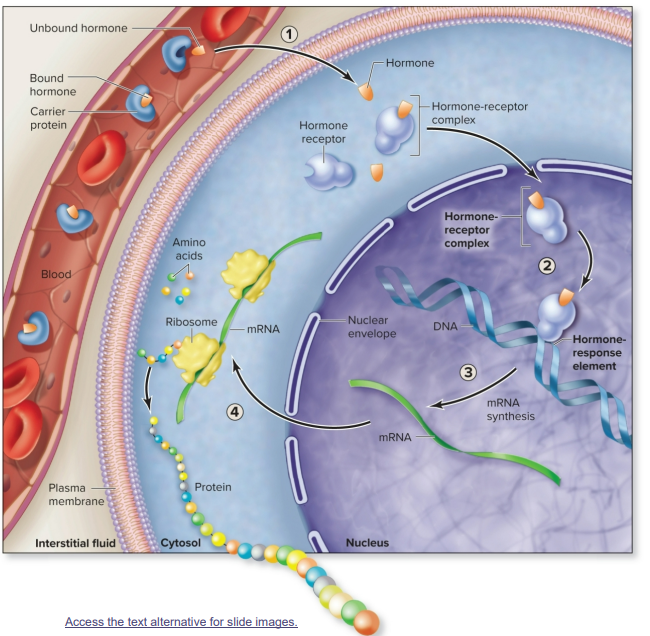
Steps of lipid-soluble hormones binding to intracellular receptors
The unbound hormone diffuses readily through the plasma membrane and binds with an intracellular receptor in the cytosol or nucleus to form a hormone-receptor complex (HRC)
The HRC binds with a specific DNA sequence called a hormone response element (HRE)
Binding of the HRC to the HRE stimulates mRNA synthesis in the nucleus
mRNA exits the nucleus and is translated by a ribosome in the cytosol. A new protein is synthesized
Unbound hormone + intracellular receptor = HRC → HRC + HRE = mRNA synthesis → mRNA + ribosome = new protein
Protein synthesis caused by a lipid-soluble hormone may result in:
An alteration in cell structure (e.g., as occurs with a greater level of sex hormones during puberty)
A shift in the target cells’ metabolic activities if the newly synthesized proteins are enzymes
Hormone-receptor complex (HRC)
Formed when a lipid-soluble hormone binds to an intracellular receptor in the cytosol or nucleus
Hormone-response element (HRE)
A specific DNA sequence that a HRC (lipid-soluble hormone + intracellular receptor) binds with
Steps of water-soluble hormones binding to membrane receptors
Signal transduction pathway (because water-soluble hormones are not polar and can’t diffuse through the membrane)
Hormone is the first messenger—it initiates events by binding to a G-protein coupled receptor and inducing a shape change
Inactive G-protein binds to the activated receptor
GDP is “bumped off” of the G-protein, and GTP binds to it instead, activating the G-protein
Activated G-protein (with GTP) is released from the receptor and moves along the inside of the plasma membrane, resulting in the formation or availability of the second messenger by activating effector proteins (adenylate cyclase or phospholipase C)

Can water-soluble hormones ever pass into the cell?
NO
The signal transduction pathway is carried out within the cell, but water-soluble hormones only ever act on plasma membrane receptors
G-protein
An internal membrane protein that binds to a guanine nucleotide
What is “bumped off” of the G-protein and replaced during the signal transduction pathway of water-soluble hormones?
GDP (guanosine diphosphate)
What binds to the G-protein to activate it during the signal transduction pathway of water-soluble hormones?
GTP (guanosine triphosphate)
Steps of G-protein activating adenylate cyclase
After hormone (for example, glucagon) binds to its receptor, G-protein is activated
Activated G protein binds to and activates plasma membrane adenylate cyclase
Adenylate cyclase converts ATP molecules to cAMP molecules
cAMP serves as the second messenger by activating protein kinase A

Protein kinase
Phosphorylating enzyme that adds phosphate to other molecules, activating or inhibiting them as a result
Glucagon > adenylate cyclase > cAMP > protein kinase A
Epinephrine > phospholipase C > PIP2 > DAG > protein kinase C OR > IP3 > Ca2+ > protein kinases
Phosphorylation
Adding phosphate to other molecules; this typically activates other molecules and continues the cascade.
This is done by protein kinases.
Dephosphorylation
Removing phosphate from other molecules; this typically deactivates other molecules and ends the cascade.
This is done by phosphatases.
What is the second messenger when adenylate cyclase is activated?
cAMP molecules
Steps of G-protein activating phospholipase C
After hormone (for example, epinephrine) binds to its receptor, G-protein is activated
Activated G protein binds to and activates plasma membrane phospholipase C
Phospholipase C splits PIP2 into two second messengers: DAG (diacylglycerol) and IP3 (inositol triphosphate)
DAG activates protein kinase C (a phosphorylating enzyme)
IP3 increases Ca2+ by stimulating its release from the endoplasmic reticulum and entry across the plasma membrane from the interstitial fluid
Ca2+ acts as a third messenger to activate protein kinase by directly binding to the enzymes or first binding to calmodulin. Ca2+ may also alter activity of ion channels within the plasma membrane by binding with them to increase the cell’s ion permeability

What are the second messengers when phospholipase C is activated?
DAG (diacylglycerol) and IP3 (inositol triphosphate)
What is the third messenger when phospholipase C is activated (IP3 route)?
Ca2+
One hormone can have multiple actions through different signal transduction pathways, including:
• Activation or inhibition of enzymatic pathways
• Growth through cellular division
• Release of cellular secretions
• Changes in membrane permeability & muscle contraction or relaxation
Significance of the intracellular enzyme cascade
• Signal is amplified at each enzymatic step (just a few hormone molecules can change many molecules within cell)
• There are many places to regulate pathway activities (ex: through hormone therapy)
Signaling pathway controls
Cells possess mechanisms to quickly inactivate intermediaries (ex: to break down second messengers)
A cell’s response to a hormone varies with:
Its number of receptors for the hormone
Its simultaneous response to other hormones
Fluctuation in receptor number
Up-regulation: increases number of receptors and sensitivity to hormone when blood levels of hormone are low (compensatory mechanism/homeostasis)
Down-regulation: decreases number of receptors and sensitivity to hormone when blood levels of hormone are high
Either occur with changes in development, cell cycle, and cell activity
Hormone interactions
Different hormones can simultaneously bind to a cell, and their effects may interact; can be synergistic, permissive, or antagonistic
Synergistic interactions
Hormones work together to produce a greater effect (ex: estrogen and progesterone’s effects on a target cell)
Permissive interactions
First hormone allows action of second hormone (ex: oxytocin’s milk ejection effect requires prolactin’s milk generating effect, so prolactin » oxytocin)
Antagonistic interactions
One hormone causes opposite effect of another hormone (ex: glucagon increases blood glucose while insulin lowers it)
Anatomic relationship of the hypothalamus and the pituitary gland
Hypothalamus controls pituitary, which controls several other endocrine organs
Pituitary gland (hypophysis)
Pea-sized
Lies inferior to hypothalamus in sella turcica of sphenoid bone
Connected to hypothalamus by infundibulum
Partitioned into anterior and posterior pituitary

Posterior pituitary (neurohypophysis)
• Smaller, neural part of pituitary gland
• Only stores and releases hormones; doesn’t make any itself
• Hypothalamic neurons project through infundibulum and release hormones in posterior pituitary
• Somas in supraoptic nucleus and paraventricular nucleus (neurosecretory cells)
• Axons in hypothalamo-hypophyseal tract of infundibulum

Hypothalamo-hypophyseal tract (posterior pituitary)
Axons of the supraoptic nucleus and paraventricular nucleus run from the hypothalamus into the posterior pituitary
Ends of these axons are within the posterior pituitary
Tract
Bundle of axon within the CNS
Nucleus
Housing structure of a group of cell bodies in the CNS
Hormones stored and released by the posterior pituitary
Antidiuretic hormone (ADH) and oxytocin (OT), which are made in hypothalamus by neurosecretory cells
• Packed in secretory vesicles, transported by fast axonal transport along unmyelinated axons to synaptic knobs in posterior pituitary
• Released into blood when neurons fire impulses
Antidiuretic hormone (ADH)/vasopressin
Made in supraoptic nucleus, secreted by hypothalamus in response to dehydration
Functions:
decrease urine production
stimulate thirst
constrict blood vessels
Oxytocin
Made in paraventricular nucleus
Functions
uterine contraction
milk ejection
emotional bonding
Anterior pituitary (adenohypophysis)
Hypothalamus hormonally stimulates anterior pituitary to release its hormones
• Hypothalamus secretes regulatory hormones
• Travel via portal blood vessels to pituitary
• Anterior pituitary secretes hormones into general circulation
Hypothalamo-hypophyseal portal system
Blood vessel network consisting of:
primary plexus: associated with the hypothalamus
secondary plexus: associated with anterior pituitary (endocrine tissue)
hypophyseal portal veins: drain primary plexus and transport to secondary plexus

Releasing hormones (hypothalamus’ regulatory hormones)
Increase secretion of anterior pituitary hormones
Include:
thyrotropin-releasing hormone (TRH)
prolactin-releasing hormone (PRH)
gonadotropin-releasing hormone (GnRH)
corticotropin-releasing hormone (CRH)
growth hormone-releasing hormone (GHRH)
Inhibiting hormones (hypothalamus’ regulatory hormones)
Decrease secretion of anterior pituitary hormones
Include:
prolactin-inhibiting hormone (PIH)
growth-inhibiting hormone (GIH)
Hormones synthesized and released by the anterior pituitary
Follicle-stimulating hormone (FSH)
Luteinizing hormone (LH)
Adrenocorticotropic hormone (ACTH)
Thyroid-stimulating hormone (TSH)
Prolactin (PRL)
Endorphins
Growth hormone (GH)
Remember FLAT PEG
Thyroid-stimulating hormone (TSH; thyrotropin)
• Release triggered by TRH from hypothalamus
• Causes release of thyroid hormone (TH) from thyroid gland
Prolactin (PRL)
• Release triggered by PRH, inhibited by PIH from hypothalamus
• Causes milk production, mammary gland growth in females
Adrenocorticotropic hormone (ACTH; corticotropin)
• Release triggered by CRH from hypothalamus
• Causes release of corticosteroids (including glucocorticoids like cortisol) by adrenal cortex
Gonadotropins: follicle-stimulating hormone (FSH) and luteinizing hormone (LH)
• Release triggered by GnRH from hypothalamus
• In female: regulate ovarian development and secretion of estrogen and progesterone
• In male: sperm development and secretion of testosterone
Growth hormone (GH; somatotropin)
• Release triggered by GHRH, inhibited by GIH
• Causes liver to secrete insulin-like growth factors 1 and 2
• GH and IGFs function synergistically to stimulate cell growth and division
What tissues do GH and IGFs act on?
Acts on all body tissues, but especially muscle, bone, cartilage, and adipose connective tissue (body fat)
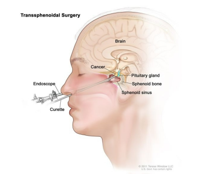
Clinical view: hypophysectomy
• Surgical removal of the pituitary gland because of tumors
• Preferred surgical approach through nasal cavity
• Various hormones need to be replaced and their levels need to be monitored
Regulation of growth hormone release
Release controlled through hormonal stimulation (GHRH and GHIH) from hypothalamus
GHRH: growth hormone-releasing hormone
GHIH: growth hormone-inhibiting hormone
Factors that impact whether the hypothalamus releases GRH
A person’s age, time of day, nutrient levels, stress, exercise
Effects of growth hormone
Stimulates release of IGFs from liver (similar functions as GH but longer half-life)
GH and IGFs stimulate
Increased protein synthesis, cell division, cell differentiation (all cells have receptors for either or both)
Release of nutrients from storage (glycogenolysis, gluconeogenesis, glycogenesis, lipolysis, lipogenesis)
Glycogenolysis
GH/IGF stimulated
Breakdown of glycogen into glucose
Gluconeogenesis
GH/IGF stimulated
Conversion of nutrients/noncarb sources/glucogenic amino acid to glucose
Glycogenesis
GH/IGF stimulated
Synthesis of glycogen from glucose
Lipolysis
GH/IGF stimulated
Breakdown of triglycerides into glycerol and fatty acids
Lipogenesis
GH/IGF stimulated
Formation of triglycerides from glycerol and fatty acids
Growth hormone release steps
Several factors influence the release of GHRH from the hypothalamus to the hypothalamo-hypophyseal portal system
The anterior pituitary releases GH
GH stimulates hepatocytes (liver cells) to release insulin-like growth factors (IGFs) into the blood
Both GH and IGF stimulate effectors (ALL CELLS, bone, muscle, liver tissue, adipose connective tissues)
Net effect: stimulates growth; nutrient release into blood
Increased GH and IGF levels inhibit GHRH release; increased GH levels inhibit further GH release
Growth hormone declines with:
Age