ANPH 1000 - Unit 3
 Call Kai
 Learn
 Practice Test
 Spaced Repetition
 Match
 Flashcards
 Knowt Play
1/80

There's no tags or description
Looks like no tags are added yet.
81 Terms
Where are mechanoreceptors found? (3)
connective tissue
sensitive, hairless areas on skin (ie. fingertips, lips, palm)
Subcutaneous tissue under skin & joints
Sensory receptor
Specialized cell that respond to specific type of stimuli
What information about the stimulus does sensory receptors tell you about? (3)
Type
Location
Intensity
Receptive field
Area monitored by 1 neuron
Sensory projection
Brain identifies where stimulus is from
Chemoreceptors
Odors, taste, chemicals
Mechanoreceptors
Change position of receptor
ie. pressure, stretch, vibration
Thermoreceptors
Change in temp
Nociceptors (Where, what, & type of stimuli)
Pain receptors w/ free nerve endings
Skin, mucous membranes, all organs except brain
Damage from trauma, heat, chemicals, lack of O2
Photoreceptors
Rods - Black & white
Cones - Color
Fast pain fibers
Sharp, localized, stabbing pain at the time of injury
Myelinated
Slow pain fibers
deep body organs, dull, aching pain
unmyelinated

Pain Pathway
Stimuli → Nociceptors
Pain → dorsal horn
SpinoTHALAMIC tract → Thalamus → Postcentral gyrus (Aware of pain) (5)
SpinoRETICULAR tract → hypothalamus & limbic (emotional & behavioural response) (6)
Referred Pain
Pain at location other than site of stimulus
Happens bcs sensory fibers from organ & the skin converge
Analgesics
Pain relievers
Warm receptors
dermis
Activated above 25*C
> 48*C → burning
Cold receptors
Epidermis
20*C - 30*C
< 10*C → less signals
Anesthetic
Pain receptors activate
Proprioceptors (Where & what)
skeletal, muscle, joints, & tendons,
Body movement, muscle stretch, position
Inner ear → balance
Gustation
Chemicals on taste buds
Papillae (What does it contain & where is it found)
Protrusions on tongue
Taste buds are found ALONG & BETWEEN papillae
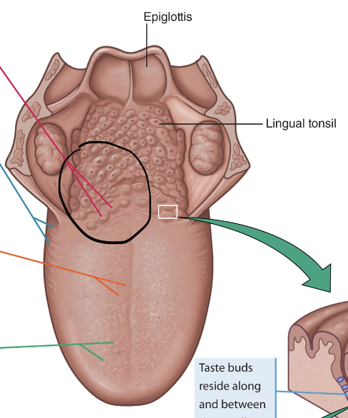
Vallate papillae
Large papillae at back of tongue
Have half of taste buds

Foliate papillae
Sides of tongue

Filiform papillae
Thread-like
Figure out texture of food
NO taste buds

Fungiform Papillae
Tip & sides of tongue

Fill in the blank
taste
saliva
stimulate
cranial nerves
facial (VII)
glossopharyngeal nerve (IX)
Vagus nerve (X)
Where are olfaction receptor cells?
Roof of nasal cavity → ethmoid bone pores → CN I
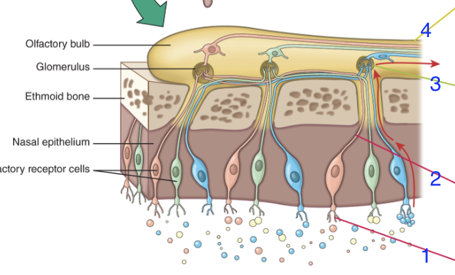
What are steps 1 to 4
Odour binds to cilia projecting from ends of receptor cells
Signal → nerve fibers in ethmoid bone pores
Fibers → glomeruli (olfactory bulbs) => partial impulse processing
Signal → primary olfactory cortex
Auricle/Pinna (What is it & function?)
Visible part of ear
Funnel sound → auditory canal

Where does the auditory canal start/end?

Temporal bone → eardrum
External auditory meatus
Outside opening of auditory canal
What is cerumen? What does it do? (2)
Secretions from canal glands + dead skin cells
Waterproof canal
Traps dirt & bacteria
How does cerumen leave the ear?
Dries & propelled by jaw movements

Auditory ossicles
Middle ear
Malleus (hammer)
incus (anvil)
stapes (stirrup)
Oval window: Where inner ear begins, connected to stapes

Tympanic membrane (2)
Separates outer & middle ear
Vibrates freely w/ sound waves

Auditory/eustachian tube (3)
Passageway from middle ear → nasopharynx
Equalize pressure on both sides of eardrum
Allows infection to spread from throat to middle ear
Bony labyrinth
temporal bone passageways
Houses inner ear
Membranous labyrinth
Lines inside bony labyrinth
Perilymph
Fluid that fills space b/w bony & membranous labyrinths
Endolymph
Fluid that fills inside membranous labyrinth
Semicircular canals
Equilibrium & balance

Vestibule
Entrance to labyrinths
Has balance organs
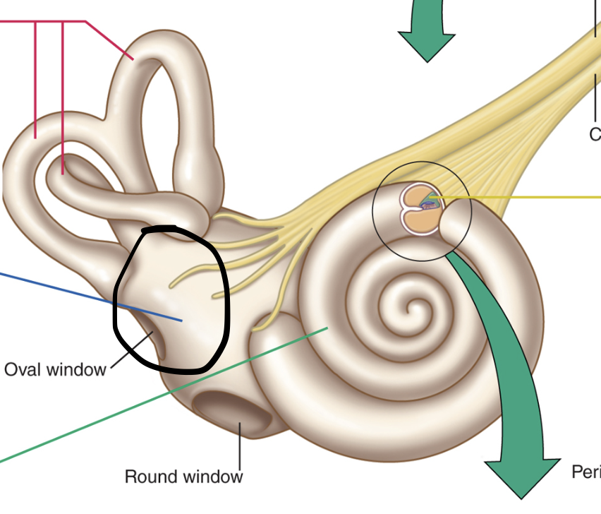

Cochlea
Hearing structures

Cochlear duct
Triangular
Filled w/ endolymph
Outer 2 compartments have perilymph
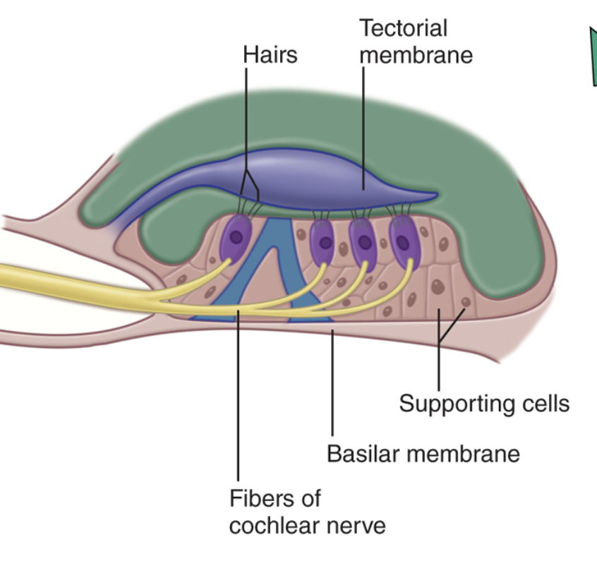
Organ of Corti
Hearing (sensory & supporting cells + hair cells)
Basilar membrane
Hair cells form CN VIII
Tectorial membrane
Gelatin membrane on top of hair cells

How does hearing work?
Sound waves → external auditory canal → tympanic membrane vibrates
Vibration → malleus, incus, & stapes
Stapes vibrates oval window → perilymph fluid shakes
Perilymph moves cochlear duct → hair cells (Organ of Corti) are stimulated to send nerve impulse → CN VIII → auditory cortex → temporal lobe
Vibration dissipate by striking round window
Why are the 3 fluid-filled semicircular canals at right angles from each other?
Each canal can be stimulated by diff head movement
Ampulla (3)
Bulb-like area at end of each canal
Has hair cells at cupula
Cupula: floats in endolymph in semicircular canals

Vestibule (3)
Utricle & saccule: sense organs
Tips of hair cells are covered by gel
Otoliths: Mineral crystals embedded in gel

Dynamic equilibrium (2)
Semicircular canals → endolymph moves & bends hair cells
Detects speed & direction of head movement
Static equilibrium (2)
Utricle & saccule → otoliths shift & bend hair cells
Detect head position when stationary & acceleration in straight line
Eyebrow (3)
Enhance facial expressions (nonverbal communication)
Keep perspiration out of eye
Shield eye from glare
Eyelids (palpebrae) (4)
Orbicularis oculi muscle + skin
Protect from foreign bodies
Block light when closed for sleeping
Periodic blinking washes out debris & moistens eyes w/ tears
Palpebral fissure
Opening b/w eyelids
Eyelashes (2)
Keep debris out of eyes
Touching lashes stimulate blink reflex
Conjunctiva (4)
Transparent mucous membrane, lines inner surface of eyelid
Covers anterior surface of eyeball EXCEPT cornea
Secretes thin mucous film → keeps eyeball moist
Vascular
Tarsal glands
tarsal plate: thickened area of eye
Secretes oil to slow evaporation of tears
Forms barrier seal when eyes are closed

Eyebrow
eyelids
palpebral fissure
eyelashes
conjunctiva
tarsal glands

Lacrimal punctum
Lacrimal apparatus
Tiny pore at end of lacrimal canals
Drains tears → lacrimal canals & nasolacrimal duct

Lacrimal gland & tears (4)
Lacrimal apparatus
Secretes tears that onto conjunctiva surface
Tears
Clean & moisten eye’s surface & deliver O2 + nutrients to conjunctiva
Have lysozyme to prevent infection

Nasolacrimal duct
Carries tears → nasal cavity
Accomodation
Curvature of lens changes so eye can focus on near object
Adaptation
↓ of sensation that occurs after continuous exposure to stimulus
Sclera (3)
Outermost layer, white part of eye
Blood vessels & nerves run through sclera
Fibrous outer layer
Cornea
Allows light → eye (No blood vessels)
Fibrous outer layer
Iris
Ring of coloured muscle
Controls # of light entering eye
Vascular Middle layer
Ciliary body
Thick extension of choroid, collar around lens
Secretes aqueous humour
Vascular Middle Layer
Choroid
Vascular tissue
Supplies O2 & nutrients → retina & sclera
Vascular Middle layer
Retina
Light-sensitive cells
Neural inner layer
CN II
Exits posterior eyeball
Sends signals → brain
Neural inner layer

Name 1-7
Sclera
Cornea
Iris
Ciliary body
Choroid
Retina
Optic Nerve
Macula lutea
Central point of retina
Fovea centralis
Most of cones → sharpest vision
No rods
Optic disc
Blood vessels enter/leave eye
Where nerve fibers join & form optic nerve
Produces blind spot (no photoreceptors)

Macula lutea
Fovea centralis
Optic disc
Anterior cavity
Space b/w lens & cornea
Anterior chamber: front of iris, filled w/ aqueous humour
Posterior humor: behind iris but in front of lens
Lens
Changes shape for near & far vision
Posterior cavity
Vitreous humor: semi-solid, keeps eyeball from collapsing