Spinal Cord and Peripheral Nervous System
 Call Kai
 Learn
 Practice Test
 Spaced Repetition
 Match
 Flashcards
 Knowt Play
1/28

There's no tags or description
Looks like no tags are added yet.
29 Terms
Attention-Deficit/Hyperactivity Disorder (ADHD)
is a neurodevelopmental condition characterized by inattention, impulsivity, and hyperactivity
Brain imaging studies have found that people with ADHD often show structural and functional differences in the thalamus
Differences in Individual with ADHD
Reduced thalamic volume: some studies show slightly smaller thalamic size in individuals with ADHD, especially in specific nuclei related to attention and sensory processing.
Altered Connectivity: The thalamus has abnormal communication with the prefrontal cortex, basal ganglia, and default mode network, all of which are crucial for executive function and attention control
EF: a set of mental skills that help us manage our behavior and cognitive abilities to achieve goals, including planning, organization, working memory, and self control
Neurotransmitter Dysregulation: the thalamus uses neurotransmitters such as dopamine and norepinephrine
Stimulant medications (like methylphenidate and amphetamines) help restore balance in these signaling pathways
Differences in ADHD Between Men and Women
ADHD symptoms experienced by males and females are more alike than different, but there are striking differences when they are asked about their levied experiences with ADHD
Women and girls with ADHD tend to have higher rates of co-existing depression and anxiety
Boys are diagnosed with ADHD 2-3 times as often as girls and women to be referred for services even when their symptom profiles are exactly the same
Researchers are investigating whether the various rates of diagnosis are due to true differences in sex or other factors, like gender bias or variations in presentation or symptoms
Solutions
Break down tasks: instead of a large goal, focus on the very next, smallest action
For example: “put dishes in the sink” vs “clean the kitchen”
Use short timers: commit to a task for a short, defined period, like 20 minutes to reduce overwhelm
Momentum may carry you forward after the timer goes off
Make it interesting: Create a more pleasant environment by making the task itself more rewarding. This could mean working in a different location or using a hot water bottle or comfortable chair to improve experience
Leverage interest: ADHD motivation is often driven by interest rather than importance. FInd ways to tie tasks to your interests
PNS
everything not brain and spinal cord
The PNS is consists of nerves and ganglia
Most of the PNS structures we discuss are connected to CNS, motor, and sensory elements
Nerves
A bundle of fibers that receives and sends messages between the body and the brain
The messages are sent by chemical and electrical changes in the cells that make up the nerves
Cranial Nerves
originate from the brain
The cranial nerves are primarily responsible for the sensory and motor functions of the head and neck
There are 12 cranial nerves; these are designated Cn I through CN XII for “Cranial Nerve,” using Roman numerals for 1 to 12
Depending on the axons that are found within, they can be classified as sensory nerves, motor nerves, or a combination
The cranial nerves are numbered for the order in which they exit the brain from anterior to posterior
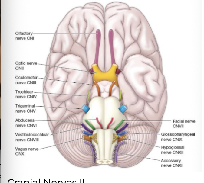
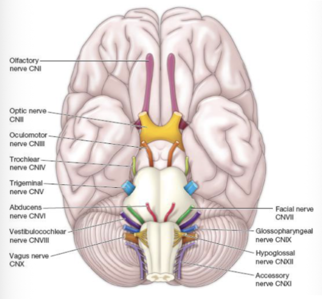
olfactory nerve (CN I) and optic nerve (CNII)
The olfactory nerve (CN I) and optic nerve (CNII) are responsible for the senses of smell and vision

oculomotor nerve (CN III)
The oculomotor nerve (CN III) is responsible for eye movements by controlling four of the extracurricular muscles. It is also responsible for lifting the upper eyelid and for constriction of the pupil

fascial nerve
The fascial nerve (CN VII) is responsible for the muscles involved in facial expressions, as well as for part of the sense of taste and the production of saliva

vagus nerve
The vagus nerve (CN X) is responsible for contributing to homeostatic control of the organs of the thoracic and upper abdominal cavities
Spinal Nerves
Originate from the spinal cord
There are 31 spinal nerves, named for the level of the spinal cord at which each one emerges
8 cervical nerves designated C1 to C8
12 thoracic nerves designated T1 to T12
5 lumbar nerves designated L1 to L5
5 sacral nerves designated S1 to S5
1 pair of coccygeal nerves
The nerves are numbered from the superior to inferior positions
Organized into plexuses: a network of nerves that combines fibers from different spinal nerves to create individual nerves that serve specific functions in the body
Plexuses
Plexuses are described as woven from spinal nerves and giving rise to certain systemic nerves, but they contain fibers that serve sensory functions or fibers that serve motor functions
Some fibers extend from peripheral sensory surfaces and send information to the CNS
Other fibers are the axons of motor neurons of the anterior horn of the spinal cord
Of the four nerve plexuses, two are found at the cervical level, one at the lumbar level, and one at the sacral level
Plexus=woven nerves

cervical plexus
branches into nerves in the posterior neck and head

brachial plexus
gives rise to the nerves of the arms, as “brachial” suggests

lumbar plexus
arises from all the lumbar spinal nerves and gives rise to nerves enervating the pelvic region and the anterior leg
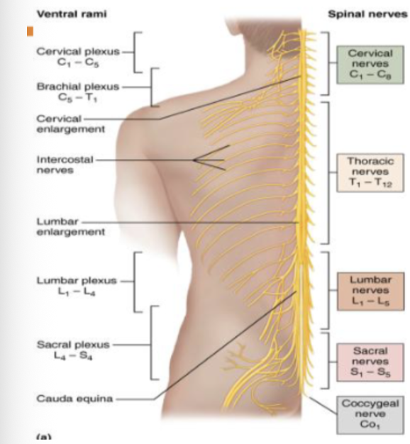
sacral plexus
contains sciatic nerve that extends across the hip joint

ganglion
A ganglion: a group of neuron cell bodies in the PNS
Present as “knot” or cluster of cell bodies outside of the brain/spinal cord
It’s the fibers (axons) from these cells cells that make up nerves themselves
Ganglia can be categorized, for the most part, as either sensory ganglia or autonomic ganglia
anterior median fissure
The anterior surface of the spinal cord is marked by the anterior median fissure
posterior median sulcus
the posterior midline is marked by the posterior median sulcus
cervical and lumbar enlargements
The cervical and lumbar enlargements occur in the regions that contain an increased number of axons entering and exiting the cord from the upper/lower limbs
The spinal cord is widest at the points of more “traffic” where more axons are coming and going from the cord
The Spinal Cord
The spinal cord consistently is shaped like a column, but it is wider at some locations than others
2 enlargements
Connects structures of the PNS to the CNS
The length of the spinal cord is divided into regions that correspond to the regions of the vertebral column
Cervical nerves
Thoracic Nerves
Lumbar nerves
Sacral Nerves
The spinal cord is shorter than the full length of the vertebral column because the spinal cord stops growing longer after the second year of life
The skeleton continues to grow through adolescence
The solid column of the spinal cord ends at the first lumbar vertebra
Last solid portion of spinal cord is the conus medullaris
The spinal cord is widest at the points of more “traffic” where more axons are coming and going from the cord

intervertebral foramina
The name of each spinal cord region corresponds to the level at which spinal nerves pass through the intervertebral foramina
Gray matter
In cross-section, the gray matter of the spinal cord has the appearance of a butterfly
Each of the four projections (wings) of gray matter are referred to as horns
poster horn
The poster horn contains the axons of sensory neurons
Their short axons enter the spinal cord through a bundle called the posterior root
anterior horn
The anterior horn contains the cell bodies of motor neurons that are sending out motor signals to the skeletal muscles
lateral horns
In the thoracic and upper lumbar regions of the spinal cord, another set of horns are visible; these lateral horns contain neurons of the autonomic nervous system

Dorsal horn
sensory (both visceral and somatic)

Ventral
somator motor
