Cardiovascular System (Part I. Blood)
 Call Kai
 Learn
 Practice Test
 Spaced Repetition
 Match
 Flashcards
 Knowt Play
1/74

There's no tags or description
Looks like no tags are added yet.
75 Terms
3 general functions of blood
transportation, regulation, protection
what does blood transport
gases, nutrients, hormones, waste products
what does blood regulate
pH, body temperature, osmotic pressure
what does blood protect
clotting, white blood cells, proteins
phrase for blood
liquid connective tissue
formed elements in blood
red blood cells, white blood cells, platelets

other names for red blood cells
RBCs, erthrocytes
other names for white blood cells
WBCs, leukocytes
other name for platelets
thrombocytes
blood plasma is made of
90% water, 10% solutes (primarily proteins)
solutes in blood plasma
albumins, clotting factors, antibodies, globulins
electrolytes, nutrients, enzymes, hormones, gases and waste products

blood is made of
red blood cells (45%), white blood cells, platelets, and plasma (55%)

red blood cells/erythocytes contain
hemoglobin
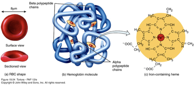
hemoglobin
oxygen-carrying protein
red blood cell shape
bioconcave disc which increases surface area

rbc plasma membrane
strong, flexible
rbcs lack
nucleus, mitochondria
surface of RBCs contain
agglutinogens
agglutinogens
genetically determined assortment of antigens
blood groups are based on the
presence or absence of various antigens
number of blood groups and antigens
at least 24 blood groups and more than 100 antigens
most important blood group and antigen classifications
ABO, Rh
antigens (Ag)
proteins that trigger immune responses
antibodies (Ab)
formed proteins of our immune response
Ab-Ag reactions protect us from
pathogens
Ab-Ag reactions can trigger
unwanted transfusion reactions
Types of ABO classifications
A, B, AB, O blood

A blood
carry the A antigen (and anti-B antibodies)
B blood
carry the B antigen (and anti-A antibodies)
AB blood
carry both A and B antigens (no antibodies)
O blood
carry both A and B antigens (no antibodies)
for transfusion compatibility
donor and recipient must be compatible
if a person with Type A blood, receives transfusion of Type B
Anti-B antibodies in recipients Type A blood will attack the RBCs of donated blood (B antigen)
Also – Anti-A in the B blood will attack the A of the recipient (but volumes are much less)
aggutination
occurs when RBCs stick together; happens when antibodies bind to incompatible antigens on RBC surfaces
hemolysis
occurs when RBCs rupture (break open); releases hemoglobin into the surrounding plasma; can happen following an incompatible blood transfusion.
agglutination and hemolysis can cause
kidney damage
Rh factor
an antigen (Rh D) found on the surface of red blood cells
Rh+ means
Rh D antigen is present on rbcs
Rh- means
Rh D antigen is absent on rbcs
A+ and AB+ indicate
the ABO blood type plus the presence of the Rh D antigen
Rh incompatibility occurs during pregnancy when
an Rh- mother carries an Rh+ fetus
during the first Rh+ pregnancy
fetal Rh+ blood may enter the mother’s blood, triggering anti-Rh antibodies
what happens in pregnancies after Rh sensitization
mother retains immune memory and can produce anti-Rh antibodies
why can a later Rh+ pregnancy be dangerous
maternal antibodies can cross the placenta and destroy fetal RBCs
hemolytic disease of newborn
destruction of fetal or newborn RBCs by maternal antibodies
how to prevent Rh-related hemolytic disease
anti-D immunoglobulin (RhoGAM) prevents Rh sensitization in eligible mothers
WBCs are part of our
circulatory and immune system
WBCs have
nuclei
WBCs do not contain
hemoglobin
WBC cell classifications
granular, agranular
granular leukoctyes
neutrophils, eosinophils, basophils

granular leukocytes structure
have dots
agranular leukocytes
lymphocytes, monocytes

general function of WBCs
immune response
WBCs non-specific immune responses (fast, general defense)
neutrophils, monoctyes, phagocytosis (engulf virus)
WBCs specific immune responses (adaptive immunity)
B and T lymphocytes (develop memory of previous infections)
WBCs allergic responses
basophils (histamine release)
platelets give us
protection when their is damage to our circulatory system (you are bleeding), hemostasis
platelet cell structure
disc-shaped with many veiscles, contain clot-forming chemicals, but no nucleus
platelet life span
5-9 days (short)
hemostasis
the body's normal physiological process that stops bleeding and prevents excessive blood loss after a blood vessel is injured.
steps of hemostasis
platelet plug
coagulation (if needed)
coagulation
blood clotting
platelet plug formation steps
damaged endothelium, platelet adhesion, secretion of inflammatory mediators, platelet aggregation

if more than platelet plug is needed…
a clot is formed
formation of fibrin strands (soluble fibrinogen → insoluble fibrin)
blood clotting factors
proteins (that get activated) and signal for coagulation cascade
blood clotting intrinsic pathway
within the damaged vessels, activation of blood-clotting factors
blood clotting extrinsic pathway
outside of the CV system (in the tissues); tissue factors help with this process
blood clotting is a
cascade effect
blood clotting pathway steps
inflammatory proteins (activation of blood clotting factors)
transform Prothrombin (II) to Thrombin (IIa)
transform Fibrinogen (I) (soluble) to Fibrin (Ia) (insoluble)
thromoembolic disorders
too much blood clotting
hemophilia
missing clotting factors (factor VIII)
plasma
draw blood
collection tube is lined with anticoagulant
clotting factors (unactivated) remain in the plasma
serum
draw blood
leave blood without anticoagulant
clotting factors are activated, form clot
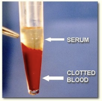
serum is plasma with no
clotting factors