Cytology of neoplasia

0.0(0)
Studied by 0 people
call kaiCall Kai
learnLearn
examPractice Test
spaced repetitionSpaced Repetition
heart puzzleMatch
flashcardsFlashcards
GameKnowt Play
Card Sorting

1/15

encourage image

There's no tags or description

Looks like no tags are added yet.

16 Terms

1
New cards

tissue cell type

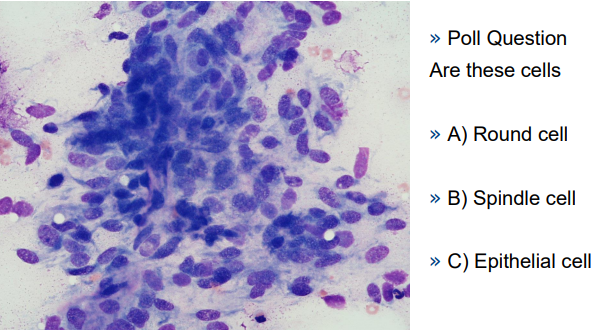
There are three basic cell types which may be seen in neoplasia,

(1) round cells-round, individual and discrete.

(2) epithelial cells--usually angular to polygonal (it may depend on their exact type) and often have tight junctions between cells.

(3) spindle cells --individual, or occasionally clustered, and may be spindle, caudate, or stellate in appearance.

<p>There are three basic cell types which may be seen in neoplasia,</p><p><strong>(1) round cells-</strong>round, individual and discrete.</p><p><strong>(2) epithelial cells-</strong>-usually angular to polygonal (it may depend on their exact type) and often have tight junctions between cells.</p><p><strong>(3) spindle cells -</strong>-individual, or occasionally clustered, and may be spindle, caudate, or stellate in appearance.</p>
2
New cards

nature of neoplasm

It is important for your patient to determine if the neoplasia is benign or malignant. This will have significant implications on prognosis.

3
New cards
<p>‘nature’ of neoplasm</p>

‘nature’ of neoplasm

Anisocytosis and Anisokaryosis-> Pleiomorphism


» Anisocytosis is variable cell size

» Anisokaryosis is variable nuclear size


» Pleiomorphism is cell variability – which may encompass a range of changes

• For the majority of mature tissues there is usually little variation in the cells


(Exceptions)

• Granulation tissue

• Lymphoid tissue

• Mesothelial cells

<p>Anisocytosis and Anisokaryosis-&gt; Pleiomorphism</p><p></p><p>» Anisocytosis is variable cell size</p><p>» Anisokaryosis is variable nuclear size</p><p></p><p>» Pleiomorphism is cell variability – which may encompass a range of changes</p><p>• For the majority of mature tissues there is usually little variation in the cells</p><p></p><p>(Exceptions)</p><p>• Granulation tissue</p><p>• Lymphoid tissue</p><p>• Mesothelial cells</p>
4
New cards
<p>nuclear atypia</p>

nuclear atypia

» Anisokaryosis- variation in nuclear size


» Multinucleation – dividing cells may have two nuclei (or four in certain cell lineages) for specific stages of cell division.

• But uneven numbers of nuclei indicate errors in cell division/replication

• (Exceptions macrophages, mesothelial cells ?)


» Nucleoli

• Prominent

• Multiple

• Large (approaching red cell size)

• Atypical shape (not round)


» Mitotic figures

» Lagging chromosomes

» Cytophagia- ‘cell cannibalism’

» Karyomegaly (large nucleus)

» Nuclear moulding (nuclear shape appears to adapt around another nucleus)

<p>» Anisokaryosis- variation in nuclear size</p><p></p><p>» Multinucleation – dividing cells may have two nuclei (or four in certain cell lineages) for specific stages of cell division.</p><p>• But uneven numbers of nuclei indicate errors in cell division/replication</p><p>• (Exceptions macrophages, mesothelial cells ?)</p><p></p><p>» Nucleoli</p><p>• Prominent</p><p>• Multiple</p><p>• Large (approaching red cell size)</p><p>• Atypical shape (not round)</p><p></p><p>» Mitotic figures</p><p>» Lagging chromosomes</p><p>» Cytophagia- ‘cell cannibalism’</p><p>» Karyomegaly (large nucleus)</p><p>» Nuclear moulding (nuclear shape appears to adapt around another nucleus)</p>
5
New cards
<p>Cytoplasmic inclusions, Perinuclear vacuolation and Cell cannibalism</p>

Cytoplasmic inclusions, Perinuclear vacuolation and Cell cannibalism

benign neoplasms

<p>benign neoplasms</p>
6
New cards
<p>benign neoplasms</p>

benign neoplasms

7
New cards
<p>malignant neoplasms</p>

malignant neoplasms

round cell tumours
<p>round cell tumours</p>
8
New cards
<p>round cell tumours-lymphoma</p>

round cell tumours-lymphoma

knowt flashcard image
9
New cards
<p>epithelial tumour - acinus and tubule</p>

epithelial tumour - acinus and tubule

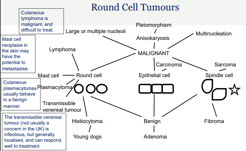
» This gives an overview of how to construct a basic concept map of skin

neoplasia in the dog.

» You may like to add in more facts that you obtain from your reading, or

construct it in a totally different manner that makes more sense to you.

» You may like to consider whether this would look different for the cat, eg would

the mast cell tumour be closer to the benign or malignant area of the map

when looking at cutaneous masses.

» You may also like to think how this might look different for the horse

<p>» This gives an overview of how to construct a basic concept map of skin</p><p>neoplasia in the dog.</p><p>» You may like to add in more facts that you obtain from your reading, or</p><p>construct it in a totally different manner that makes more sense to you.</p><p>» You may like to consider whether this would look different for the cat, eg would</p><p>the mast cell tumour be closer to the benign or malignant area of the map</p><p>when looking at cutaneous masses.</p><p>» You may also like to think how this might look different for the horse</p>
10
New cards
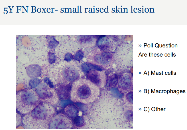
<p>5Y FN Boxer- small raised skin lesion</p>

5Y FN Boxer- small raised skin lesion

knowt flashcard image
11
New cards
term image
12
New cards
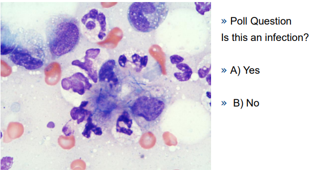
<p>2Y ME Springer Spaniel- swelling between digits- right fore</p>

2Y ME Springer Spaniel- swelling between digits- right fore

knowt flashcard image
13
New cards
term image

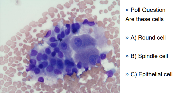
9Y ME Boxer- perianal raised mass

<p>9Y ME Boxer- perianal raised mass</p>
14
New cards
<p>9Y ME Boxer- perianal raised mass</p>

9Y ME Boxer- perianal raised mass

knowt flashcard image
15
New cards
<p>5Y MN Labrador – small thoracic mass</p>

5Y MN Labrador – small thoracic mass
knowt flashcard image
16
New cards
term image