Clinical chemistry lab
 Call Kai
 Learn
Learn Practice Test
 Spaced Repetition
 Match
Match Flashcards
 Knowt Play
1/81

There's no tags or description
Looks like no tags are added yet.
Name | Mastery | Learn | Test | Matching | Spaced | Call with Kai | Chat |
|---|
No analytics yet
Send a link to your students to track their progress
82 Terms
Light - based analytical methods: Absorbance spectrophotometry
light absorbed by the reaction product; absorbance increases with concentration when beer’s law applies
Light - based analytical methods: Fluorometry
emitted light after excitation
Light - based analytical methods: Nephelometry
Light scattered by particles, typically measured at an angle
Light - based analytical methods: Reflectance
light reflected from a reaction surface or dry chemistry layer
Light - based analytical methods: Chemiluminescence
Light produced by a chemical reaction; best sensitivity compared to other methods discussed
Light - based analytical methods: Monochromator
isolates the desired wavelength from polychromatic or white light
Light - based analytical methods: Beer’s law
depends on molar absorptivity and path length remaining constant
Light - based analytical methods: Bichromatic analysis
can help correct for background or instrument - related absorbance interference
Light - based analytical methods: A method
May fail even when the optics are fine if the reagent, blank, temperature, or mixing is abnormal
Light - based analytical methods: White optical diffuser
reflects light in reflectance spectrophotometry
ISE / Potentiometry
Respond preferentially to selected anions or cations
Na⁺, K⁺, Cl⁻
Electrode deterioration, membrane contamination, bubbles, bad calibration, wrong specimen additive
Amperometry
measures electrical current produced by an oxidation or reduction reaction
PO₂
Membrane damage/interference, diffusion problem, electrode problem, incorrect applied potential
Dye-binding absorbance
Negatively charged dye (coumassie blue) is dissolved in acid and binds to positively charged proteins
Albumin with bromocresol green
Dye degradation/contamination, incorrect pH, reagent blank problem, poor mixing
Colorimetric reaction
Creatinine by Jaffe reaction
Reagent deterioration, timing/pH errors, interfering chromogens, poor blank correction
Coupled enzymatic absorbance
BUN; total CO₂ uses coupled enzymatic chemistry
Cofactor/enzyme degradation, abnormal reagent blank, temperature or mixing problems
Scatter / emitted-light methods
Nephelometry, fluorometry, chemiluminescence
Dirty optics, wrong wavelength/filter, reagent instability, signal quenching or background
Purpose of analytical methods
The point is to recognize the analytical principle and the kinds of failures that can affect any assay using that principle.
Analyzer configurations: Random access
Different tests can be run on different specimens as needed
Analyzer configurations: Continuous flow
Specimens travel through a common flowing stream
Analyzer configurations: Centrifugal
specimen / reagent mixtures are held in rotor chambers
Analyzer configurations: Discrete
Each reaction has its own vessel or reaction space
Analyzer configurations: The ACE Axel
is a discrete, random-access analyzer
Specimen handling: Barcodes
identify specimens and connect them to test orders
Specimen handling: Primary tubes & Secondary cups
Primary tubes reduce manual transfer; secondary cups may be needed for low volume
Specimen handling: Affects
Tube size, gel separator position, dead volume, and aspiration can all affect testing
Specimen handling: loading
Continuous loading supports high-throughput workflows
Sample / fluid-path problems 1
Carry over can transfer analyte or reagent into the next reaction
Ex.) a very high concentration from one sample causing a falsely high value in subsequent sample
Sample / fluid-path problems 2
Probe contamination, inadequate washing, or incomplete rinsing can bias results
Sample / fluid-path problems 3
Air bubbles can interfere with aspiration or electrode contact
Sample / fluid-path problems 4
Insufficient sample volume can cause aspiration errors
Poor mixing can produce unusual QC or patient results
Reaction - environment problems: 1
Incorrect temperature changes reaction rates and enzyme activity
Reaction - environment problems: 2
Reusable cuvettes require adequate cleaning and maintenance
(remember that contamination can cause bias results)
Reaction - environment problems: 3
Reagent storage and onboard stability matter
|
V
Incorrect reagent lot information, expired material, or
calibration mismatch can cause systematic shifts
Reaction - environment problems: 4
Humidity and ambient conditions may affect analyzer performance within vendor limits
Reagent problems: Degraded enzyme or cofactor
Might see: Weak reaction, low rate, abnormal blank, poor recovery
Examples: NADH/NADPH-based coupled assays; enzymatic total CO₂; other enzyme-rate methods
Reagent problems: Contaminated or deteriorated color reagent
High/low blank, unexpected absorbance, bias in patient results
Dye-binding or colorimetric methods such as albumin or creatinine chemistry
Reagent problems: Incorrect pH / buffer condition
Altered color development or enzyme activity
Dye-binding, Jaffe, and enzyme-based reactions
Reagent problems: Improper storage / expired reagent
Drift, low activity, instability, repeated QC failure
Can affect essentially any chemistry reagent
Reagent problems: New lot not aligned with calibration
QC shift or systematic patient bias
Recalibration / lot verification may be required
Reagent problems: Inadequate mixing or delivery
Erratic precision, incomplete reaction, unusual QC
Pump/syringe/probe delivery or mixing problem
Reagent problems: Carryover / wash failure
Next sample biased after a high sample or concentrated reagent
Look for sequence-dependent error, not just one isolated value
Potentiometry (Electrochemistry)
Measures voltage (potential difference) between electrodes with essentially no current flow
Used for pH and ion-selective electrodes
Reference electrode supplies a stable potential
Indicator/ISE electrode changes potential in response to the target ion
Calibration converts electrode response into a reportable concentration

Amperometry (Electrochemistry)
A fixed voltage is applied to drive oxidation or reduction
The resulting current is measured.
Current is proportional to analyte concentration
Classic clinical application: PO₂ measurement
Working electrode = site of redox reaction; reference electrode stabilizes potential

Electrodes to recognize: 1
Na⁺ (sodium): glass membrane
Electrodes to recognize: 2
K⁺ (potassium): valinomycin neutral ion carrier (ionophore)
Electrodes to recognize: 3
Cl⁻ (chloride): liquid ion-exchange / electroactive salt membrane
Electrodes to recognize: 4
pH: glass membrane responsive to H⁺ activity.
Electrodes to recognize: 5
PO₂ (partial pressure of oxygen): amperometric oxygen sensor with a gas- permeable membrane
Selectivity concepts
SE membranes respond preferentially—not perfectly— to a target ion
Interfering ions are described by a selectivity coefficient
Lower selectivity coefficient = better selectivity for the intended ion
The liquid junction / salt bridge completes the electrical circuit while separating the sample from the reference electrode
PCO₂ measurement depends on CO₂ diffusion causing a change in H⁺ within an internal electrolyte layer
pO₂ electrode = amperometry
Also called a Clark-type oxygen electrode
Ag/AgCl anode + platinum wire cathode
Fixed negative potential is applied; course wording uses −0.65 V
O₂ diffuses through a selectively permeable membrane to the platinum cathode.
O₂ is reduced at the cathode; the resulting current is proportional to pO₂
Recognition clue: current + platinum cathode + membrane → pO₂, not Na⁺, pCO₂, or pH
pCO₂ electrode: gas-sensing
CO₂ diffuses through a membrane into a thin internal electrolyte layer
Internal electrolyte contains bicarbonate and chloride salts
CO₂ forms carbonic acid, then dissociates and increases H⁺ concentration
The internal pH electrode detects the pH/H⁺ change
Key relationship: higher pCO₂ → higher H⁺ in the internal layer
Recognition clue: pCO₂ is tied to H⁺ concentration, not current, resistance, or chloride ion concentration
Blood gas electrodes: pO₂ and pCO₂
pO₂ = amperometric current; pCO₂ = CO₂-driven pH/H⁺ change
pH / H⁺ measurement
pH is measured potentiometrically as hydrogen ion activity
A pH indicator electrode includes a H⁺-sensitive glass membrane with an internal Ag/AgCl element
Course/exam wording may identify Ag/AgCl in the pH electrode system
Reference electrode gives the stable comparison point; calomel/Hg-HgCl₂ is a classic reference electrode option
Do not confuse the stable reference electrode with the H⁺-responsive indicator system
Cl⁻ electrode membrane
Chloride uses a liquid ion-exchange membrane
Selective membrane contains arsonium salt / electroactive salt in the course wording
It responds to chloride activity through the membrane phase
A Cl⁻ electrode problem can distort electrolyte patterns
If Cl⁻ is overestimated, the calculated anion gap becomes falsely low
Na⁺ / Cl⁻ correlation
Na⁺ is the major extracellular cation; Cl⁻ is the major extracellular anion
They often move together to preserve electroneutrality and osmotic balance
Unexpected Na⁺/Cl⁻ mismatch can be a clue to specimen, calibration, or electrode problems
Do not expect K⁺ or HCO₃⁻ to track with Na⁺ in the same simple way
What happens during measurement
Sample is delivered to the measuring chamber
Na⁺, K⁺, and Cl⁻ electrodes contact the specimen
Each selective membrane develops an electrical potential related to ion activity
The analyzer compares each signal with the reference electrode
Stored calibration converts the electrical signal into a concentration
Calibration logic
Known calibrators establish expected electrode response and slope
A bad slope can reflect electrode deterioration, contamination, calibrator problems, or fluid-path issues.
Calibration does not replace QC.
Calibration logic 2
A new reagent lot or new test method requires calibration
Persistent QC shift or trend (e.g., 10x violation) should trigger troubleshooting and calibration, not automatic reporting
WESTGARD rules
1₂s = warning; investigate the pattern before rejecting solely on this rule
1₃s = reject.
2₂s = reject.
4₁s = reject.
10x = reject; points to systematic error.

Pattern recognition: Shift
Results suddenly move to a new side / level of the mean

Pattern recognition: Trend
Results progressively rise or fall

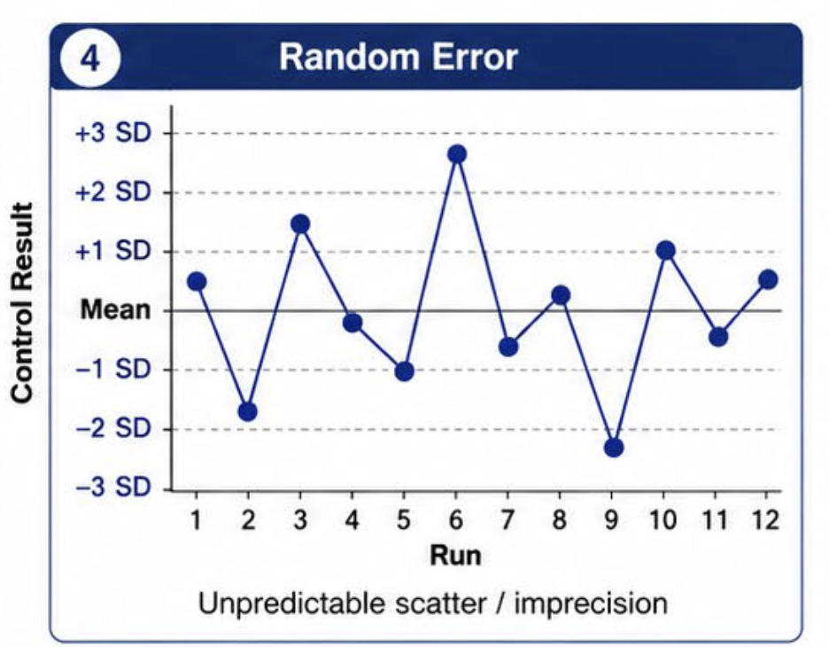
Pattern recognition: Random error
Unpredictable scatter
Pattern recognition: Systematic error
Consistent directional bias

Na⁺
Reference range: 135–145 mmol/L
Critical values: <120 or >160
Method of measurement: ISE
K⁺
Reference range: 3.5–5.0 mmol/L
Critical values: <3.0 or >7.5
Method of measurement: ISE
Cl⁻
Reference range: 98–107 mmol/L
Critical values: <90 or >110
Method of measurement: ISE
Total CO₂ / HCO₃⁻
Reference range: 23–30 mmol/L
Critical values: <10 or >40
Method of measurement: Enzymatic
Specimen issues with K⁺
Delayed separation from cells can falsely increase
potassium.
Hemolysis can markedly increase potassium and may
make the result unacceptable
Specimen issues with Na⁺
A sodium-containing anticoagulant can falsely increase
sodium.
Specimen issues: Total CO₂ / HCO₃⁻
Total CO₂ is especially sensitive to handling because
CO₂ can be lost before analysis
Specimen issues: general
Wrong tube, insufficient volume, contamination, or
aspiration problems can create misleading results.
Questions before reporting
Is QC acceptable?
Was the correct specimen/tube used?
Was the specimen separated and stored appropriately?
Is hemolysis or another visible interference present?
Does the result agree with related analytes and prior
results?
Is this a one-patient issue or a batch-wide pattern?
Anion gap
Formula: Na⁺ − (Cl⁻ + HCO₃⁻).
Reference range: : 8–16 mmol/L
High gap = unmeasured anions / added acids
Low gap = uncommon and may be clinical or analytical
Batch logic
A single low gap can be a real patient finding.
Many low gaps in unrelated patients suggest a
system problem
Potential analytical causes include Na⁺
underestimation or Cl⁻ / HCO₃⁻ overestimation
Look at QC, calibration, electrode condition, recent
maintenance, and lot changes
Result acceptability: a repeatable decision process
1. Check QC: if the run is out of control, patient results are not reportable.
2. Check specimen integrity: collection tube, hemolysis, delayed separation, volume, and handling.
3. Check analytical limits: reportable range, flags, and critical values.
4. Evaluate internal consistency: do Na⁺, Cl⁻, HCO₃⁻, K⁺, BUN, and creatinine make sense together?
5. Compare with prior results when available.
6. Decide whether to report, repeat, troubleshoot the analyzer, or recollect the specimen.
Azotemia patterns: Pre Renal
Problem before the kidney: decreased renal perfusion with initially intact renal tissue.
BUN often rises more than creatinine because urea reabsorption increases when flow is reduced.
BUN:creatinine ratio is often >20:1.
Common settings: dehydration/volume loss, hemorrhage, reduced cardiac output.
May improve rapidly if perfusion is restored early.
Azotemia patterns: Intrinsic Renal
Problem within the kidney: glomerular, tubular, interstitial, or vascular injury.
BUN and creatinine both rise; creatinine tends to rise more proportionally.
BUN:creatinine ratio is often about 10–15:1.
Tubular injury reduces normal urea reabsorption.
Electrolyte or acid–base abnormalities may accompany renal dysfunction.
Azotemia patterns: Post Renal
Problem after the kidney: obstruction to urine flow.
BUN and creatinine can both increase; ratio is variable
Early obstruction may resemble pre- renal; prolonged obstruction can
resemble intrinsic renal injury.
Consider urinary obstruction and reduced urine output
Clinical context and imaging are often needed.
Potentiometry & amperometry
Potentiometry = voltage
Amperometry = current
amperometric Clark electrode
pO₂: amperometric Clark electrode; Ag/AgCl anode + platinum cathode + membrane; current reflects O₂ reduction
fundamental pH measurement system
pH/H⁺ systems use a H⁺-sensitive glass membrane with Ag/AgCl internal element; classic references include calomel/Hg-HgCl₂.
clinical chemistry and laboratory troubleshooting
Cl⁻ electrode membrane contains arsonium/electroactive salt; Cl⁻ overestimation lowers anion gap.
Na⁺ and Cl⁻ should generally correlate; repeated mismatch suggests specimen/analyzer review