Lab Practical 2
 Call Kai
 Learn
 Practice Test
 Spaced Repetition
 Match
 Flashcards
 Knowt Play
1/342

There's no tags or description
Looks like no tags are added yet.
343 Terms
epimysium tissue and function
dense irregular connective tissue
surrounds entire skeletal muscle

perimysium (perimeter) tissue and function
areolar connective tissue surrounding a fascicle
separates muscle cells/fibers into bundles

what is a fascicle
bundle of muscle fibers

endomysium (inside) tissue and function
areolar connective tissue surrounding each separate muscle cell/fiber
carry blood vessels/sources

muscle fiber/cell
each cell stretches the length of muscle, surrounded by a plasma membrane and a layer of connective tissue called endomysium
- bundles of myofibrils
- composed of several myofibrils wrapped together by a sarcolema and sarcoplasm

myofibril
long, filamentous organelles found within muscle fibers/cells that has a banded appearance
- propagate muscle contraction

myofilaments
filaments of myofibrils that are constructed from proteins and are responsible for muscle contraction
- actin (thin)
- myosin (thick)

agonist or prime mover
primary responsible for producing a particular movement
- the muscle that does most of the work during a movement
antagonist
a muscle that has the opposite function to another muscle
- works in the opposite direction during a movement and stabilizes the other muscle
- keeps body in stable position
synergist
muscle that aids a prime mover in a movement and helps prevent rotation
origin of a muscle
muscle end attached to the bone that stabilizes the movement and does not move when the muscle contracts
- proximal attachment point
insertion of a muscle
muscle end attached to the bone that drives the movement and moves when the muscle contracts
- distal attachment point
flexion
bending a joint by decreasing the angle

extension
straightening of a joint by increasing the angle

hyperextension
extension beyond anatomical position

dorsiflexion
bending of the foot or the toes upward

plantar flexion
bends the foot downward toward the ground at the ankle

abduction
movement away from the midline of the body

adduction
movement toward the midline of the body

rotation
movement where bone rotates on its long axis/fixed point
circumduction
the circular movement at the far end of a limb

supination
movement that turns the palm up

pronation
turning the palm downward

elevation
raising a body part

depression
lowering a body part

protraction
moving a body part forward and parallel to the ground

retraction
moving a body part backward and parallel to the ground

deltoid action/antagonist/origin/insertion
a: abducts arm at shoulder
ant: latissimus dorsi & pec major
o: clavicle and scapula
i: humerus

infraspinatus
lateral rotation at shoulder
- origin: scapula
- insertion: humerus

subscapularis
medially rotates arm, stabilizes shoulder joint

teres major
medial rotation of shoulder joint
- origin: scapula
- insertion: humerus

teres minor
lateral rotation of shoulder joint
- origin: scapula
- insertion: humerus

biceps brachii short head
flexes and supinates forearm
- origin: scapula
- insertion: radius
- antagonist: triceps brachii

biceps brachii long head action/antagonist/origin/insertion
a: flexes and supinates forearm
ant: triceps brachii
o: scapula
i: radius

brachialis
flexes forearm at elbow
- origin: humerus
- insertion: ulna
- antagonist: triceps brachii

brachioradialis
flexes forearm at elbow
- origin: humerus
- insertion: radius
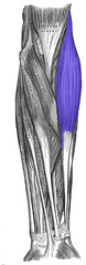
triceps brachii
extends forearm at elbow joint
- origin: scapula and humerus
- insertion: ulna
- antagonist: biceps brachii and brachialis

anterior forearm
muscles that flex the hand at the wrist

posterior forearm
muscles that extend the hand at the wrist

supinator (posterior)
supination of forearm
- origin: humerus and proximal ulna
- insertion: proximal end of radius

pronator teres
pronation of forearm
- origin: humerus
- insertion: radius
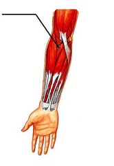
palmaris longus
flexes the wrist
- origin: humerus
- insertion: metacarpals

flexor carpi radialis
flexes the wrist
- origin: humerus
- insertion: metacarpals

flexor carpi ulnaris
flexes the wrist
- origin: humerus
- insertion: metacarpals

another view of flexor carpi radialis and ulnaris

extensor carpi radialis longus (posterior)
extends the wrist
- origin: humerus
- insertion: metacarpals
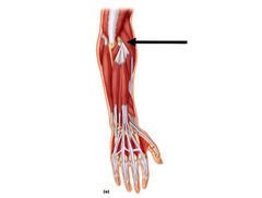
extensor carpi ulnaris (posterior)
extends the wrist
- origin: humerus
- insertion: metacarpals

flexor digitorum (anterior)
flexes wrist and digits
- origin: ulna
- insertion: metacarpals

extensor digitorum (posterior)
extends wrists and digits
- origin: humerus
- insertion: metacarpals

anconeus (posterior)
extension of elbow
- origin: humerus
- insertion: radius

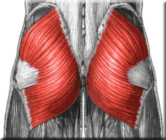
gluteus maximus
extends thigh at hip
- origin: ilium, sacrum and coccyx
- insertion: femur
gluteus medius
abducts and medially rotates thigh; steadies pelvis
- origin: ilium
- insertion: femur

tensor fascia latae
abducts and medially rotates thigh
- origin: ilium
- insertion: tibia
- antagonist: gluteus maximus (all gluteal muscles/hip abducting muscles)

adductor muscles
adduct and flex thigh at the hip

adductor brevis
adducts and flexes thigh at hip
- origin: pubic bone
- insertion: femur
- antagonist: gluteus maximus (all gluteal muscles/hip abducting muscles)

adductor magnus
adducts and flexes thigh at hip
- origin: pubic bone
- insertion: femur
- antagonist: gluteus maximus (all gluteal muscles/hip abducting muscles)

adductor longus
adducts and flexes thigh at hip
- origin: pubic bone
- insertion: femur
- antagonist: gluteus maximus (all gluteal muscles/hip abducting muscles)

another view of adductor longus

gracilis
adducts and flexes thigh at hip
- origin: pubic
- insertion: femur
- antagonist: gluteus maximus and adductor magnus

pectineus
adducts and flexes thigh at hip
- origin: pubic bone
- insertions: femur
- antagonist: gluteus maximus
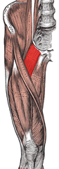
iliacus
flexes thigh at hip
- origin: ilium
- insertion femur
- antagonist: gluteus maximus and hamstrings

psoas major
flexes thigh at hip
- origin: vertebrae
- insertion: femur
- antagonist: gluteus maximus and hamstrings

sartorius
flexes thigh at hip and flexes leg at knee; longest muscle in the body
- origin: ilium
- insertion: tibia

gastrocnemius
plantar flexes foot and flexes leg at knee
- origin: femur
- insertion: calcaneus
- antagonist: tibialis anterior

soleus
plantar flexes foot at ankle
- origin: tibia and fibula
- insertion: calcaneus
- antagonist: tibialis anterior

tibialis anterior
dorsiflexes and inverts foot
- origin: tibia
- insertion: metacarpals

fibularis longus action/antagonist/origin/insertion
a: plantar flexes and everts foot
ant: tibialis anterior
o: fibula
i: metatarsals

rectus femoris
extends knee and flexes thigh at hip
- origin: anterior inferior iliac spine
- insertion: tibia
- antagonist: hamstrings

vastus lateralis action/antagonist/origin/insertion
a: extends leg
ant: hamstrings
o: femur
i: patella

vastus intermedius
extends leg at knee
- origin: femur
- insertion: patella
- antagonist: hamstrings

vastus medialis
extends leg at knee
- origin: femur
- insertion: patella
- antagonist: hamstrings

biceps femoris
flexes leg at knee and extends thigh at the hip

biceps femoris long head
flexes leg at knee extends thigh at the hip
- origin: pubic bone
- insertion: fibula
- antagonist: rectus femoris

biceps femoris short head
flexes leg at knee
- origin: femur
- insertion: fibula
- antagonist: rectus femoris
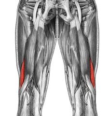
semitendinosus
flexes leg at the knee and extends thigh at the hip
- origin: pubic bone
- insertion: tibia
- antagonist: rectus femoris

semimembranosus
flexes leg at the knee and extends thigh at the hip
- origin: pubic bone
- insertion: tibia
- antagonist: rectus femoris

central nervous system (CNS)
brain and spinal cord; integration and command center
peripheral nervous system (PNS)
the sensory and motor neurons that connect the central nervous system (CNS) to the rest of the body
soma or cell body of a neuron contains?
contains nucleus and organelles

axon function
a part of a neuron that carries impulses away from the cell body

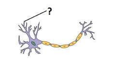
dendrites function
a neuron's bushy, branching extensions that receive messages and conduct impulses toward the cell body
axon terminal function
the endpoint of a neuron where neurotransmitters are stored

what is a unipolar neuron
a neuron with one process extending from its cell body

what is a bipolar neuron
a neuron with one axon and one dendrite attached to its soma

what is a multipolar neuron
a neuron with one axon and many dendrites attached to its soma

what is a sensory neuron
neurons that carry incoming information from the sensory receptors to the brain and spinal cord

what is a motor neuron
a neuron that sends an impulse to a muscle or gland, causing the muscle or gland to react
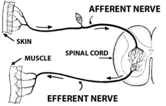
what is a interneuron
a nerve cell that relays messages between nerve cells, especially in the brain and spinal cord; carries nerve impulses from one neuron to another

what is epineurium
dense connective tissue that surrounds entire nerve including fascicles and blood vessels

what is perineurium
coarse connective tissue that bundles fibers into fascicles

what is endoneurium
loose connective tissue that surrounds nerve fibers and encloses axons and their myelin sheaths

what is a myelin sheath and its function
a fatty tissue layer segmentally encasing the axons of some neurons; enables vastly greater transmission speed as neural impulses hop from one node to the next

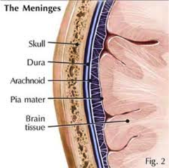
what is the dura mater
thick, outermost layer of the meninges surrounding and protecting the brain and spinal cord

what is the subdural space
the mater between the dura mater and the arachnoid space

what is the arachnoid mater
weblike middle layer of the three meninges

what is the subarachnoid space
a space in the meninges beneath the arachnoid membrane and above the pia mater that contains the cerebrospinal fluid

what is the pia mater
the delicate innermost membrane enveloping the brain and spinal cord
what is the anterior median fissure
a groove along the anterior midline of the spinal cord that divides it into symmetrical halves

what is the posterior median sulcus
a shallow vertical groove dividing the spinal cord throughout its whole length


what is this and its function
central canal
a tiny, fluid-filled channel found within the spinal cord