Bones and Joints of the Distal Limb
 Call Kai
 Learn
 Practice Test
 Spaced Repetition
 Match
 Flashcards
 Knowt Play
1/41

Earn XP
Description and Tags
M.8R, W.2, L.4
42 Terms

What are the arrows pointing at in the image?
carpus

List the species each of these are from
Left to right:
Canine
Equine
Bovine
Pig
What type of bones is the carpus composed of and what type of bones are they?
Carpal bones
Short bones
List the components of the canine carpus
Proximal Row
Fused Radial Carpal Bone + Intermediate Carpal Bone (named RCB)
Ulnar Carpal Bone
Accessory carpal bone
Distal Row
1st, 2nd, 3rd, and 4th carpal bones

What bone is palpable on the canine carpus?
Accessory carpal bone
How many centers of ossification do short bones USUALLY have? List the two short bones that are the exception and how many centers of ossification they have
SINGLE center of ossification
Exceptions:
Fused Radial Carpal Bone + Intermediate Carpal Bone (RCB) - 3 centers
Accessory carpal bone (ACB) - 2 centers
List the 3 joints in the carpus, where they are located, which has the most + least movement
Antebrachio-carpal joint
between antebrachium + proximal row of carpal bones
most movement
Middle carpal joint
proximal + distal rows of carpal bones
Carpo-metacarpal joint
distal row + metacarpal bones
least movement

What kind of joints are between bones and what type of joints are they?
Intracarpal Joints
Fibrous joints (not a lot of movement)
When are joint spaces in the carpus palpable? and why is that clinicall relevant?
in FLEXION
helps with carpal joint fluid sampling
What does the Accessory Carpal Bone act as for the distal limb?
acts as a LEVER for distal limb

What major muscles insert on the carpus and what ligament counteracts this?
major carpal FLEXOR muscle
counteraction by PALMAR LIGAMENT

Describe anatomically where the single joint capsule of the synovial joint extends. What kind of communication is there between compartments?
Extends:
Proximally between radius + ulna
Distally between metacarpal bones
Poor communication
List the 3 structures that provide the carpus with joint stability
Collateral Ligaments
Lateral: ulna (lat. Styloid process) to 5th metacarpal
Medial: radius (medial styloid process) to 2nd metacarpal
Cartilage plate
palmar aspect of joint capsule
Retinaculum
List long bones of the hindlimb
metacarpal / metatarsal bones
Digits:
Proximal phalanx
Middle phalanx
Distal Phalanx
List sesamoid bones of the hindlimb
Proximal sesamoids
all species
Dorsal sesamoids
Distal sesamoid
List synovial joints of the hindlimb
Metacarpo / metatarso-phalangeal joint
Proximal Interphalangeal joint (PIP)
Distal Interphalangeal joint (DIP)
Why do dogs and cats shed the 1st digit?
for speed
List the 3 different types of stances, give examples of species, what do they bear weight on, and what protects those structures
Plantigrade
Eg. rabbits, bears, humans
Bear weight on
Digits + metatarsals + tarsal bones
Digitgrade
Eg. Dogs, cats
Bear weight on
Digits / phalanges
Protected by foot pads
Unguligrade / Ungulates
Eg. Ruminant, horses
Bear weight on distal phalanx only (tippy toes)
Protected by hoof
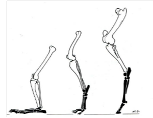
what is usually similar between digitigrade species and unguligrade species?
distal fore & hind limb

How many centers of ossification do metacarpals & digits have and what is the exception?
2 centers of ossification
Except:
Distal phalanx = 1

Why is it important to know the position of growth plates?
to be able to distinguish from joint spaces

Describe the Equine Metacarpal Bones (name, number metacarpal, purpose, articular surfaces
CANNON Bone
3rd metacarpal / metatarsal
weight bearing
Distal Articular Surface = sagittal ridge
Describe the 2nd and 4th MC / MT (name, function, palpable?)
Splint Bones
Non-weight bearing (vestigial)
Palpable - ‘button’ at distal end

What kind of joints and which ligament are between the splints and 3rd MT
Fibrous joints
Interosseous ligament
Describe sesamoid bones in all species (single or paired, location, embedded in what ligament, what do they protect)
paired
palmaro-distal to every MC3 /MT3
embedded in suspensory ligament
Protect:
Superficial Digital Flexor Tendons (SDFT)
Deep Digital Flexor Tendon (DDFT)
Describe the dorsal sesamoid bones in the canine ( single or paired, location, embedded in and protect what?)
Single
Dorsal aspect to every MCP / MTP joint
Embedded in & protect: extensor tendons
Proximal Phalanx
Long Pastern Bone + P1
Raised V on palmar surface
oblique distal sesamoidean ligament
List the components of the metacarpo-phalangeal (MCP) / metatarso-phalangeal (MTP) joint
3rd metacarpal / metatarsal bone
Proximal phalanx
Proximal sesamoid bones
Typical synovial joint
Large palmar pouch - incorporates proximal sesamoids
Large dorsal pouch - cushions extensor tendons
Collateral ligaments
Sagittal ridge / groove
Movement:
limited to flexion & extension
‘High motion’ joint
What is the natural weight bearing position of the equine distal limb and what position is the distal limb forced into during locomotion?
Extension
Hyperextension
originates proximal palmar MC3 / MT3
Divides into two
fuses with Common Digital Extensor tendon
List the function of the Suspensory Ligament
acts like a sling
returns joint to resting position
relies on stability of proximal sesamoids
Inter-sesamoidean ligament
Fibrocartilage plate between proximal sesamoid bones
Smooth passage of DDFT and SDFT
B/ Collateral sesamoidean ligaments:
Goes from Proximal sesamoids to MC / MT 3 and PP
Medial & lateral
C/ Series of Distal sesamoidean ligaments:
1. Cruciate - PP
2. Short - PP
3. Oblique ligament - PP
V-shaped trough on PP
4. Straight ligament - MP
Inserts with SDFT
List 4 things that the function of the Suspensory Apparatus relies on
Relies on interaction between:
Suspensory ligament
Proximal sesamoids
Sesamoidean ligaments
Common digital extensor tendon
Describe the middle phalanx (MP) - (name, number phalanx,
Short pastern bone
p2
saggital ridge
saggital groove
bony bumps
Describe the Distal Phalanx
Pedal Bone / P3
Extensor process
Attachment Common Digital Extensor tendon (CDE)
Palmar Processes:
Support lateral cartilages
Solar surface
Attachment Deep Digital Flexor Tendon (DDFT)
Dorsal surface
Describe the distal interphalangeal joint
“Coffin” joint - buried within hoof
Components:
Middle & distal phalanges
Distal sesamoid & distal phalanx
Distal sesamoid & middle phalanx
Typical synovial joint
Extensive joint capsule
Forms dorsal bursa to cushion extensor tendon
Accessible for injection into jointStability:
Collateral ligaments
Movement :
flexion & extension
Some rotation to accommodate uneven ground
Describe the Equine Distal Sesamoid
“Navicular” Bone
2 smooth articular surfaces
Cancellous centre with vascular channels
Absent in dog & cat
has a navicular / podotrochlear bursa
Fluid filled pocket between distal sesamoid and DDFT
Cushions DDFT
Separated from joint space by impar ligament
Navicular disease syndrome
Forelimb lameness
Erosion visible on radiographs
Poor correlation between radiographic changes and clinical signs
Which bone(s) of the forelimb has least contribution to the weight bearing function?
A. Carpal Bones
B. Humerus
C. Radius
D. Ulna
Ulna
In the horse, which ligament serves as a sling to support the proximal sesamoid bones and prevent the over-extension of the MCP joint?
A. Collateral Sesamoidean Ligament
B. Cruciate Ligament
C. Distal Sesamoidean Ligament
D. Suspensory Ligament