W3 Non-Traumatic Red Eye
 Call Kai
 Learn
Learn Practice Test
 Spaced Repetition
 Match
Match Flashcards
 Knowt Play
1/35

There's no tags or description
Looks like no tags are added yet.
Name | Mastery | Learn | Test | Matching | Spaced | Call with Kai | Chat |
|---|
No analytics yet
Send a link to your students to track their progress
36 Terms
What does red eye indicate?
External inflammation- think common like conjunctivitis but also rare like acute glaucoma
What should be done before examination?
Use flourescein dye- AVOID pupil dilation
What secondary symptoms are associated with red eye?
Reduced vision, pain, photophobia
What can red eye also indicate outside the eye?
Eyelid disorder, eye disease
Which examinations are required for red eye?
Slit lamp anterior chamber examination excludes iritis or acute glaucoma
Reduced vision, pain or photophobia may indicate infectious corneal ulcer in CL wearer
If symptoms are unexplained this could be serious eye disease- same day referral
If px has reduced vision or pain what should also be done?
Conduct IOP measurements to exclude acute glaucoma- above 40 indicates acute, 22-40 indicates chronic
What are symptoms of bacterial conjunctivitis?
2 red eyes, pus-like discharge, no reduced vision, pain or photophobia- antibiotic drops given
What question do we ask for referral of wet AMD?
Is this recent or have you always had it
What questions do we ask for secondary symptoms?
Reduced/blurred vision
Eye soreness/irritation- bacterial conjunctivitis
Pain mild/moderate/severe- episcleritis/scleritis
Sensitive to bright light- iritis/AACG
Itchy/gritty eyes- allergic conjunctivitis/bacterial conjunctivitis
Foreign body sensation- corneal ulcer
Do you see haloes- acute glaucoma
What do we ask for regarding ocular history?
Do you wear CL- allergic conjunctivitis, dirty CL leading to corneal ulcer
Has something hit your eye- recurrent corneal erosion
Have you ever had eye surgery
Have you ever had red eye with blurred vision- iritis, corneal ulcer
What do we ask for regarding medical history?
Smoking
Cold- viral conjunctivitis
Gh blood pressure- subconjunctival haemmorahge
Inflammatory bowel disease
Rthritis
Asthma, hay fever or eczema
Which tests do we conduct?
Visual acuity
Pinhole test- if distance vision reduced
Pupil reactions

Which methods do you perform for external examination?
Direct opthalmoscopy
Slit lamp
Flourescein
Tear test
Anterior chamber depth
Lid eversion
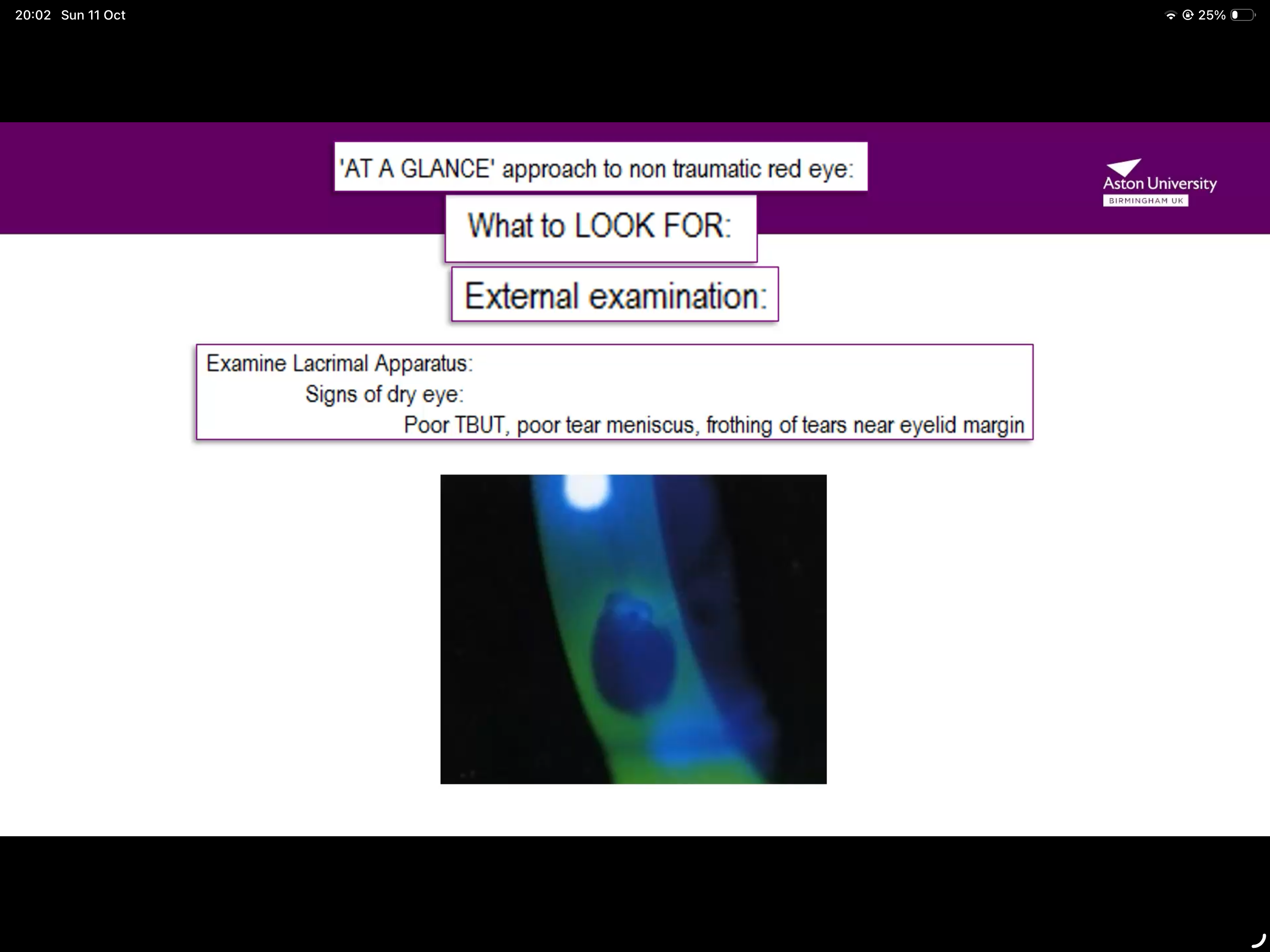
When examining the lacrimal apparatus what signs might we see?
Poor TBUT (tear break up time- should not break up before 10 secs)
Poor tear meniscus
Frothing of tears near eyelid margin
All these indicate dry eye
What do we examine in the conjunctiva?
Redness, discharge, papillae and folds
What does redness in the conjunctiva indicate?
Subconjunctival haemorrhage
Bulbar redness- concerning if vessels do not move over sclera, indicates scleritis
Limbal redness- serious eye disease like iritis and acute glaucoma (unreliable sign)

What does discharge indicate when examining conjunctiva?
Yellow and pus like bacterial cause
Watery and clear viral/allergic cause

What are noticeable observations when observing papillae?
Stain with flouresein and observe any roughness under everted lid- indicates bacterial/allergic conjunctivitis

What do you look for when observing follicles?
Observe under everted lids- indicates viral conjunctivitis

What are characteristics of viral conjunctiva?
Watery discharge which becomes mucoid
Associated with cold symptoms/fever
Seen on follicles
Treated with cool compress/artificial tears
What are features of allergic conjunctiva?
Watery discharge
Associated with itching, redness and puffy eyelids
Seen on papillae
Treat with antihistamines drops
What are features of bacterial conjunctiva?
Mucoid, crusty discharge
Associated with redness and sticky eyelids
Seen on papillae
Treated with antibiotic eye drops/ointments
Whats the difference between papillae and follicles?
…

Whats papillae?
Raised, found in palpebral conjunctiva, caused by allergic conjunctivitis, bacterial infections or CL intolerance
Whats follicles?
Dome shaped, found in lower fornix, caused by viral infections
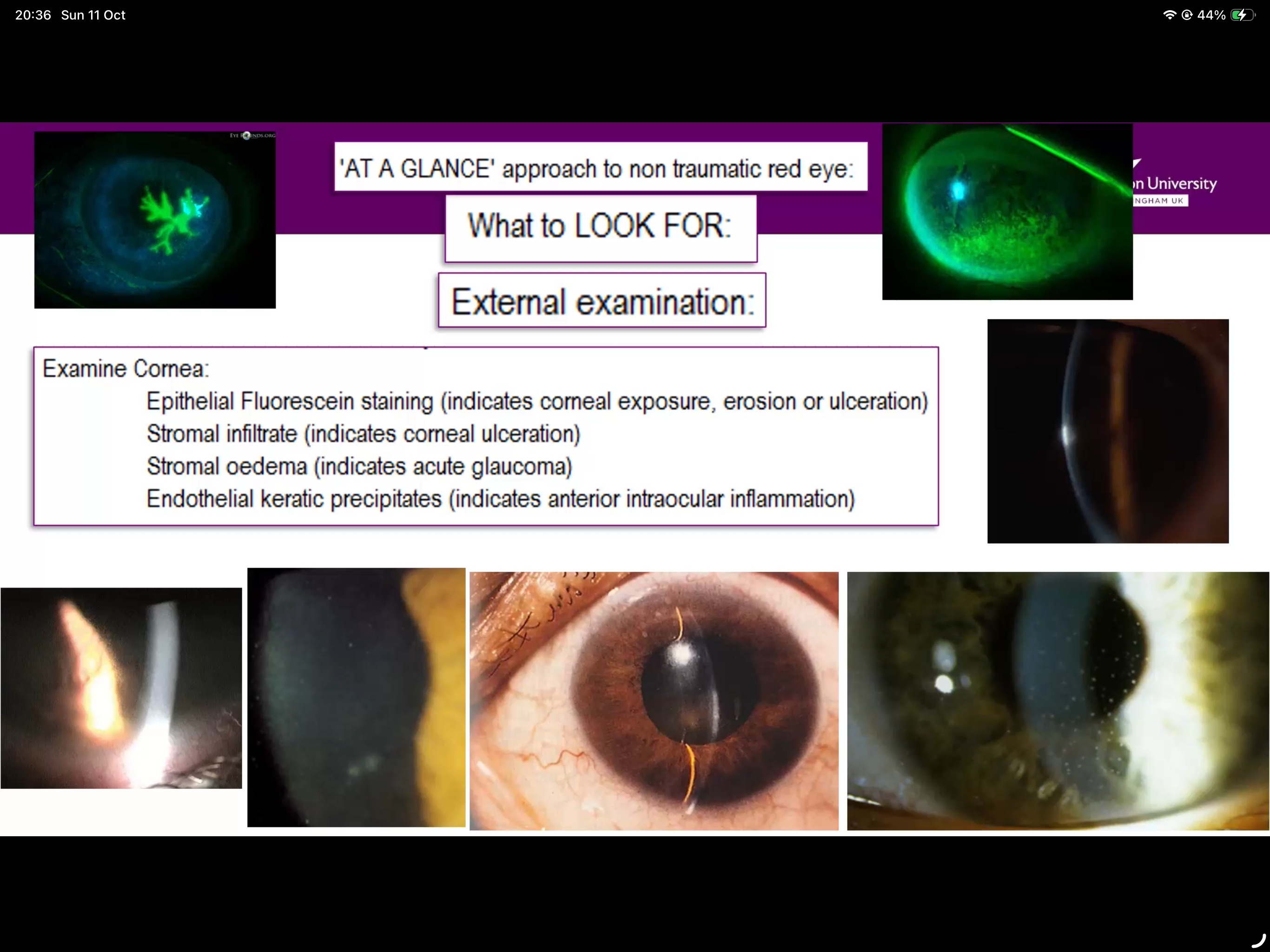
What do you look for when examining the cornea?
Staining indicates erosion/ulceration (top pictures)
Stromal infiltrate- indicates corneal erosion (central bottom picture)
Stromal oedema indicates acute glaucoma (bottom right)
What can you see when you observe anterior chamber?
Aqueous flare and cells (indicates anterior intraocular inflammation)
Hypopyon indicates anterior intraouclar inflammation
Closed/shallow anterior chamber indicates acute glaucoma
What do you look for when conducting an internal examination via opthalmoscopy?
Tonometry- above 40mmHg indicates acute glaucoma

How can you differentiate between the 4 corneal ulcers?
Wearing CL wear and smoking makes it infectious
Non-infectious if they don’t smoke, no CL wear but they have photophobia
Marginal keratitis if they have no photophobia and no epithelial opacity
Recurrent corneal erosion if no watering of the eye

What are key differences between scleritis and episcleritis?
Episleritis recent onset- scleritis unknown onset
Iscelitis has a clear cornea, scleritis has AC cells and flare
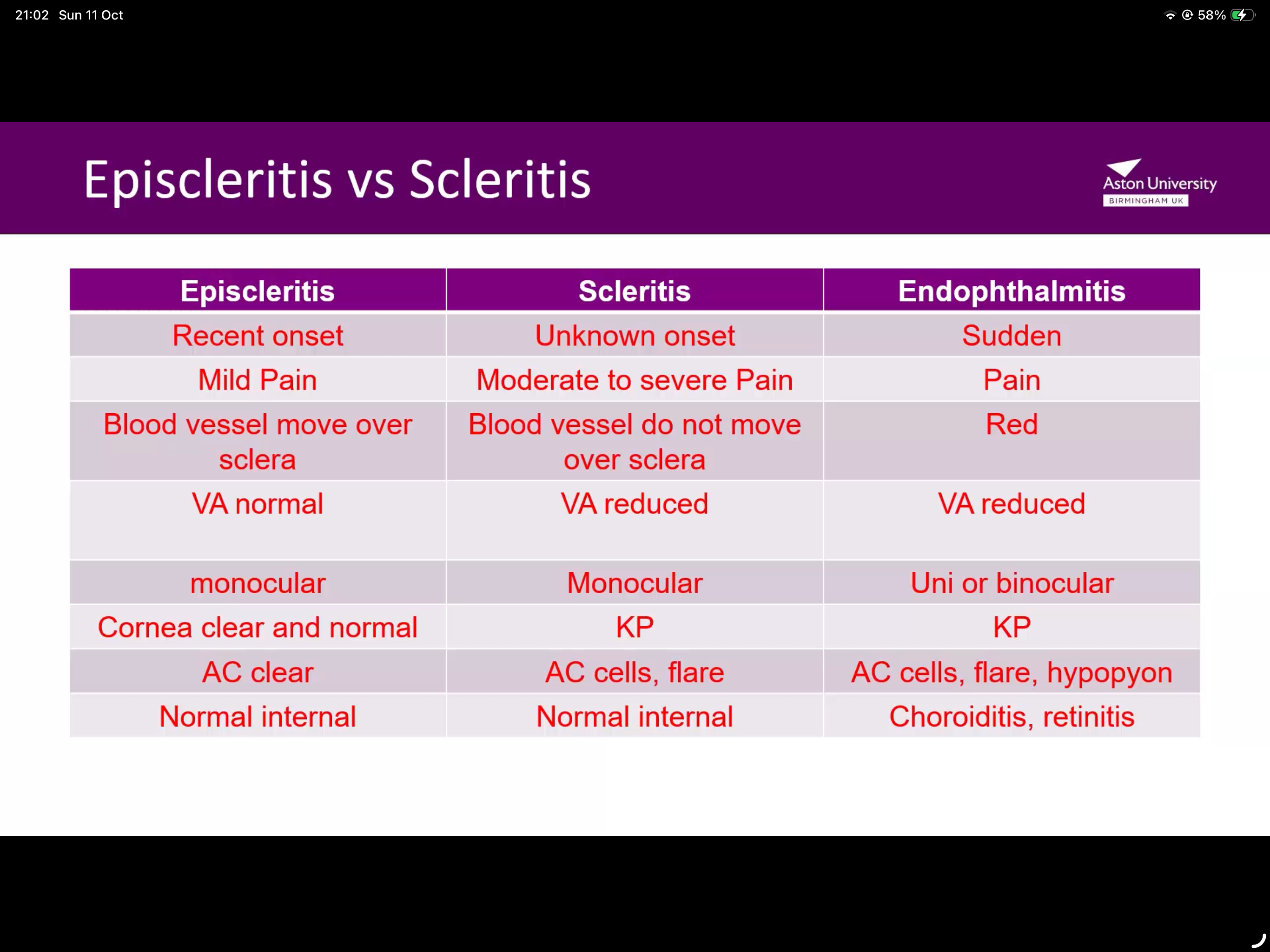
What are features of endophthalmitis?
sudden
Painful
Red sclera
VA reduced
Binocular/uni
Flare, AC cells
Caused by choroiditis or retinitis
What calls for immediate referral?
Acute glaucoma
Corneal ulcer
What calls for same day referral?
unexplained red eye with reduced vision, photophobia or pain
Marginal keratitis
Recurrent corneal erosion
Iritis
What calls for an urgent (1 week) referral?
Persistent allergic and bacterial conjunctivitis
Viral conjunctivitis with corneal involvement
Scleritis
Dry eye (ONLY IF PX IS SEVERE AND HAS RHEUMATOID ARTHRITIS- sjordgens syndrome)
When is a routine referral needed?
recrruent spontaneous subconjunctival haemorrhage
Persistent episcleritis
Dry eye
Which conditions would you simply advise?
conjunctivitis s
Subconjucntival spontanous haemorrhage
Episcleritis
Dry eye
Bacterial and allergic conjunctivitis with recent onset- recommended antibiotics/antihistamines