ch. 27: disorders of cardiac function, heart failure, and circulatory shock -
 Call Kai
 Learn
 Practice Test
 Spaced Repetition
 Match
 Flashcards
 Knowt Play
1/131

There's no tags or description
Looks like no tags are added yet.
132 Terms
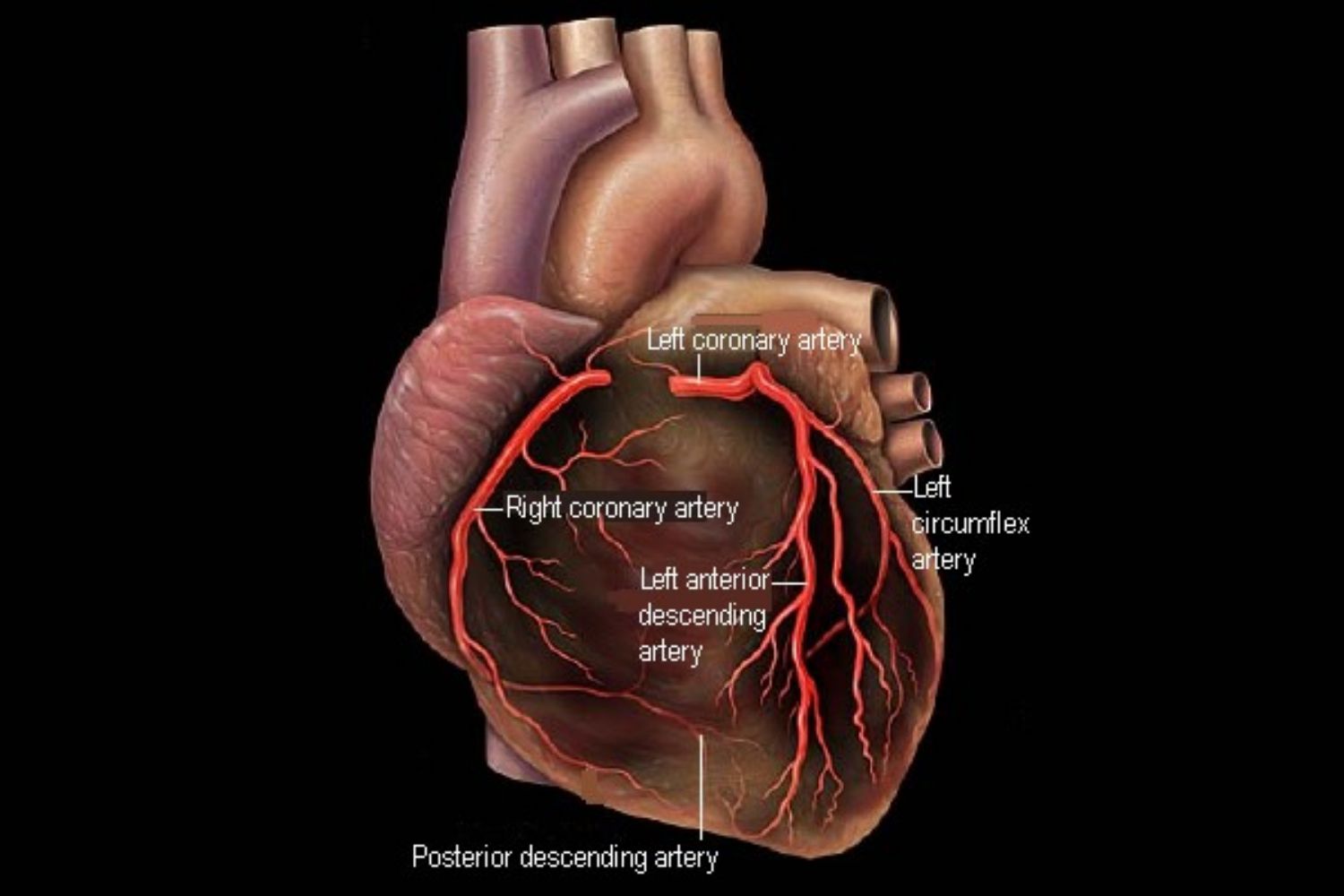
what makes up the systemic circulation that delivers oxygen-rich blood to the myocardium?
the coronary arteries
what does the L coronary artery divide into?
the left anterior descending or inter ventricular artery
left circumflex artery

what does the R coronary artery divide into?
right marginal artery
posterior inter ventricular artery
what is coronary artery disease (CAD)?
atherosclerotic plaques have built up in the coronary arteries —> a narrowed lumen —> reduced blood flow to heart muscle
does coronary artery disease (CAD) have an immediate onset
not necessarily, can remain silent for years until blood is significantly reduced
what are possible manifestations of CAD?
angina attacks
myocardial infarction
arrhythmias
heart failure
sudden death
what are 2 catergories of CAD?
chronic ischemic heart disease
acute coronary syndrome
what are manifestations of chronic ischemic heart disease (CIHD)?
stable angina
variant angina
silent myocardial ischemia
what are manifestations of acute coronary syndrome?
more severe
periods of no ST-segment elevation, causing:
unstable angina
non-ST-segment AMI (acute myocardial infarction)
ST-segment elevation can lead to ST-segment elevation, causing:
Q-wave AMI
non-ST-segment AMI (acute myocardial infarction)
when does chronic ischemic heart disease (CIHD) occur?
when there is a deficit of oxygen supply to meet myocardial needs
what is a classic symptom of CIHD?
angina
recurrent episodes of substernal chest pain
triggered by emotional or physical stress
how is CIHD classified?
by the kind of angina occurring
what is stable angina?
angina triggered by physical exertion or emotional stress (when O2 demand increases)
what is variant angina?
cyclical spasms in coronary arteries without an increased workload
pain occurs at rest, often in early morning or at night
what is unstable angina?
attacks last longer than in stable angina
less responsive to medication
prolonged pain at rest may precede acute coronary syndrome
what is silent myocardial ischemia?
reduced blood flow & oxygen supply to the heart, without noticeable symptoms
occurs with no pain
where does angina frequently occur?
chest
upper abdomen
back
beck
jack
left shoulder & arm
what does screening for CIHD do?
hopefully identifies asymptomatic stages to reduce risk of complications
what does a stress test monitor?
heart activity & coronary perfusion during exercise
how do you treat an angina attack?
rest, stopping the activity
sublingual nitroglycerin
dilates coronary arteries —> improves blood flow to myocardium
what causes acute coronary syndrome?
sudden, severe reduction in blood flow to myocardium because of severe narrowing or complete blockage
what is the most common cause of acute coronary syndrome?
atherosclerosis with a plaque rupture
thrombus at site of ruptured plaque —> blockage
what are manifestations of acute coronary syndrome?
chest pain at rest
chest pain is not relieved by vasodilators
substernal, crushing, radiating, pressure, burning pain in chest
sudden SoB
what is ST elevation associated with?
complete arterial blockage
requires immediate surgery
how does ischemia affect an ECG?
inadequate blood supply alters electrical activity of myocardial cell
what should you assess in an ECG?
the ST segment
when the ventricles are fully depolarized and contracting to eject blood
any ECG changes in this segment indicate high probability of MI
what does ST-segment elevation (STEMI) indicate?
likely that a coronary thrombus occluding artery

what do abnormal Q waves indicate?
no depolarizing current conducting from necrotic tissue
what are ECG changes that indicate intermediate or low probability of MI?
an NSTEMI
T-wave inversion
what does an NSTEMI indicate?
lack of ST segment elevation
clot only partly occludes coronary artery
what does a T-wave inversion indicate?
repolarization of ventricle altered because of myocardial ischemia or MI

how do you treat a STEMI?
medical emergency to prevent irreversible damage
percutaneous coronary intervention (PCI)
balloon angioplasty to widen arteries
stent placement to keep artery open
thrombolytics
dissolve blood clots
anticoagulants
how do you treat a NSTEMI?
pharm treatment to reduce myocardial workload
beta blockers
anticoagulants
heparin
monitor ECG & check serum markers
angioplasty if not stable
what defines a myocardial infarction?
evidence of myocardial necrosis caused by acute myocardial ischemia
what is the time limit in which irreversible damage and necrosis can occur with a severe coronary artery occlusion?
within 20-40 mins
what are cardiac markers of myocardial infarction?
blood levels of cardiac troponin increase
why is troponin I a preferred cardiac market?
highly sensitive & specific
may rise within 3-6 hours after symptom onset
when does myoglobin rise after a myocardial injury is sustained?
within 2 hours
not specific to heart though, also in skeleton muscle
when does creatine kinase-MB rise after a myocardial injury is sustained?
6-8 hours afterwards
what are complications of MI?
sudden death
cardiogenic shock
cardiac tamponade
thromboembolism
ventricular aneurysm
bulging of weakened ventricular wall
what is cardiac tamponade?
a rupture of necrotic heart tissue with blood accumulation in the pericardium
what is the cardiac cycle?
the events occurring in one complete contraction and relaxation of the heart
define cardiac output (CO):
amount of blood pumped by one ventricle in one minute
what’s the average CO for an adult?
5 L/min
define stroke volume (SV):
amount of blood ejected by one ventricle during each contraction
what’s the average SV for an adult?
70 mL
define preload:
the initial stretching of cardiac muscle inside the ventricles at the end of diastole, just before systole
increased preload = increased SV
define afterload:
resistance the ventricles must overcome to eject blood during contraction
determined by arterial pressure & peripheral vascular resistance
increased afterload —> decreases SV
what is an ejection fraction (EF)?
percentage of blood that is pumped by the L ventricle with each heart beat/contraction
what is cardiac reserve?
the ability to increase CO in order to meet body’s increased demands during physical activity or stress
what is heart failure (HR)?
clinical condition in which heart cannot pump our sufficient blood to meet metabolic needs for O2 and nutrients
usually complication of another cardiopulmonary condition
what happens when heart cannot maintain pumping capability during heart failure?
CO or SV decreases
less blood reaches organs
mild acidosis develops
backup and congestion in the affected side of heart since output is less than inflow
what are the 2 ways heart failure is classified?
functional problem
systolic or diastolic
side of heart affected
left or right
what is systolic heart failure?
heart has difficulty contracting & ejecting blood because of weakness and dilation of a ventricle
what is diastolic heart failure?
heart cannot relax & fill with blood because of stiffness or hypertrophy of a ventricle
what is left-sided heart failure?
left ventricle cannot pump blood effectively into systemic circulation
what is right-sided heart failure?
right ventricle cannot pump blood effectively into pulmonary circulation
describe the pathophysiology of left-sided heart failure:
left ventricle weakens, cannot empty —> decreased CO to body —> decreased renal blood flow, activates RAAS system —> blood backs up in the pulmonary vein —> high pressure in pulmonary capillaries —> pulmonary congestion or edema
describe the pathophysiology of right-sided heart failure:
right ventricle weakens & cannot empty —> decreased CO to body —> decreased renal blood flow, activates RAAS system —> blood backs up into systemic circulation (in the vena cava) —> increased venous pressure —> edema in legs, liver, and abdominal organs —> neck vein distention & cerebral edema
define the forward effects of HF:
the consequences of reduced blood flow from the heart to the body’s tissues
similar in left and right sided HF
what are the forward effects of HF?
reduced oxygen supply
cells receive insufficient oxygen to meet their metabolic needs
dyspnea
shortness of breath
impaired cellular function
reduced oxygen availability decreases cellular energy production
fatigue
weakness
define compensatory mechanisms?
events activated to maintain BP and blood flow in the event of HF
what are compensatory mechanisms of HF?
tachycardia
trying to maintain systemic blood flow
heart muscle begins working much harder, increasing its own oxygen demand
more oxygen is delivered during diastole, which is now shortened, so the heart ends up starving itself of oxygen
RAAS activation
due to decreased kidney perfusion
triggers cellular stress, inflammation, and cell death
stiffens heart walls, impairs relaxation, and dilates chambers
what are backup effects of left-sided heart failure?
pulmonary congestion
because blood backs up in pulmonary circulation
what are clinical manifestations of backup effects of left-sided heart failure?
dyspnea
orthopnea
apnea
lung crackles
cough
pink, frothy sputum (severe cases)
what are backup effects of right-sided heart failure?
edema
blood backs up in systemic circulation, increasing hydrostatic pressure, and forcing fluid into tissues
what are clinical manifestations of backup effects of right-sided heart failure?
dependent edema in feet, ankles
jugular venous distention
hepatomegaly
splenomegaly
ascites
what causes left-sided HF?
infarction of left ventricle
aortic valve stenosis
hypertension
hyperthyroidism
what causes right-sided HF?
infarction of right ventricle
pulmonary valve stenosis
pulmonary disease
what are basic effects of left-sided HF?
pulmonary congestion
decreased CO
what are basic effects of right-sided HF?
systemic congestion
edema of legs & abdomen
decreased CO
what are forward effects of left-sided HF?
fatigue
weakness
dyspnea
exercise intolerance
cold intolerance
what are compensations effects of left-sided HF?
tachycardia
pallor
secondary polycythemia
daytime oliguria (low urine output)
what are compensations effects of right-sided HF?
same as with the L side
tachycardia
pallor
secondary polycythemia
daytime oliguria (low urine output)
what are forward effects of left-sided HF?
same as with L side
fatigue
weakness
dyspnea
exercise intolerance
cold intolerance
what are congenital heart diseases?
structural defects of the heart that develop during the first 8 weeks of embryonic development
what are valvular congenital heart defects?
abnormality in 1 or more of heart’s 4 valves, disrupting normal blood flow
what are septal congenital heart defects?
defect in septum
what are blood vessel congenital heart defects?
abnormality in large arteries
what are cyanotic congenital heart defects?
cyanosis caused by structural defect that reduces O2 in blood
what are acyanotic congenital heart defects?
heart problems present at birth that do not interfere with the amount of O2 in the blood or cause cyanosis
what are common manifestations of congenital heart disease (CHD)?
pallow
cyanosis
tachycardia
heart murmurs
what is the risk of leaving a child with CHD untreated?
child can develop heart failure
what are commonly affected valves in valvular heart defects?
aortic & mitral
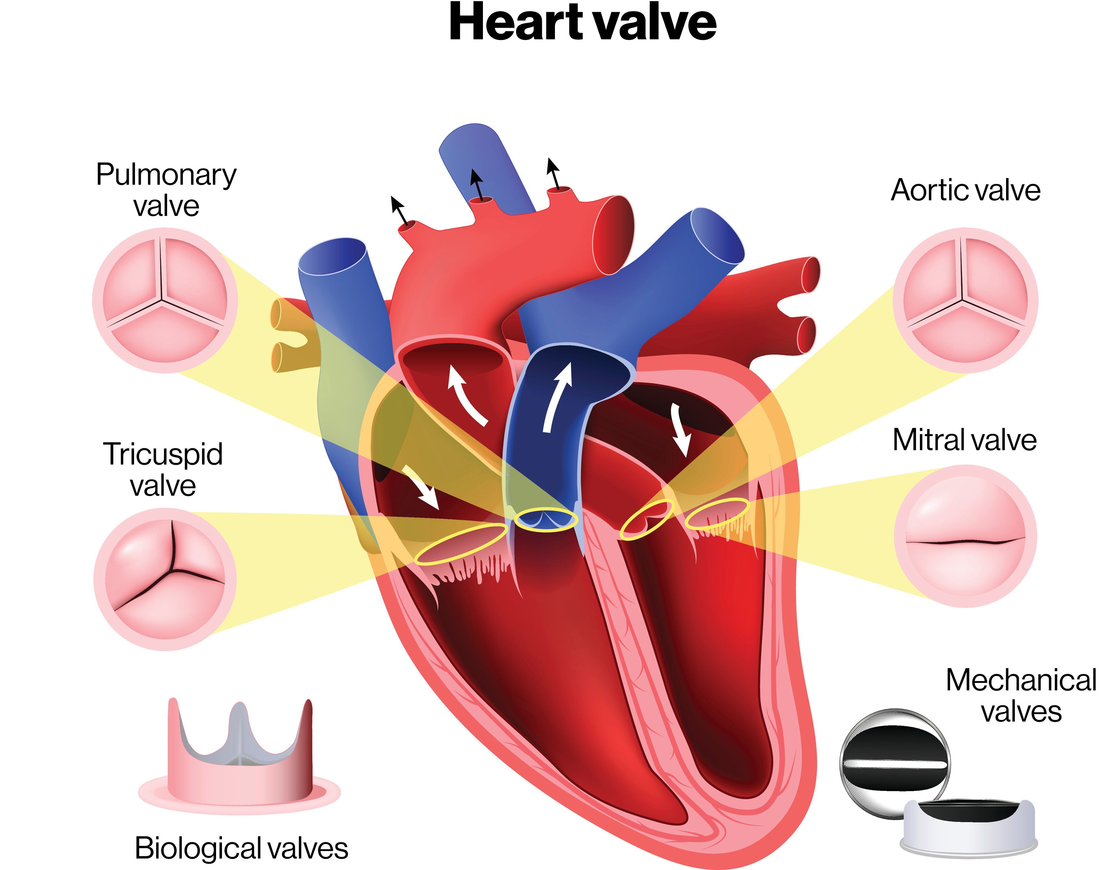
what are the classifications of valvular defects?
stenosis
valvular incompetence
valve prolapsa
what is stenosis? what does this cause, and what causes the murmur?
narrowing
cause: harder for blood to flow through
murmur: blood forced throw narrow opening = whooshing sound
nutshell: less blood flows through, but there is no backflow
what is valvular incompetence? what does this cause, and what causes the murmur?
regurgitation
cause: valve leaks when it should be closed
murmur: abnormal heart sound caused by blood leaking backward through the valve
nutshell: blood flows freely forward, but blood regurgitates backwards through the leaky valve
what is valve prolapse?
floppy valve
one or more of the leaflets bulge backward into the preceding chamber
what are the structural defects in tetralogy of fallot?
a R —> L shunt congenital heart condition
what are the hemodynamic alterations in tetralogy of fallot?
shunt bypasses pulmonary circulation —> cyanosis
pressure in heart changes —> alters blood flow
right heart —> lungs, but less blood traveling here —> deoxygenated blood goes to the left heart —> deoxygenated blood goes to the body —> back to the right heart
what are the 4 structural abnormalities in tetralogy of fallot?
pulmonary stenosis
narrowed pulmonary valve
ventricular septal defect (VSD)
hole in the ventricular septum
dextroposition of the aorta
aorta positioned over the VSD
right ventricular hypertrophy
thickened wall of right ventricle because of increased workload

define pericarditis:
inflammation of pericardium, the double layered membrane surrounding the heart
what is pericarditis usually a result of?
secondary to another condition or injury
open heart surgery
MI
rheumatic fever
SLE
cancer
what are signs and symptoms of pericarditis?
distended neck veins
faint heart sounds
fluid surrounding heart muffles sounds
cardiac tamponade
pressure around heart limits ability to fill with blood
describe the pathogenesis of pericarditis:
fluid around heart compresses heart wall —> heart cannot expand to fill —> blood backs up into systemic circulation —> decreased blood flow to lungs —> decreased output to body
define chronic pericarditis:
long-term inflammation of pericardium
describe pathologic changes associated with chronic pericarditis:
long-term inflammation of pericardium —> formation of adhesions in the pericardial cavity —> adhesions restrict heart’s movement —> reduced CO
what are symptoms of chronic pericarditis?
symptoms associated with systemic venous congestion & decreased CO
SoB
edema
ascites
what is rheumatic fever?
an acute systemic inflammatory condition from an abnormal immune reaction after an untreated infection of group A beta-hemolytic streptococcus
within what age group is rheumatic fever most common?
5-15 y/o