Bio Test 2: Cells
 Call Kai
 Learn
Learn Practice Test
 Spaced Repetition
 Match
Match Flashcards
 Knowt Play
1/96

There's no tags or description
Looks like no tags are added yet.
Name | Mastery | Learn | Test | Matching | Spaced | Call with Kai | Chat |
|---|
No analytics yet
Send a link to your students to track their progress
97 Terms
the central dogma of molecular biology
describes the way in which information flows in a cell (DNA→RNA→protein)
transcription
occurs in nucleus, DNA sequence is copied to a complementary RNA sequence, will produce a pre-mRNA molecule that must be processed

gene
is a sequence of DNA that contains the information to make one polypeptide
promoter
site of transcription initiation of gene
terminator
site of transcription terminator of gene
exons
coding sequence, ends up in RNA
introns
non-coding sequence, removed during RNA processing
RNA polymerase
enzyme, synthesizes RNA using a strand of DNA as a template, reads from the 3’ to 5’ direction and new nucleotides are added to the 3’ carbon end of the growing mRNA, the RNA polymerase binds to the promoter and unwinds the DNA (hydrogen bonds broken), RNA polymerase and the RNA transcript are released from the DNA when the terminator site is reached
Messenger RNA (mRNA)
one strand of DNA is copied to a complementary mRNA strand by RMA polymerase
template strand of DNA
used by RNA polymerase to generate mRNA and is complementary to the DNA, a template for transcription
coding strand of DNA
has the same polarity and sequence as mRNA (except mRNA has uracil instead of thymine)
mRNA processing
occurs in nucleus, introns are spliced out by proteins, exons are joined to make mature mRNA, the mRNA is capped (5’: a chemically modified guanosine triphosphate (GTP) which facilitates binding to a ribosome and protects mRNA from being digested)(3’: 3’ poly A tail that is a long sequence of adenine nucleotides which helps export mRNA from nucleus and stabilizes mRNA)

alternative splicing
allows different mRNAs, and therefore different polypeptides, to be made from one gene, different exons are spliced together to create different mature mRNAs from the same pre-mRNA strand
translation
occurs in cytosol, the RNA sequence is the template for an amino acid sequence
ribosomes
composed of ribosomal RNA (rRNA) and protein, has a large and small subunit, reads mRNA in the 5’ to 3’ direction to produce a protein, puts amino acids on a growing polypeptide chain as it translates along the mRNA
codon
sequence of three bases in mRNA that specifies a particular amino acid (signals for start-AUG codes for methionine- and stop-have no amino acid- are codons as well)
genetic code
specifies which amino acids will be used to build a protein, it is redundant, it is not ambiguous, and it is nearly universal
transfer RNA (tRNA)
links mRNA codons with specific amino acids, they bind to particular amino acids through a covalent attachment, tRNA becomes charged when it carries an amino acid, they bind to the mRNA through the anticodon, which is complementary to the mRNA codon for the amino acid the tRNA carries
A site
binds with the anticodon of charged tRNA
P site
where the tRNA adds its amino acid to the growing chain
E site
where the uncharged tRNA (which has given up its amino acid) sits before being released from the ribosome
during translation initiation
small ribosomal subunit binds to its recognition sequence on the mRNA, methionine-charged tRNA binds to the start codon, the large ribosomal subunit joins the complex and the methionine charged tRNA is in the P site
during translation elongation
a charged tRNA enters the A site; the anticodon of the tRNA binds to the codon at the A site, a polypeptide bond is formed between the amino acids in the P site and the A site, the ribosome shifts down one codon, a new charged tRNA enters the A site; the uncharged tRNA is released from the E site, the polypeptide is transferred to the amino acid on the A site tRNA, the ribosome shifts down one codon (the process repeats)

during translation termination
when a stop codon enters the A site, a release factor binds, binding of the release factor disconnects the polypeptide from the complex, the mRNA and ribosomal subunits separate
cell theory
unifying principle of biology, cells are fundamental units of life, all living organisms are composed of cells, all cells come from pre-existing cells, modern cells evolved from a common ancestor
light microscopes
can be used to visualize living cells and internal structures, glass lenses and visible light are used to form an image (ex: phase-contrast microscopy, stained bright-field microscopy, fluorescence microscopy)

electron microscopes
can be used to visualize the many details of subcellular structures, electromagnets are used to focus an electron beam that are directed at a digital camera (ex: transmission electron microscopy, scanning electron microscopy)
cell membrane
outer boundary of every cell, is a phospholipid bilayer with proteins embedded, transport of nutrients and waste occurs there, encloses cells (intake of nutrients and release of waste happens faster with a higher surface area to volume ratio)

cytoplasm
everything in the cell except for the nucleus
cytosol
fluid cytoplasm not contained inside another cellular compartment
prokaryotic cells
have no membrane-enclosed internal compartments, transcription and translation happen at the same time and place (ex: bacteria, archaea) (includes: capsules, nucleoids, flagellums, cell walls)
nuceloid
DNA is stored in the nucleoid region of prokaryotes (not compartment like nucleus)
cell wall
rigid cell wall of most prokaryotes that is outside of cell membrane, made of peptidoglycan (polymers of sugars that are linked by short peptides
capsule
some prokaryotes have a slimy capsule outside of the cell wall made of polysaccharides , keeps cells from drying out, involved in adhesion to other cells, protects cells from detection by host immune systems
flagella
some prokaryotes swim using flagella, made of the protein flagellin, connected by motor that spins the flagella

pili
some prokaryotes, hairlike structures projecting from the cell surface, they help to adhere to other cells
eukaryotic cells
have membrane enclosed compartments called organelles that carry out specific functions
nucleus
DNA stored here, surrounded by the nuclear envelope (composed of two membranes), nuclear pores in the envelope control movement of molecules across envelope, the outer membrane is continuous with the endoplasmic reticulum
the endoplasmic reticulum (ER)
is a network of membranes in the cytoplasm with a large surface area

rough endoplasmic reticulum (RER)
is “rough” because ribosomes attached to the membrane, newly made proteins enter the RER and are modified-folded-and transported to other areas of the cell
smooth endoplasmic reticulum (SER)
lacks ribosomes, synthesis of lipids and steroids, chemically modifies small molecules, site of glycogen degradation in animal cells, stores calcium ions
the Golgi Apparatus
is composed of cisternae (flattened membranous sacs that make up the Golgi Apparatus) and receives proteins from the RER and modifies-packages-and transports them, 3 regions (cis face-same side of ER, medial face, and trans face-opposite side of ER)

Lysosomes
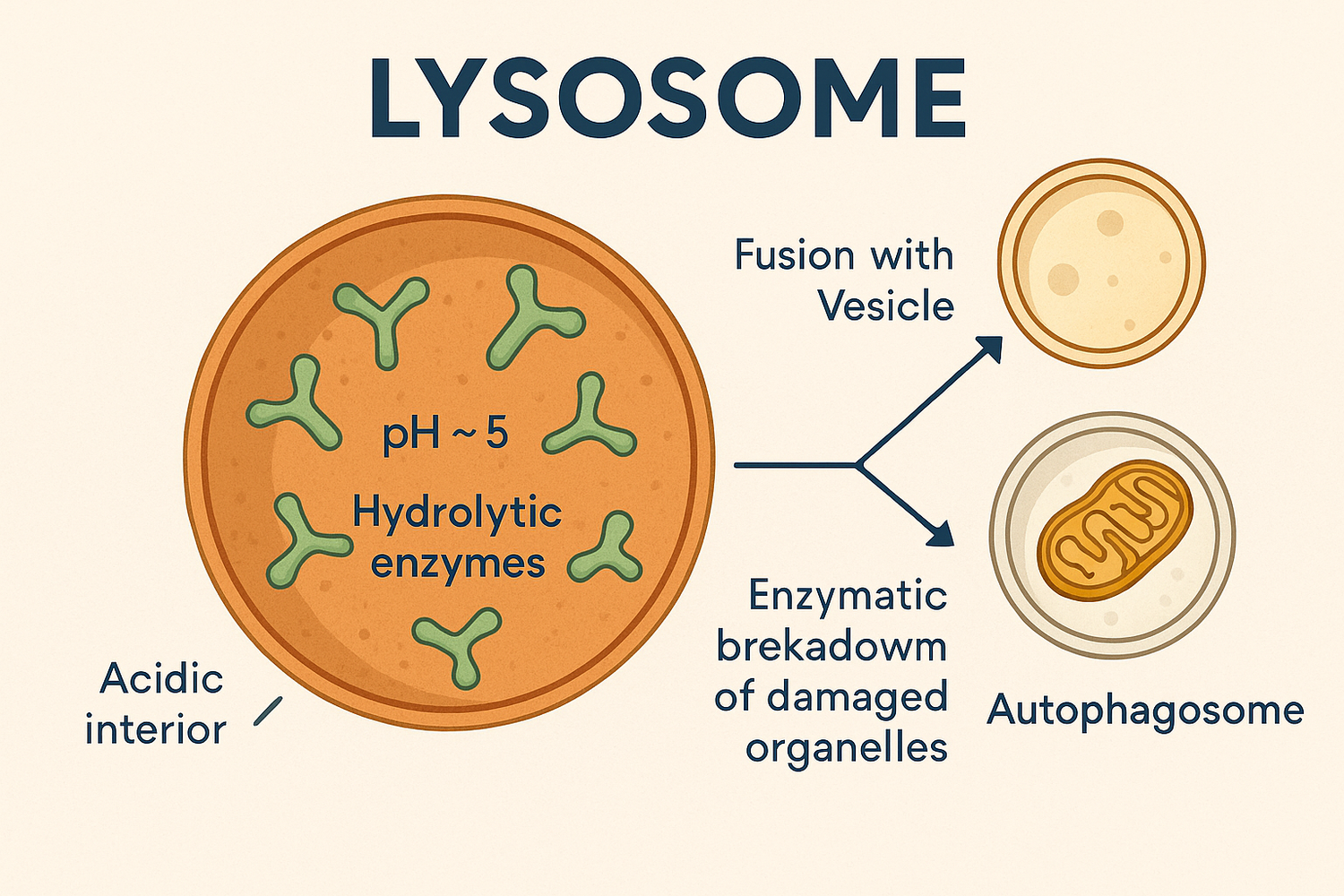
contain digestive enzymes that hydrolyze macromolecules into monomers, interior is acidic with a pH of 5, primary lysosomes originate from the Golgi, secondary lysosomes form from the fusion of the primary lysosome with the phagosome (phagocytosis: process where a cell uses its cell membrane to enclose and engulf a macromolecule or particle in the extracellular environment to form a small vesicle called a phagosome
mitochondria
harvest chemical bond energy in the form of adenosine triphosphate (ATP) from glucose, cells that require a lot of ATP have many mitochondria, have their own DNA, can divide independently of nucleus, two membranes (inner membrane folds inward to form cristae- creates large surface area for the embedded proteins involved in making ATP), the mitochondrial matrix contains DNA and ribosomes to make proteins, very dynamic

chloroplasts
help provide plant cells with energy, have their own DNA, can divide separately from nucleus, convert light energy to ATP through photosynthesis (chlorophyll: green pigment found in chloroplasts)

endosymbiotic theory
mitochondria and chloroplasts were formerly small prokaryotes that were engulfed by larger cells
plant vacuoles
provide structure, aid in digestion, and store water or waste, plants have large vacuoles, sometimes animals have small vacuoles
plant cell wall
is made of cellulose and protects and provides structure to the cell
free ribosomes
not associated with any membrane and translate proteins in the cytoplasm, protein synthesis always begins on free ribosomes in the cytosol, signal sequences on proteins determine if the ribosome remains in the cytosol or attaches to the RER
signal sequence
a short stretch of amino acids attached to a polypeptide (usually the N-terminus) that directs the protein to a particular organelle (ex: a nuclear localization signal directs a protein to the nucleus)
from the RER
when the signal sequence emerges from the ribosome, the entire polypeptide-ribosome-mRNA complex is transported to the membrane of the RER, elongation of the polypeptide continues as it is channeled to the RER, destinations after the RER is RER, Golgi Apparatus, lysosomes, cell membrane, or secreted from the cell
endomembrane system
is an interconnected system of membrane-enclosed compartments (including: nuclear envelope, ER, Golgi apparatus, lysosomes, cell membranes)
vesicles
are small, membrane-enclosed compartments that can carry molecules between components of the endomembrane system, they bud off one compartment carrying cargo molecules, then they fuse with another compartment releasing cargo molecules
going through the Golgi apparatus
the vesicles from the RER fuse with the cis face of the Golgi apparatus, then they may bud off the trans face of the Golgi apparatus (destination could be lysosome, cell membrane, or secreted from cell)
released or fused to cell membrane
molecules in the vesicle can be released outside, molecules embedded in the membrane of the vesicle can be embedded in the cell membrane
cytoskeleton
is a network of different protein filaments in the cytosol that provides cells with structure and facilitates their movement, supports and maintains cell shape, holds organelles in position, moves organelles, interacts with extracellular structures to hold the cell in place, three types of filaments (actin monomers or microfilaments, intermediate, microtubules), cytoskeletal polymers are dynamic, can reorganize the cytoskeletal structure, polymers assemble from monomer subunit and undergo self-assembly
microfilaments
are long coiled chains of the protein actin, maintain cell shape, move cells via muscle contraction or crawling (actin and the motor protein myosin interact to cause movement), dynamic (cells can adapt a variety of shapes), divide animal cells in two, move organelles and cytoplasm in plants, fungi, and animals, actin monomers have a plus and minus side so the microfilament polymer also has a plus or minus, actin monomers bind together head-to-tail, actin and myosin form a contractile ring that separates two newly divided cells, microfilaments form a meshwork just inside the cell membrane that provides structural support (intestinal cells)

motor proteins
proteins that bind to cytoskeletal filaments and use ATP as energy to move along them when the myosin head attaches to actin and uses ATP to move, the actin filament slides (drives muscle contractions)
intermediate filaments
are rope-like structures made up of intermediate filament proteins, many different kinds and each has a different composition (like keratins or lamins), anchoring cell structures in place and resisting tension, maintain cell shape, don’t have distinct ends, very stable

microtubules
are long hollow cylinders (polymers) made of dimers of the protein tubulin, form a rigid internal skeleton, act as “train tracks” for motor proteins (kinesin and dynein) to move vesicles or organelles from one part of the cell to the other (move cargo to other parts of the cell), move chromosomes during cell division, have plus and minus ends with alpha tubulin and beta tubulin ends, kinesin moves toward the plus end of the microtubule, dynein moves toward the minus end of the microtubule

fluid mosaic model
proteins suspended in the phospholipid bilayer move freely in the plane of the bilayer, the mosaic refers to the phospholipids-proteins-and carbohydrates that make up the membrane

membrane fluidity
determines how easily lipids and proteins can move laterally in the plane of the membrane, flip-flop movement is a phospholipid moving from inside to outside of the cell membrane or vice versa
increasing membrane fluidity
more unsaturated fatty acids because they are not as densely packed and rigid as saturated fatty acids
more shorter fatty acids than longer fatty acids
only animals: lower cholesterol is more fluid because of the less van der Waals forces
higher temperature or kinetic energy makes it more fluid
integral membrane protein
are at least partially embedded in the bilayer, inside of cell and outside is hydrophilic domains, with phospholipid tails is hydrophobic domain, some are transmembrane proteins, transmembrane proteins extend all the way through the phospholipid bilayer, a transmembrane domain is a part of a protein that extends all the way through the phospholipid bilayer (expect to see hydrophobic R groups)

anchored membrane proteins
are covalently attached to fatty acids or other lipids

peripheral membrane proteins
lack exposed hydrophobic regions and do not penetrate the phospholipid bilayer, interact with the membrane surface or an integral membrane protein so that the peripheral membrane protein can travel to the other side

carbohydrate attachments
carbohydrates can be added to protein or lipid components of membranes (glycoprotein: carbohydrate + protein)(glycolipid: carbohydrate + lipid)
cell recognition
one cell specifically recognizes and binds to another cell of a certain type
cell adhesion
the connection between the two cells is strengthened, (homotypic: the same molecule extends from both cells and bind to each other) (heterotypic: the cells have different molecules that bind together), in both cases the exposed surfaces of the molecules fit together and have a chemical affinity for one another
cell junctions
are specialized structures that hold cells together
tight junctions
form a tight seal between adjacent cells, this blocks the movement of materials through the space between them

desmosomes
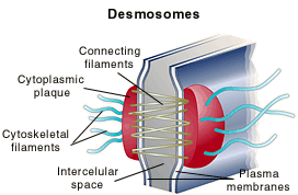
hold cells together while still allowing materials to move around in the intercellular space between them
gap junctions
form tunnels between adjacent cells so they can communicate by exchanging small molecules

the extracellular matrix (ECM)
is a macromolecule-rich gel outside of cells
integrins
are transmembrane proteins that attach to the ECM outside of the cell (extracellularly) and actin microfilaments inside of the cell (intracellularly), cells can move within a tissue by the binding and reattaching of integrin receptors to the ECM
cell membrane selective permeability
some substances can pass through but not others, some cross the membrane through diffusion with net movement from higher concentrations to lower concentrations
diffusion depends on
concentration gradient (more with a steep gradient)
temperature (more with higher temperatures)
molecule size (more with smaller molecules)
surface area (more with more area of membrane)
diffusion distance (more with thinner membrane)
membrane permeability (more with more permeable membrane)
passive transport
a way to cross a membrane that does not require energy input and transports substances down a concentration gradient
simple diffusion
small molecules pass through the lipid bilayer
osmosis
water diffusing across a membrane (ex: isotonic, hypotonic, and hypertonic)
isotonic
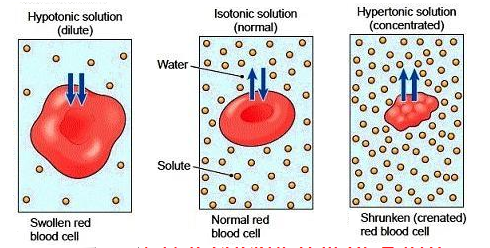
equal solute concentrations inside and outside the cell (net movement of water is 0)
hypotonic
lower solute concentration outside the cell (more water diffuses into the cell) (animal cells can burst)

hypertonic
higher solute concentration outside the cell (more water diffuses to the outside of the cell) (animal cells can shrink)

facilitated diffusion
is carried out by protein channels or carriers that increase the rate of diffusion
channel proteins
are integral membrane proteins that form channels across the membrane through which certain substances can pass (ex: aquaporins allow water to pass through the membrane)

ion channels
are integral membrane proteins that have hydrophilic pores, most are gated (can be opened or closed to ion diffusion), the gate opens when the protein is stimulated to change conformation (shape), change in shape can be stimulated by binding of a chemical signal, a difference in electrical charge, or a mechanical signal

carrier proteins
bind substances and speed up their diffusion through the phospholipid bilayer, transport polar molecules, such as glucose, across membranes in both directions, causes molecules to change conformation

active transport
to cross membranes requires energy to move substances against a concentration and/or electrical gradient, energy often comes from ATP hydrolysis, it is directional (ex: uniporter, symporter, and antiporter)
uniporter
moves one substance in one direction

symporter
moves two substances in one direction

antiporter
moves two substances in opposite directions

primary active transport
requires direct hydrolysis of ATP to drive movement of specific ions against their concentration gradient
secondary active transport
energy comes from an ion concentration gradient that is established by primary active transport
endocytosis and exocytosis
for molecules that are too big to cross the membrane, eukaryotic cells may take up and release fluids, large molecules, and smaller cells via endocytosis or exocytosis
phagocytosis
molecules or entire cells are engulfed
pinocytosis
a vesicle forms to bring small dissolved substances or fluids into a cell
receptor-mediated endocytosis
molecules at the cell surface recognize and trigger uptake of specific molecules (highly specific), molecules to be moved bind to receptor proteins-integral membrane proteins located on the cell membrane