ch 18, 19, 20 Cardiovascular
 Call Kai
 Learn
 Practice Test
 Spaced Repetition
 Match
 Flashcards
 Knowt Play
1/75

There's no tags or description
Looks like no tags are added yet.
76 Terms
Blood is:
connective tissue
sample of whole blood consits of
plasma and formed elements
plasma compositition
water
plasma proteins
other solutes
plasma proteins
albumins
globulins
fibrinogen
regulatory proteins
other solutes of plasma
electrolytes
organic nutrients
organic wastes
albumins
major contributors to osmotic pressure of plasma; transport lipids, steroid hormones
globulins
transport ions, hormones, lipids; immune function
fibrinogen
essential component of clotting system; can be converted to insoluble fibrin
regulatory proteins
enzymes, proenzymes, hormones
electrolytes
ions contribute to osmotic pressure of body fluids
organic nutrients
used for ATP production; growth and cellular maintenance; include lipids, carbs, and amino acids
organic wastes
carried to sites for breakdown or excretion; include urea, uric acid, creatinine, bilirubin, ammonium ions
formed elements of plasma
red blood cells
white blood cells
platelets
leukocytes under microscope
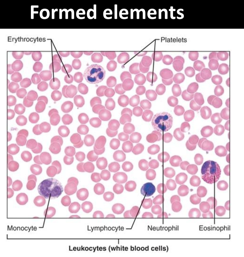
Two groups of Leukocytes
are granulocytes and agranulocytes.
granulocytes
neutrophil
eosinophil
basophil
agranulocytes
monocytes and lymphocytes
neutrophil
multilobed nucleus, pale red and blue cytoplasmic granules

eosinophil
bilobed nucleus, red cytoplasmic granules
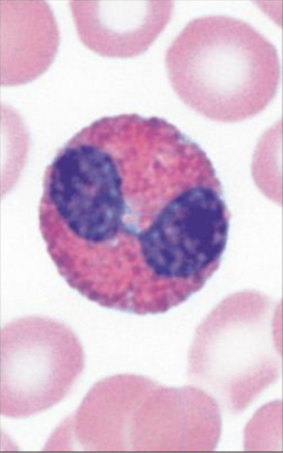
basophil
bilobed nucleus, purplish-black cytoplasmic granules

lymphocyte
large spherical nucleus, thin rim of pale blue cytoplasm

monocyte
kidney-shaped nucleus, abundant pale blue cytoplasm

Leukocytes size
7000
5,000-10,000 μL
Platelets size
350, 000
150,000-500,000 μL

Erythrocytes
have no organelles or nuclei
are packed with oxygen-carrying
hemoglobin
• Most numerous of the formed
elements
females RBCs
Females: 4.3–5.2 million cells/cubic
millimeter
males RBCs
Males: 5.1–5.8 million cells/cubic
millimeter
Oxidation of iron atoms
Oxidation of iron atoms of
hemoglobin molecules gives blood its
red color
Hematopoiesis
100 billion new blood cells
formed each day!!
Where is hematopoiesis?
blood marrow, which is protected by spongy bone

heart anatomy diagram

The Heart is located
between
two pleural cavities.
It is surrounded by the pericardial cavity.
Located near the anterior chest wall, posterior to the sternum, anterior to the vertebral column.

The ____ cavity surrounds the heart
The pericardial cavity surrounds the heart

Fluid in the pericardial cavity
Pericardial cavity contains 50 ml of pericardial fluid; protects the heart
What forms the heart wall?
The heart wall is formed by three layers: the epicardium (visceral layer of pericardium), myocardium, and endocardium.

Three membranes enclosed the heart
pericardium, myocardium, and endocardium

Pericardium
The membrane enclosing the heart, consisting of two layers: the fibrous pericardium and the serous pericardium.
(serous membrane)
formed by two layers
epicardium
parietal and visceral
Mesothelium and areolar
tissue
Endocardium
Endothelium and areolar tissue
Myocardium
Cardiac muscle

Four chambers
of the heart, consisting of the left and right atria, and left and right ventricles, which facilitate blood circulation throughout the body.

Atrium
receives blood returing to the heart
superior chambers
Ventricle
pumps blood out into the blood vessels (Thicker walls)
inferior chambers
Borders of internal chambers
Sulci or grooves show approximately borders of the external chambers
surrounded by fat

Areas of fat in the heart
coronary sulcus
anterior and posterior interventricular sulcus

Location of coronary sulcus
Located between the atria and ventricles, it encircles the heart and contains coronary arteries.

Location of anterior and posterior interventriculus sulci
The anterior interventricular sulcus is located on the anterior surface of the heart, while the posterior interventricular sulcus is situated on the posterior aspect, both defining the boundaries between the left and right ventricles.

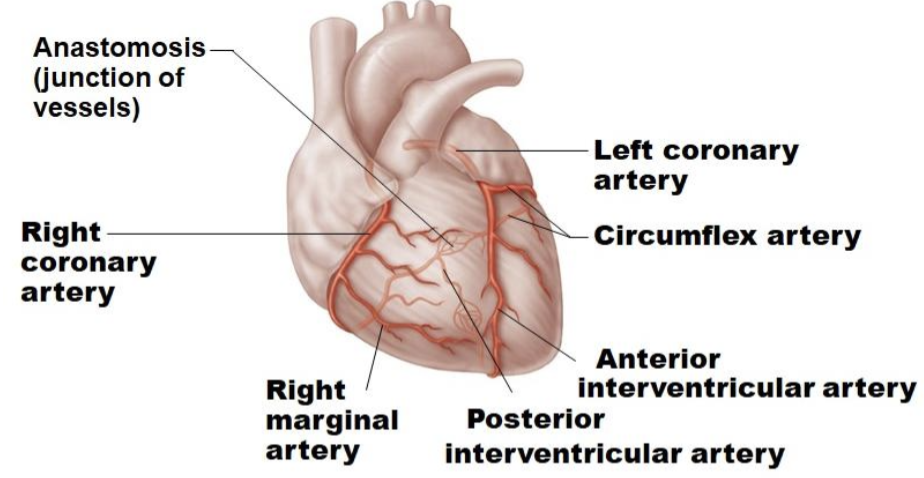
anastomosis
junction of vessels
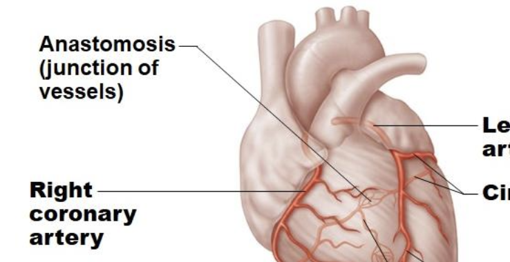
Coronary arteries
left and right coronary artery
circumflex artery
anterior and posterior interventricular artery
right marginal artery
Cardiac veins
anterior cardiac veins
small cardiac vein
great cardiac vein
coronary sinus
middle cardiac vein

How do recirculate blood flow when arteries are clogged?
grafts: surgically created connections that bypass blocked arteries, restoring blood flow.
bypass: a procedure to reroute blood around blocked arteries.
stents: devices placed in arteries to keep them open and improve blood flow.
epicardium aka
the visceral layer of pericardium

structures of the heart: ventricles and chambers

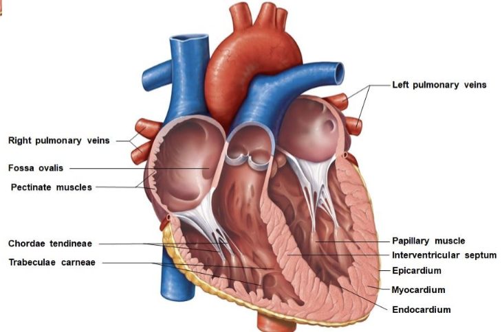
structures of the heart: layers

Two sets of heart valves:
Atrioventricular valves
Semilunar valves.
Atrioventricular valves
AV valves.
(between the atria and ventricles).
Tricuspid (R) and Mitral (L)

Semilunar valves
(between ventricles and arteries).
Aortic and Pulmonary

Cardiac valves are folds of ______.
Cardiac valves are folds of endocardium.
heart valve function
Heart valves prevent the backward flow of blood

AV valves open

AV valves close

Semilunar valves open

Semilunar valves close
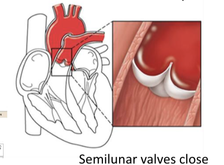
Ventricular contraction:
semilunar valves and AV valves
semilunar: open
AV: closed

Ventricular relaxation:
semilunar valves and AV valves
semilunar: closed
AV: open

The Atria contract together, then Ventricles contract

Digram of blood flow

ventricular contraction

Follow the order of a drop of blood flow, starting from the vena cava and ending at the aorta
vena cava
right atrium
tricuspid AV valve
right ventricle
pulmonary valve
pulmonary artery
lungs (gas exchange)
pulmonary veins
left atrium
mitral AV valve
left ventricle
aortic valve
aorta

Pulmonary circuit
Transports blood from the heart to the lungs.

What structures are involved in pulmonary circuit primarily?
pulmonary trunk
left pulmonary arteries
left pulmonary veins

pulmonary circuit
Oxygen-poor blood leaves the right ventricle→
Oxygen-poor blood leaves the right ventricle→
passing through the pulmonary valve→
left and right pulmonary arteries→
alveolar capillaries→
pulmonary veins→
left atrium.
Systemic circuit
Transports blood between the heart and all other tissues.

systemic circuit: blood and oxygen
Blood on the left side of the heart enters the systemic
circulation.
Oxygenated blood leaves the left ventricle by passing through the aortic valve
right side of heart uses what type of circulation?
pulmonary circulation

left side of heart uses what type of circulation?
systemic circulation
